Review
. 2018 Apr;8(1):10.1128/ecosalplus.ESP-0004-2017.
doi: 10.1128/ecosalplus.ESP-0004-2017.

Molecular Epidemiology of Extraintestinal Pathogenic Escherichia coli

Affiliations
Review

Molecular Epidemiology of Extraintestinal Pathogenic Escherichia coli

James R Johnson et al. EcoSal Plus. 2018 Apr.

Abstract

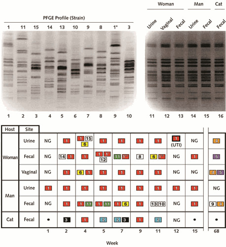
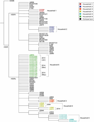
Extraintestinal pathogenic Escherichia coli (ExPEC) are important pathogens in humans and certain animals. Molecular epidemiological analyses of ExPEC are based on structured observations of E. coli strains as they occur in the wild. By assessing real-world phenomena as they occur in authentic contexts and hosts, they provide an important complement to experimental assessment. Fundamental to the success of molecular epidemiological studies are the careful selection of subjects and the use of appropriate typing methods and statistical analysis. To date, molecular epidemiological studies have yielded numerous important insights into putative virulence factors, host-pathogen relationships, phylogenetic background, reservoirs, antimicrobial-resistant strains, clinical diagnostics, and transmission pathways of ExPEC, and have delineated areas in which further study is needed. The rapid pace of discovery of new putative virulence factors and the increasing awareness of the importance of virulence factor regulation, expression, and molecular variation should stimulate many future molecular epidemiological investigations. The growing sophistication and availability of molecular typing methodologies, and of the new computational and statistical approaches that are being developed to address the huge amounts of data that whole genome sequencing generates, provide improved tools for such studies and allow new questions to be addressed.


Figures

Figure 1
Figure 1
PCR analysis of pap operon. Open boxes represent genes within the pap operon (including papA, structural subunit; papC, usher; papEF, minor tip pilins; and papG, adhesin). Forward and reverse primers (right- and left-pointing black triangles, respectively, above and below the pap operon) are used in combinations as shown to yield the indicated PCR products (thin rectangles, below pap operon). Heavily striped rectangles, papA and papG allele PCR products. Solid black rectangles, pap gene PCR products. Finely striped rectangles, long PCR operon fragments (as generated using either flanking or internal allele-specific papG reverse primers, as illustrated for allele I-I′). Different papA and papG variants are associated with specific lineages, hosts, and clinical syndromes. Intraoperonic deletions that yield a null phenotype (which may be associated with compromised or asymptomatic hosts) can be detected as a truncated long-PCR product. Reprinted from reference , with permission.
Figure 2
Figure 2
Dendrogram based on pulsed-field gel electrophoresis (PFGE) profiles of 33 clinical and fecal isolates of Escherichia coli sequence type 131 (ST131) from the members of 6 households. Profiles are diverse, despite all isolates deriving from the same ST, which reflects the superior resolving power of PFGE over MLST. Isolates from a given household cluster together, consistent with intrahousehold strain sharing. Scale is % profile similarity. All isolates were fluoroquinolone-resistant. H30, clonal subset within the ST131-H30 clade (R1 = H30R1, Rx = H30Rx). Abbreviations: ESBL, extended-spectrum β-lactamase production; HH, household; ID, identifier; PFGE, pulsotype. Reprinted from reference , with permission.
Figure 3
Figure 3
Random amplified polymorphic DNA (RAPD) analysis of E. coli strains 536, NU14, and RS218. RAPD profiles generated by using primer 1247 (12) show E. coli O18:K1:H7 strains NU14 (cystitis: lane 3) and RS218 (neonatal meningitis: lane 4) to be indistinguishable from one another, but distinct from strain 536 (O6:K15:H31, pyelonephritis: lane 2), illustrating both the broad syndrome capability of certain ExPEC lineages and the clonal diversity of urinary tract infection-causing ExPEC strains. M (lanes 1 and 5), 100-bp marker. Reprinted from reference , with permission.
Figure 4
Figure 4
RAPD-based phylogenetic and clonal analysis of Escherichia coli isolates. Genomic profiles (shown in computer reconstruction), as generated for each isolate by using RAPD primers 1247, 1254, 1281, and 1283, were concatenated for cluster analysis. Pyelonephritis isolates (n = 10; “Py” strain designations) are labeled in bold if from E. coli clonal group A (CGA) (n = 5) and in lightface italic if non-CGA (n = 5). CGA isolates (bold) are bracketed and labeled as to syndrome (CY, cystitis; PY, pyelonephritis) and serogroup (O11/O17/O77) (right), with the corresponding cluster shown in bold (left). The two E. coli O15:K52:H1 control strains are bracketed and labeled by serotype. Reference strains from the E. coli Reference (ECOR) collection (bold) are identified as to phylogenetic group (right). The depth of the molecular weight ladder cluster (brackets; MW) reflects the intrinsic variability inherent in gel electrophoresis and image analysis, independent of amplification. The CGA isolates cluster together irrespective of clinical syndrome (pyelonephritis, cystitis) and geography (UCB: Berkeley, California; UMN: Minneapolis, MN; Py: multiple centers around the U.S.). Reprinted from reference , with permission.
Figure 5
Figure 5
Time-scaled whole genome sequence (WGS)-based phylogeny of ST131 Escherichia coli (n = 215). Meta-data include allelic variants of blaCTX-M (extended-spectrum beta-lactamase gene) and fimH (type-1 fimbriae adhesin gene), plus mutations in the quinolone resistance-determining region (QRDR) of gyrA (WT, wild-type). Brackets identify defined ST131 clonal subsets. Branch tips are colored by geographic region, per the key. T, bla plasmid transformant generated for strain; *, cases with putative deletions in the assembled bla gene. Geographic clustering is evident, some of it linked with specific accessory gene variants; e.g., within the C1/H30R clonal subset, the Southeast Asian isolates (green) are largely confined to a specific clade that consists of two subclades, one characterized by blaCTX-M-14 and the other by blaCTX-M-27. Reprinted from reference , with permission.
Figure 6
Figure 6
Phylogenetic distribution of extraintestinal pathogenic Escherichia coli (ExPEC)-associated virulence traits. Dendrogram at left depicts phylogenetic relationships for the 72 members of the E. coli Reference (ECOR) collection, as inferred based on multilocus enzyme electrophoresis (67). The four traditional major E. coli phylogenetic groups, i.e., A, B1, B2, and D (now split into groups D and F). The nonaligned (“non”) strains (now called group E) are bracketed and labeled. Bullets at right indicate presence of putative virulence genes (papA, P fimbriae; kpsMT, group II capsule synthesis; sfa/foc, S and F1C fimbriae; iutA, aerobactin system; traT, serum resistance; and fimH, type 1 fimbriae). Horizontal bars at right indicate the 10 ECOR strains isolated from humans with symptomatic UTI. The remaining strains, except for one asymptomatic bacteriuria isolate, are fecal isolates from healthy human or animal hosts. Note the concentration of (chromosomal) ExPEC-defining virulence genes papA, kpsMT, and sfa/foc within phylogenetic groups B2 and D, but their occasional joint appearance also in distant lineages, consistent with coordinate horizontal transfer, giving rise to ExPEC strains in historically non-ExPEC lineages. The more scattered phylogenetic distribution of iutA (ExPEC-defining) and traT is consistent with these two genes’ typically plasmid location, although iutA also can be chromosomal. fimH is nearly universally prevalent, consistent with its presence in other species of Enterobacteriaceae, presumably reflecting an origin in a shared enterobacterial ancestor. Note the concentration of UTI isolates within phylogenetic groups B2 and D and the concentration of virulence genes among UTI isolates. Note also the appreciable minority of fecal isolates with multiple virulence genes, reflecting a fecal reservoir of ExPEC. Reprinted from reference , with permission.
Figure 7
Figure 7
Genome map of ExPEC strain 536. The map is based on the chromosome of E. coli MG1655 (K-12). Pathogenicity islands (PAIs) are indicated according to their chromosomal insertion sites next to tRNA-encoding genes. Contents, by PAI, include: PAI I (α-hemolysin, F17-like fimbriae, CS12-like fimbriae); PAI II (α-hemolysin, P fimbriae with papG III); PAI III (S fimbriae, iro siderophore system, Tsh-like hemoglobin protease); PAI IV (yersiniabactin system). Many additional smaller DNA insertions compared to K-12 are present (not shown). Linkage of virulence genes in PAIs contributes to statistical associations between different virulence genes and between specific virulence genes and the lineages within which the corresponding PAIs tend to occur. Reprinted from reference , with permission.
Figure 8
Figure 8
Maps of pathogenicity-associated islands (PAIs) from ExPEC strain 536. Known or putative open reading frames (ORFs) are grouped according to the following characteristics: blue, functional, known ORFs; green, truncated ORFs with a start codon and a stop codon; gray, as-yet-unidentified ORFs without homologues on the DNA level. Nonfunctional ORFs (e.g., due to internal stop codons or frameshifts) are indicated by hatched symbols. ORF numbers are indicated below the corresponding ORF symbols. Functional or truncated tRNA-encoding genes are marked in red. Direct repeat (DR) structures flanking PAIs are indicated. Thick black lines below the PAIs represent regions that were detected by PCR. Several PAI-specific PCRs were grouped into PAI regions. The molecular epidemiology of novel ORFs that are discovered through sequence analysis of PAIs can be investigated in subsequent studies. Reprinted from reference , with permission.
Figure 9
Figure 9
Receptor binding specificity of type 1 fimbrial adhesin (FimH) variants. In vitro binding of isogenic recombinant strains expressing the Ala-62 or Ser-62 FimH variants (from strains NU14 and 536, respectively) to (A) a trimannose substrate (bovine RNAse B), (B) human collagen type IV, and (C) a monomannose substrate (yeast mannan). Both variants bind equally well to trimannose, but the Ala-62 variant exhibits stronger type IV collagen and monomannose binding than does the Ser-62 variant. (Commensal-associated FimH variants exhibit equally strong trimannose binding but minimal binding to type IV collagen or monomannose [not shown].) Open columns, bacteria incubated without α-methyl mannoside (αmM); solid columns, bacteria incubated with 50 mM αmM. Data are mean + SEM (n = 4) of number of bacteria bound per well. Molecular epidemiological studies can be used to elucidate the likely clinical relevance of such genetic and phenotypic variation within different virulence factors. Adapted from reference , with permission.
Figure 10
Figure 10
Pulsed-field gel electrophoresis (PFGE) profiles and colonization patterns of Escherichia coli isolates from three household members (man, woman, and pet cat). (Top panel) PFGE profiles. Lane numbers are shown below gel images. Lanes 1 through 10, profiles of nine of the unique strains, with strain designations shown above gel lanes, plus subtype 1″) (lane 9). Lanes 11 through 16, profiles of independent isolates of strain 1, as recovered from various anatomical sites from the woman (lanes 11–13), man (lanes 14 and 15), and cat (lane 16). (Bottom panel) Distribution of 14 unique E. coli strains over time (week of sampling shown below grid), as recovered from various anatomical sites from the three household members. NG, no growth; •, no sample. Strains isolated more than once appear in colored boxes, with a unique color for each strain. Strains isolated only once appear in colorless boxes. Week 12, which coincided with symptoms of acute urinary tract infection (UTI) in the woman, yielded strain 1 from the woman’s urine specimen (boldface box). There is no strain 7. Strain 1, the woman’s UTI strain, was the most extensively shared and persistent strain, and had the most virulence genes of the 14 strains. Reprinted from reference , with permission.
Figure 11
Figure 11
Bootstrapped consensus core genome phylogeny for 33 household isolates and 47 comparison isolates of Escherichia coli sequence type 131 (ST131). The tree was based on 1000 bootstrapped maximum likelihood trees, retaining only those nodes that appeared in >70% of trees, and was rooted with strain CD306. Branch lengths are meaningless. Household isolates (as in Fig. 2) are color-coded by household (–6); comparison isolates are shown in black. For the household isolates: boldface indicates clinical isolates; regular font indicates fecal isolates; underlining indicates fecal isolates from a clinical isolate’s source host; and asterisks indicate the 6 household isolates that were included in Price et al (11), i.e., JJ1886, JJ1887, JJ2547, CU758, CU799, and CD364 (which in Price et al was labeled as CD449). Dates are shown for the 2 households that underwent serial sampling (households 4 and 6). Clustering by household supports within-household transmission (strain sharing); near identify of clinical and fecal isolates within each household supports the fecal reservoir as the source for infection-causing strains; and variation in a given strain within its source household over time (households 4 and 5) suggests microevolution during long-term host colonization. Reprinted from reference , with permission.

References

    1. Russo TA, Johnson JR. 2000. Proposal for a new inclusive designation for extraintestinal pathogenic isolates of Escherichia coli: ExPEC. J Infect Dis 181:1753–1754. [PubMed] - PubMed
    1. Russo TA, Johnson JR. 2003. Medical and economic impact of extraintestinal infections due to Escherichia coli: an overlooked epidemic. Microbes Infect 5:449–456. - PubMed
    1. Lipsky BA. 1989. Urinary tract infections in men. Epidemiology, pathophysiology, diagnosis, and treatment. Ann Intern Med 110:138–150. - PubMed
    1. Andriole VT, Patterson TF. 1991. Epidemiology, natural history, and management of urinary tract infections in pregnancy. Med Clin North Am 75:359–373. - PubMed
    1. Rothman KJ, Greenland S. 1998. The Emergence of Modern Epidemiology, 2nd ed. Lippincott-Raven, Philadelphia.

Publication types

Substances

LinkOut - more resources