

Magnitude of cone beam CT image artifacts related to zirconium and titanium implants: impact on image quality
- PMID: 29668300
- PMCID: PMC6196050
- DOI: 10.1259/dmfr.20180021
Magnitude of cone beam CT image artifacts related to zirconium and titanium implants: impact on image quality
Abstract
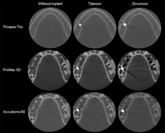
Objectives: To evaluate the magnitude of artifacts related to titanium and zirconium implants at different distances and angulations and their impact on cone beam CT(CBCT) image quality.
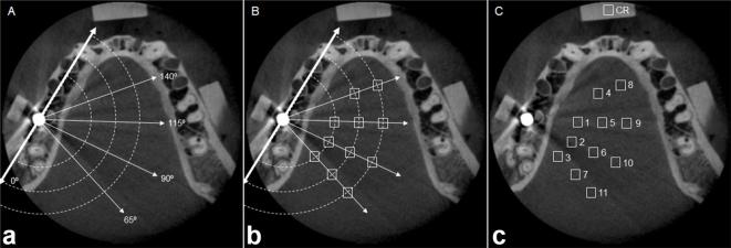
Methods: CBCT images were obtained before and after the insertion of titanium and zirconium implants in a mandible on different CBCT units: Picasso Trio, ProMax 3D and 3D Accuitomo 80. Artifact was assessed by measuring the standard deviation (SD) of gray values and contrast-to-noise ratio (CNR) of 11 regions of interest (ROIs) at different distances (1.5 cm, 2.5 cm and 3.5 cm) and angulations (65°, 90°, 115° and 140°) from implant region.
Results: For titanium images, SD values did not differ from those of images without implant in all ROIs; however, some effect occurred in Picasso images as higher values were observed in ROIs closer to the implant (p < 0.05). Zirconium images showed higher SD values than the others in some ROIs for Picasso and ProMax (p < 0.05). In ProMax, the difference was observed even in the farthest ROIs from the implant. CNR values were not influenced by the ROI in Picasso, but presented lower values in ROIs closer to the zirconium implant for ProMax and Accuitomo.
Conclusions: The quantity and magnitude of artifacts in CBCT are influenced by the type of implant and CBCT unit. Although they are more pronounced in regions closer to the implant and located at 90° in relation to the mandibular long axis, they can reach as far as 3.5 cm from the artifact-generator object.
Figures
References
-
- Tyndall DA, Price JB, Tetradis S, Ganz SD, Hildebolt C, Scarfe WC. . Position statement of the American academy of oral and maxillofacial radiology on selection criteria for the use of radiology in dental implantology with emphasis on cone beam computed tomography. Oral Surg Oral Med Oral Pathol Oral Radiol 2012; 113: 817–26. doi: 10.1016/j.oooo.2012.03.005 - DOI - PubMed
-
- SEDENTEXCT Project. European commission. Radiation protection Nº. 172 sedentexCT. guidelines on CBCT for Dental and Maxillofacial Radiology. Luxembourg: The British Institute of Radiology.; 2012.
-
- Harris D, Horner K, Gröndahl K, Jacobs R, Helmrot E, Benic GI, et al. . E.A.O. guidelines for the use of diagnostic imaging in implant dentistry 2011. a consensus workshop organized by the European association for osseointegration at the medical university of Warsaw. Clin Oral Implants Res 2012; 23: 1243–53. doi: 10.1111/j.1600-0501.2012.02441.x - DOI - PubMed
MeSH terms
Substances
LinkOut - more resources
Full Text Sources
Other Literature Sources
