

Metastatic epithelioid trophoblastic tumor of the lung: A case report
- PMID: 29668580
- PMCID: PMC5916664
- DOI: 10.1097/MD.0000000000010306
Metastatic epithelioid trophoblastic tumor of the lung: A case report
Abstract
Rationale: Epithelioid trophoblastic tumor (ETT) is a very rare form of gestational trophoblastic disease (GTD) which arises from neoplastic proliferation of intermediate trophoblasts. Metastatic ETT of the lung is extremely rare in postmenopausal women.
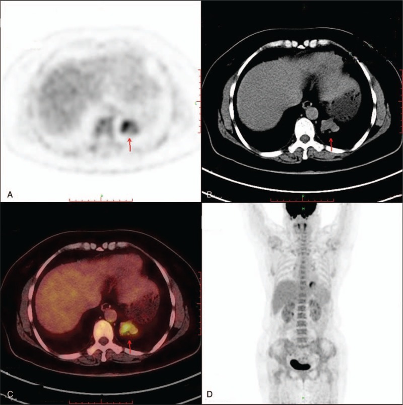
Patient concerns: Here we describe a 50-year-old woman with a metastatic ETT of the lung showing increasing tracer uptake at PET/CT.
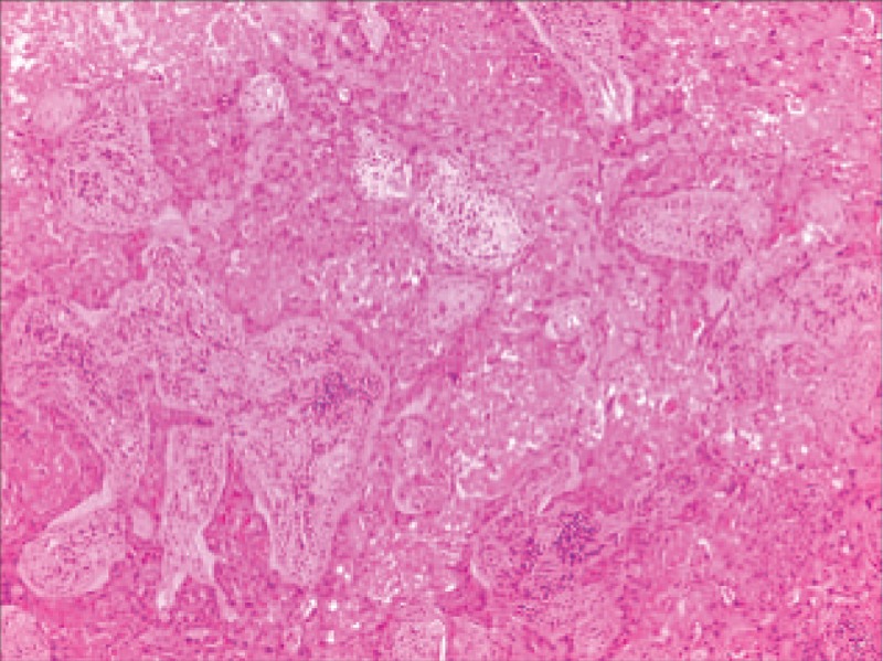
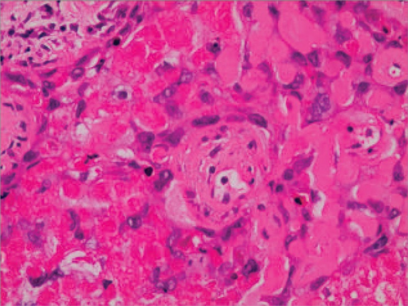
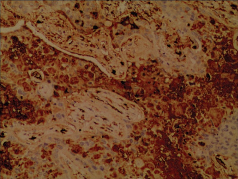
Diagnosis: Hematoxylin-eosin staining showed a tumor composed of nests of epithelioid cells with necrotic debris and peritumoral hyaline-like material. Immunohistochemical staining of the tumor cells was positive for human chorionic gonadotropin (HCG) and cytokeratin 18.
Interventions: The patient underwent thoracoscopic lower left lobectomy combined with mediastinal lymphadenectomy. At surgery, a solid mass (size 3.0 × 3.0 cm) was found in the left lower lung.
Outcomes: The patient was discharged on the tenth day postsurgery, following an uneventful recovery. Three months postsurgery, the patient was asymptomatic and is currently being managed with close follow-up.
Lessons: Metastatic ETT of lung is a very rare disease. Complete surgical resection and chemotherapy may be the critical therapeutic option.
Conflict of interest statement
The authors have no conflicts of interest to disclose.
Figures
References
-
- Shih IM, Kurman RJ. Epithelioid trophoblastic tumor: a neoplasm distinct from choriocarcinoma and placental site trophoblastic tumor simulating carcinoma. Am J Surg Pathol 1998;22:1393–403. - PubMed
-
- Chohan MO, Rehman T, Cerilli LA, et al. Metastatic epithelioid trophoblastic tumor involving the spine. Spine 2010;35:E1072–5. - PubMed
-
- Shih IM, Kumaran RJ. The pathology of intermediate trophoblastic tumours and tumour like lesions. Int J Gynecol Pathol 2001;20:31–47. - PubMed
Publication types
MeSH terms
Substances
LinkOut - more resources
Full Text Sources
Other Literature Sources
Medical
