. 2018 Jul 6;46(12):6344-6355.
doi: 10.1093/nar/gky267.

HITS-CLIP in various brain areas reveals new targets and new modalities of RNA binding by fragile X mental retardation protein

Affiliations

HITS-CLIP in various brain areas reveals new targets and new modalities of RNA binding by fragile X mental retardation protein

Thomas Maurin et al. Nucleic Acids Res. .

Abstract

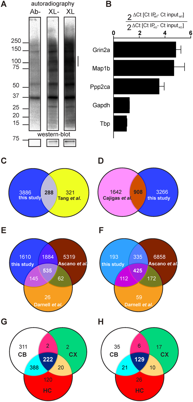
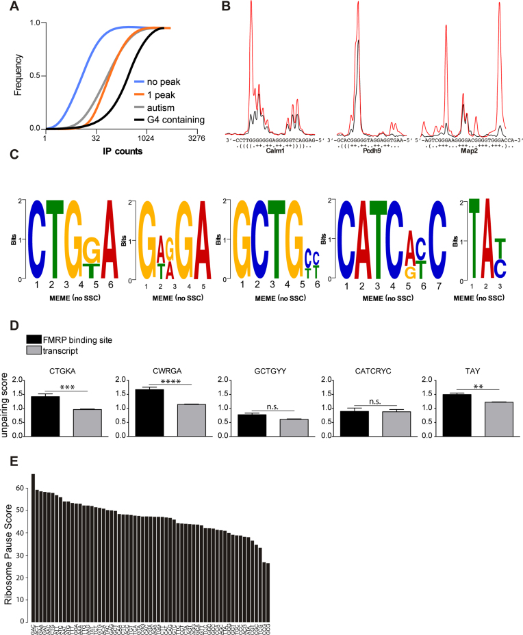
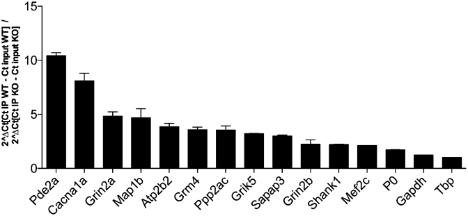
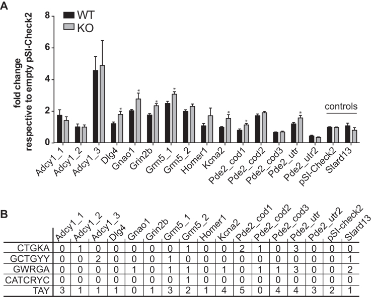
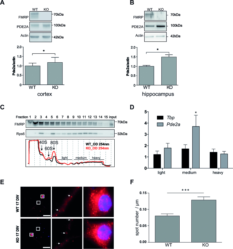
Fragile X syndrome (FXS), the most common form of inherited intellectual disability, is due to the functional deficiency of the fragile X mental retardation protein (FMRP), an RNA-binding protein involved in translational regulation of many messenger RNAs, playing key roles in synaptic morphology and plasticity. To date, no effective treatment for FXS is available. We searched for FMRP targets by HITS-CLIP during early development of multiple mouse brain regions (hippocampus, cortex and cerebellum) at a time of brain development when FMRP is most highly expressed and synaptogenesis reaches a peak. We identified the largest dataset of mRNA targets of FMRP available in brain and we defined their cellular origin. We confirmed the G-quadruplex containing structure as an enriched motif in FMRP RNA targets. In addition to four less represented motifs, our study points out that, in the brain, CTGKA is the prominent motif bound by FMRP, which recognizes it when not engaged in Watson-Crick pairing. All of these motifs negatively modulated the expression level of a reporter protein. While the repertoire of FMRP RNA targets in cerebellum is quite divergent, the ones of cortex and hippocampus are vastly overlapping. In these two brain regions, the Phosphodiesterase 2a (Pde2a) mRNA is a prominent target of FMRP, which modulates its translation and intracellular transport. This enzyme regulates the homeostasis of cAMP and cGMP and represents a novel and attractive therapeutic target to treat FXS.


Figures

Figure 1.
Figure 1.
Identification of mRNA targets of FMRP. (A) Upper panel: audioradiography of immunoprecipitated UV-crosslinked RNPs from 13 PND male mouse brain cortex extracts after separation on a SDS-PAGE gel and then transferred onto a nitrocellulose membrane. Ab-: immunoprecipitation in the absence of the polyclonal anti-FMRP antibody Rb#11; XL-: Immunoprecipitation in the presence of Rb#11, but without UV-crosslink; XL: Immunoprecipitation in the presence of both Rb#11 and UV cross-link. Bottom panel: the presence of FMRP on the nitrocellulose membrane was revealed by immunoblot using the monoclonal anti-FMRP 1R antibody. On the left, the apparent molecular weights (kDa) of proteins and RNPs are indicated. On the right, the presence of RNPs coimmunoprecipitated with FMRP in a dose-dependent manner but absent in the control lane (Ab-) is indicated by a vertical line. (B) Levels of RNA targets co-immunoprecipitated with FMRP were measured by qRT-PCR. Grin2a, Map1b, Ppp2ca, were the positive controls of this CLIP and showed a significant enrichment according to one sample t-test. Gapdh and Tbp, two RNA that are not targeted by FMRP were used as negative controls. (C–E) Schematic representation of the shared identified FMRP targets between our HITS-CLIP dataset and (C) proteins that were previously described to be differentially localized in cortical synaptosomes between WT and Fmr1-KO mice and (D) mRNAs localized at the neuropil of rat CA1. (E) FMRP targets previously identified by CLIP analysis using polyribosome-associated RNAs and RNAs obtained by HEK-293 cells expressing an inducible and tagged FMRP. (F) Schematic representation of the shared identified FMRP targets between our HITS-CLIP restricted (only 50 counts) dataset and FMRP targets previously identified by CLIP analysis using polyribosome-associated RNAs and RNAs obtained by HEK-293 cells expressing an inducible and tagged FMRP. (G) Schematic representation of shared FMRP targets (restricted dataset) in the cortex (CX), hippocampus (HC) and cerebellum (CB). (H) Schematic representation of shared regulated pathways identified by Ingenuity Pathways analysis of FMRP targets in the cortex (CX), hippocampus (HC) and cerebellum (CB). See Supplementary Table S5 for details.
Figure 2.
Figure 2.
Preferred motifs bound by FMRP. (A) Cumulative distribution of counts in immuno-precipitated transcripts. RNAs harboring one binding FMRP binding site, RNAs from SFARI database (07/2016) and predicted G-4-containing transcripts are enriched in the immuno-precipitated material. Genes that are not conserved across species and the RNA that were not detected in the IP were removed from the analysis. (B) RNAs from FMRP bound regions in Calm1, Pscdh9 and Map2 were produced and folded in vitro in the presence of K+ (red line) or Li+ ions (blue line). G-4 forming structures were mapped by fluorescent primer extension, fragments were separated by capillary electrophoresis and signals processed with QuShape (20). The sharp increase of the red over the blue signal highlights strong RT-stops in the presence of K+ ion. (C) DREME analysis of FMRP binding sites in its target mRNAs shows a significant enrichment for the CTGKA (E-value = 7.7e−40); GCTGYY (E-value = 9.4e−28); GWRGA (E-value = 2.4e−25); CATCRYC (E-value = 1.9e−17) and TAY (E-value = 2.0e−16) motifs. (D) FMRP recognizes unpaired motifs presented in stem-loop structures. Each nucleotide of a given transcript containing an FMRP binding site was attributed a Watson–Crick pairing score according to the Guo and Bartel dataset (18). Scores were computed for each motif when present in a FMRP binding site or outside in the corresponding transcript. Results are presented as mean ± SEM. Statistical significance was assessed by the Wilcoxon matched-pairs signed rank test; n.s.: not significant; **P < 0.005; ***P < 0.0005; ****P < 0.0001. (E) FMRP binds preferentially to coding sequences enriched in GAC codon. The ribosome pause score is computed based on the read coverage of each codon and normalized to its frequency in the ORF considering the expression and length of the ORF.
Figure 3.
Figure 3.
Validation of target mRNAs of FMRP. FMRP was immunoprecipitated from two independent UV-crosslinked assays. FMRP-associated RNAs were quantified by RT-qPCR. All RNAs except P0, Gapdh and Tbp (these mRNAs are not a target of FMRP) showed a significant enrichment according to one sample t-test.
Figure 4.
Figure 4.
Role of RNA motifs bound by FMRP in translational regulation. (A) Influence of FMRP on luciferase reporter gene assay bearing motifs bound by FMRP or an unrelated control. pSI-CHECK-2 plasmids expressing Renilla carrying the various peak sequences in its’ 3′-UTR were transfected in STEK cells expressing or not FMRP. At least three-independent experiments with two biological replicates, for each transfection were quantified. For each transfection Renilla luciferase activity was normalized with Firefly luciferase activity. Ratios for all conditions were divided by the mean of the ratio measured for the empty vector (pSI-CHECK-2). Results are presented as the mean ±SEM (t-test, *P < 0.05). Empty vector and pSI-CHECK-2 carrying a sequence not bound by FMRP are named ‘controls’. (B) Summary Table reporting the motifs present in the various sequence analyzed.
Figure 5.
Figure 5.
Characterization of Pde2A mRNA as predominant target of FMRP. (A) Synaptoneurosomes from 13 days old WT and Fmr1-KO cortices and (B) hippocampi were purified and proteins extracted as described in the ‘Material and Methods’ section. The presence of FMRP and PDE2A was revealed by western blot. Upper panels/representative western-blot of FMRP, PDE2A and β-Actin protein levels in synaptosomal extracts from cortex (A) and hippocampus (B). Densitometric quantification of immunoblots reveal that the absence of FMRP (KO) leads to a significant increase in PDE2A protein levels relative to controls (WT). Data are presented as mean ± SEM of n = 4 independent samples (Mann–Whitney test * P < 0.05). (C) Cortical protein extracts from PND 13 WT and Fmr1-KO male mice were analyzed by sedimentation velocity through a 20–50% sucrose gradient. Fifteen fractions were generated. The integrity and distribution of polyribosomes were based on the 254 nm UV profile (in the lower panel representative profiles of two sucrose gradients preparations from WT and Fmr1-KO cortices, respectively, are shown). The distribution of polyribosomes was also verified by the presence of ribosomal protein S6 (rpS6), a core protein of the small ribosomal subunit. The presence of rpS6 and FMRP in the various fractions was detected by immunoblot (top panels) using specific antibodies for these two proteins. The name of the protein is indicated on the left while the molecular weight is indicated on the right of each immunoblot. RNAs purified from the indicated fractions were pooled and the abundance of Pde2a mRNA measured by qRT-PCR. (D) Fold changes in Tbp or Pde2a mRNA levels in the pooled fractions were measured as described in the ‘Materials and Methods’ section. Mean ± SEM from three independent experiments (three WT and three Fmr1-KO brain were used) are shown. One sample t-test was performed for each mRNA in the pooled fractions, *P < 0.05 (E) Left panels: representative pictures of cells expressing the Pde2a mRNA detected by smFISH in WT and Fmr1-KO cultured cortical neurons. Scale bar: 50 μm. Images of cell bodies and dendrites boxed in this panel are enlarged (zoom 900×) in middle and right panels, respectively. Each dot corresponds to a single RNA molecule. White arrows indicate examples of individual mRNA molecules. (F) Number of transported mRNAs (spots) localized in the distal segment of dendrites of 17 days in vitro WT (n = 44) or Fmr1-KO (n = 60) neurons. Mean ± SEM is shown. Statistical significance was calculated using the unpaired t-test, ***P < 0.0005.

References

    1. Maurin T., Zongaro S., Bardoni B.. Fragile X syndrome: from molecular pathology to therapy. Neurosci. Biobehav. Rev. 2014; 46:242–255. - PubMed
    1. Bassell G.J., Warren S.T.. Fragile X syndrome: loss of local mRNA regulation alters synaptic development and function. Neuron. 2008; 60:201–214. - PMC - PubMed
    1. Sidorov M.S., Auerbach B.D., Bear M.F.. Fragile X mental retardation protein and synaptic plasticity. Mol. Brain. 2013; 6:15. - PMC - PubMed
    1. Erickson C.A., Davenport M.H., Schaefer T.L., Wink L.K., Pedapati E.V., Sweeney J.A., Fitzpatrick S.E., Brown W.T., Budimirovic D., Hagerman R.J. et al. . Fragile X targeted pharmacotherapy: lessons learned and future directions. J. Neurodev. Disord. 2017; 9:7. - PMC - PubMed
    1. van Karnebeek C.D., Bowden K., Berry-Kravis E.. Treatment of neurogenetic developmental conditions: from 2016 into the future. Pediatr. Neurol. 2016; 65:1–13. - PubMed

Publication types

Substances