. 2018 Jun 20;46(11):5470-5486.
doi: 10.1093/nar/gky259.

DNA-mediated dimerization on a compact sequence signature controls enhancer engagement and regulation by FOXA1

Affiliations

DNA-mediated dimerization on a compact sequence signature controls enhancer engagement and regulation by FOXA1

Xuecong Wang et al. Nucleic Acids Res. .

Abstract

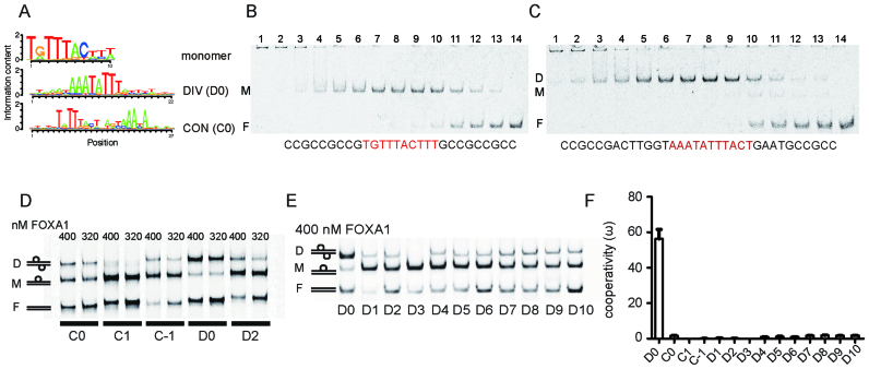
FOXA1 is a transcription factor capable to bind silenced chromatin to direct context-dependent cell fate conversion. Here, we demonstrate that a compact palindromic DNA element (termed 'DIV' for its diverging half-sites) induces the homodimerization of FOXA1 with strongly positive cooperativity. Alternative structural models are consistent with either an indirect DNA-mediated cooperativity or a direct protein-protein interaction. The cooperative homodimer formation is strictly constrained by precise half-site spacing. Re-analysis of chromatin immunoprecipitation sequencing data indicates that the DIV is effectively targeted by FOXA1 in the context of chromatin. Reporter assays show that FOXA1-dependent transcriptional activity declines when homodimeric binding is disrupted. In response to phosphatidylinositol-3 kinase inhibition DIV sites pre-bound by FOXA1 such as at the PVT1/MYC locus exhibit a strong increase in accessibility suggesting a role of the DIV configuration in the chromatin closed-open dynamics. Moreover, several disease-associated single nucleotide polymorphisms map to DIV elements and show allelic differences in FOXA1 homodimerization, reporter gene expression and are annotated as quantitative trait loci. This includes the rs541455835 variant at the MAPT locus encoding the Tau protein associated with Parkinson's disease. Collectively, the DIV guides chromatin engagement and regulation by FOXA1 and its perturbation could be linked to disease etiologies.


Figures

Figure 1.
Figure 1.
FOXA1 cooperates on the DIV motif with strict constraints on half-site spacing. (A) Sequence logos representing position weight matrices (PWMs) of the FOXA1 monomer motif as well as logos for the composite DIV (D0) and CON (C0) motifs. The source PWMs are in Supplementary Table S1. (B, C) EMSAs using 1 nM DNA probes with a monomeric FOXA1 element (B) or the composite DIV element (C) and a concentration series of the FOXA1 DBD. Lanes 1–14 are marked and contain decreasing FOXA1 DBD concentrations. 1: 2500 nM; 2: 1250 nM; 3: 625 nM; 4: 312.5 nM; 5: 156.3 nM; 6: 78.1 nM; 7: 39 nM; 8: 19.5 nM; 9: 9.8 nM; 10: 4.9 nM; 11: 2.4 nM; 12: 1.2 nM; 13: 0.6 nM and 14: no protein). DNA sequences are shown with core binding sites colored red. Dimer (D), monomer (M) and free DNA bands (F) are indicated. (D) EMSAs using DNA probes with DIV and CON motifs and mutants with inserted spacers (D2, C1 and C−1). 100 nM of each Cy5-labelled double stranded DNA was incubated with 400 or 320 nM of the FOXA1 DBD protein. (E) EMSAs using 100 nM DIV DNA (D0) and 1 to 10 base-pair spacers (D1 to D10) performed with 400 nM FOXA1 DBD protein. (F) Cooperativity values (ω) calculated after quantifying dimer, monomer and free DNA fractions from EMSAs (38,39). The mean ± SD of at least five measurements is shown (raw values in Supplementary Table S1).
Figure 2.
Figure 2.
Structural model of dimeric FOXA1/DNA complexes. Structural models were constructed using FOXA1 homology models generated using the FOXA3/DNA crystal structures (PDB ID 1VTN) as template. The modeling strategy is outlined in Supplementary Figure S1A–C. The interaction of Asn165 with an Adenine is a critical mediator of the DNA recognition of forkhead DBDs. Based on the alignment with binary forkhead DBD/DNA structures Asn165 is expected to interact with A4’ in both 5′-T1A2T3T4T5-3′/3′-A’1T’2A’3A’4A’5-5′ DIV half sites (A, D0_M1). We surmised that in the context of a homodimeric complex Asn165 could switch Adenines leading to alternative models where Asn165 contacts A5’ (B, model D0_M4) or A3’ (C, model D0_M7). Left panels show overviews and right panels are zoomed in views highlighting amino acids V229 and A232 exposed to the neighboring molecule that could mediate the dimer formation. The DNA is shown as gray tube, protein helices, sheets or loops are in red, blue and yellow cartoons, respectively. The molecular surface is shown in transparent green. Selected amino acids are labeled and shown as ball-and-sticks. The DNA sequence used to construct the models is shown and nucleotides (or their reverse-complement) contacted by Asn165 are in red.
Figure 3.
Figure 3.
FOXA1 strongly binds to DIV sequences in the context of chromatin. (A) FOXA1 expression measured by the FANTOM 5 consortium in 562 cell and tissue types. Each dot represents a cell or tissue type and selected samples with highest FOXA1 expression are marked. (B) Schematic how ChIP-seq peaks were categorized based on the absence/presence of monomer, DIV or control dimer (DIV1-10) motifs. (C) Boxplot to compare ChIP-seq scores (ENCODE narrowPeak signal values) in the four FOXA1 ChIP-seq peak categories defined in (B) using data from T47D, HepG2 and MCF7 cells. P-values are calculated using pairwise comparisons with the unpaired Wilcoxon rank sum test (R function pairwise.wilcox.test) and adjusted using the Holm method (***P < 0.001). (D) ChIP-seq peaks were ranked by signal values and divided into deciles (top decile = 1, bottom decile = 10, shown as boxplots) and the fractional counts of the four binding categories per decile are shown as proportional barplots.
Figure 4.
Figure 4.
FOXA1/DIV binding regulates gene expression in cancer cells. (A) FOXA1 ChIP-seq peaks in MCF7 cells (accession numbers indicated) at five DIV loci near genes with potential roles in oncogenesis (see also Supplementary Figure S4A and B). (B) EMSAs using Cy5 labeled DNA elements derived from the five endogenous DIV loci. (C) EMSAs where the five DIV DNA elements were mutated to monomer binding sites by adding a 3 base-pair spacer destroying dimeric binding but leaving monomeric binding intact (‘Monomer’). (D) EMSA where both half sites of the DIV elements were mutated abolishing binding (‘No binding’). (E) Homodimer cooperativity value (formula image) shown as mean ± SD from nformula image 3 measurements. (F) Total GFP fluorescence quantified by FACS analysis using Tol2 constructs containing DIV enhancers or the two types of mutated DIV sites (‘Monomer’ or ‘No Binding’) integrated into the genomes of MCF7 cells. FACS plots and photographs of cells are in Supplementary Figure S4D. (G) Dual Luciferase reporter assay using the T47D cell line endogenously expressing FOXA1. (H) Dual Luciferase assay in HCT116 cells co-transfected with FOXA1 expression plasmids (filled bars) or the pcDNA3 control (empty bars). Empty vector is the luciferase reporter without inserted DIV enhancer. Reporter signals in (F) and (G) were normalized to ‘Monomer’ values. Luciferase and Tol2 reporters were constructed as outlined in Supplementary Figure S4C. The mean ± SD of three biological replicates is shown in (F), (G) and (H). P-values were calculated using the unpaired two-tailed Student's t-test (***P< 0.001; **P< 0.01;*P< 0.05).
Figure 5.
Figure 5.
FOXA1 bound DIV locations are associated with chromatin dynamics. (A, B) Boxplot of ChIP-seq scores (summit height of fragment pileup) from FOXA1 data in T47D cells comparing peak categories for the subsets containing DIV motifs (D), control motifs (C), monomer motifs (M) and no FOXA1 motif (N) (see Figure 2B). T47D cells were exposed to DMSO (A) or to the PI3K pathway inhibitor BYL719 (B) (67). (C). Heatmap of ATAC-seq reads as well as FOXA1 ChIP-seq reads under DMSO or BYL719 treatment conditions in three categories of accessibility patterns: PO (permanently open in DMSO and BYL), CO (closed in DMSO but open in BYL) and OC (open in DMSO but closed in BYL). (D) ATAC-seq read heatmaps under DMSO or BYL719 treatment conditions facetted by the four FOXA1 ChIP-seq peak categories defined in the DMSO condition. The lower panels are aggregate pileups of ATAC-seq signals. (E) Boxplot of the ratio between ATAC-seq read counts between BYL719 treated T47D cells and non-treated (DMSO) T47D cells in the four categories of FOXA1 binding sites. (F) Fractional barplots showing the relative proportions of FOXA1 peak categories and sites not bound by FOXA1 (‘Unbound’) associated with PO, OC or CO sites defined according to accessibility patterns measured by ATAC-seq (see panel C). (G) Genome browser plot examples of ChIP-seq and ATAC-seq signals at FOXA1/DIV sites near the PVT1/MYC and KAT6B loci. Black boxes mark locations with DIV motif while green box show alternative location showing a disappearance of ATAC-seq signals. (H) Bar plot of the Luciferase/Renilla signal measured in T47D cells treated with DMSO (red) or BYL719 (blue) using DIV element containing luciferase reporter from the PVT1/MYC and KAT6B loci and mutated ‘no binding’ controls. The mean ± SD of 3 biological replicates is shown. P-values in (H) were calculated using the unpaired two-tailed Student's t-test (***P< 0.001; **P< 0.01; *P< 0.05). P-values in (A), (B) and (E) are calculated using Wilcoxon rank sum test and adjusted using the Holm method ( ***P< 0.001).
Figure 6.
Figure 6.
Disease-associated SNPs perturb dimerization and gene expression. (A) Flowchart to select SNPs for functional evaluation. (B) EMSAs for the rs2097744 locus where the minor allele completely disrupts dimerization. (C) ChIP-seq profiles of histone marks at the rs2941742 locus in various cell lines. (D) EMSA comparing dimer formation for major and minor alleles of rs2941742. (E) Cooperativity factor for EMSA in D (mean ± SD, n = 5). (F) Dual luciferase assay with exogenously supplied FOXA1 in HCT116 cells using both alleles of rs2941742 (mean ± SD, n = 5). (G) FOXA1 ChIP-seq profiles from several human cancer cell lines at the rs5414555835 locus (an alternative ID of the same locus is rs67668514). (H) EMSAs using the major and the minor allele of rs5414555835. I. Dual luciferase assay for the two alleles of the rs5414555835 locus in HCT116 cells. Filled bars are for exogenously provided full length FOXA1 and empty bars for the pcDNA3 vector controls (mean ± SD, n = 3 biological replicates). P-values were calculated using the unpaired two-tailed Student's t-test (**P< 0.01).

References

    1. Cirillo L.A., Lin F.R., Cuesta I., Friedman D., Jarnik M., Zaret K.S.. Opening of compacted chromatin by early developmental transcription factors HNF3 (FoxA) and GATA-4. Mol. Cell. 2002; 9:279–289. - PubMed
    1. He H.H., Meyer C.A., Shin H., Bailey S.T., Wei G., Wang Q., Zhang Y., Xu K., Ni M., Lupien M. et al. Nucleosome dynamics define transcriptional enhancers. Nat. Genet. 2010; 42:343–347. - PMC - PubMed
    1. Sekiya T., Muthurajan U.M., Luger K., Tulin A.V., Zaret K.S.. Nucleosome-binding affinity as a primary determinant of the nuclear mobility of the pioneer transcription factor FoxA. Genes Dev. 2009; 23:804–809. - PMC - PubMed
    1. Shim E.Y., Woodcock C., Zaret K.S.. Nucleosome positioning by the winged helix transcription factor HNF3. Genes Dev. 1998; 12:5–10. - PMC - PubMed
    1. Zaret K.S., Carroll J.S.. Pioneer transcription factors: establishing competence for gene expression. Genes Dev. 2011; 25:2227–2241. - PMC - PubMed

Publication types

MeSH terms