. 2018 Apr 17;48(4):799-811.e9.
doi: 10.1016/j.immuni.2018.03.026.

An Antibody Targeting the Fusion Machinery Neutralizes Dual-Tropic Infection and Defines a Site of Vulnerability on Epstein-Barr Virus

Affiliations

An Antibody Targeting the Fusion Machinery Neutralizes Dual-Tropic Infection and Defines a Site of Vulnerability on Epstein-Barr Virus

Joost Snijder et al. Immunity. .

Abstract

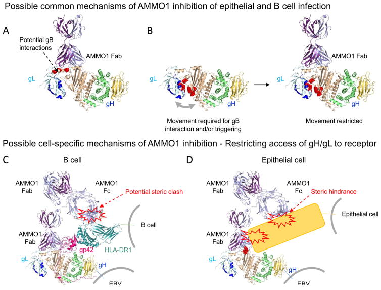
Epstein-Barr virus (EBV) is a causative agent of infectious mononucleosis and is associated with 200,000 new cases of cancer and 140,000 deaths annually. Subunit vaccines against this pathogen have focused on the gp350 glycoprotein and remain unsuccessful. We isolated human antibodies recognizing the EBV fusion machinery (gH/gL and gB) from rare memory B cells. One anti-gH/gL antibody, AMMO1, potently neutralized infection of B cells and epithelial cells, the two major cell types targeted by EBV. We determined a cryo-electron microscopy reconstruction of the gH/gL-gp42-AMMO1 complex and demonstrated that AMMO1 bound to a discontinuous epitope formed by both gH and gL at the Domain-I/Domain-II interface. Integrating structural, biochemical, and infectivity data, we propose that AMMO1 inhibits fusion of the viral and cellular membranes. This work identifies a crucial epitope that may aid in the design of next-generation subunit vaccines against this major public health burden.


Conflict of interest statement

Declaration of Interests

A.T.M discloses that he has filed provisional patents, 62/504,447 and 62/560,061 related to this work.

Figures

Figure 1
Figure 1. Isolation of anti-EBV antibodies from antigen specific B cells
(A) Class switched B cells were stained with SA-PE, or SA-PE conjugated to gH/gL, gB, gp350, or gp42 as indicated. (B) Peripheral blood mononuclear cells were stained with a decoy protein conjugated to SA-PE-DL650 and SA-PE alone, or SA-PE conjugated to gH/gL or gB as indicated. A positive magnetic enrichment using anti-PE microbeads was performed and then cells were stained as in A. PE-DL-650−/PE+ class switched B cells are shown. Numbers indicate the % of PE+ class switched B cells in each panel in A and B. (C) Binding of the AMMO1, AMMO2, AMMO3, AMMO4, and AMMO5, antibodies cloned from B cells sorted using the approach in B to gB, gp42, gp350 and gH/gL by BLI as indicated. SA-PE: streptavidin-phycoerythrin SSA: side-scatter area in A and B. See also Figures S2 and S3.
Figure 2
Figure 2. Isolated MAb neutralizes EBV infection in epithelial cells and B cells
Serial dilutions of the indicated antibodies were evaluated for their ability to neutralize (A) AKTA-GFP EBV infection of epithelial (SVKCR2) cells or (B) B95.8/F EBV infection of B (Raji) cells. Anti-gH/gL antibodies are shown in shades of blue, anti-gB antibodies are shown in shades of red, and the anti-gp350 antibody (72A1) is shown in gray in B. The human or murine origins of the antibodies are indicated in the legend. Data points are mean ± SD of duplicate wells. Representative curves from 2 to 6 replicates are shown.
Figure 3
Figure 3. AMMO1 targets a unique discontinuous epitope on gH/gL
(A) CryoEM reconstruction of gH/gL-gp42-AMMO1 complex at 4.8 Å resolution. (B) 90° rotation from A: AMMO1 heavy chain is shown in dark purple, AMMO1 light chain in light purple, gL in light blue, gH D-I in dark blue, gH D-II in wheat, gH D-III in green, gH D-IV in yellow and gp42 in cherry. (C–D) Ribbon diagram of the gH/gL/gp42-AMMO1 atomic model rendered with the same colors as panels A and B. (E) Zoomed-in view of the AMMO1 epitope with regions of interest labeled. (F) AMMO1 footprint on the gH/gL-gp42 complex. Only the AMMO1 CDR loops are shown interacting with gH/gL-gp42 rendered in surface representation. Residues that have been identified as being important for AMMO1 binding (K73 and Y76) are shown in dark red. (G) The gH N60 glycan is shown in stick representation in the corresponding region of cryoEM density to highlight the putative contacts made with AMMO1. The star shows the position of the gH KGD motif. See Figures S4 and S5.
Figure 4
Figure 4. AMMO1 and CL40 share partially overlapping epitopes
Ribbon diagrams of the gH/gL/gp42-AMMO1 (A) and gH/gL/gp42-CL40 complexes (B, PDB 5W0K). gp42 is omitted from A and B for clarity. (C) gH/gL residues experiencing a change in accessible surface area upon AMMO1 or CL40 binding are colored purple, or light pink, respectively. Areas that experience a change in accessible surface area upon binding of either AMMO1 or CL40 are shown in dark pink. The binding of AMMO1 (D), CL40 (E), CL59 (F), or E1D1 (G) to gH/gL alone, or gH/gL pre-complexed with the indicated antibodies were measured by BLI. BLI traces from one experimental replicate are shown in DG.
Figure 5
Figure 5. An AMMO1-gp42 N173 glycan clash displaces the gp42 CTD
(A) gH/gL residues that experience a change in accessible surface area upon AMMO1 binding are colored light purple, those that experience a change in accessible surface area upon gp42 binding are colored cherry. Asn 240 (colored in magenta) experiences a change in accessible surface area upon binding to AMMO1 and to gp42. (B) Zoomed-in view of the region around gH Asn 240. Ribbons are rendered with the same color scheme as in Figure 3 and amino acid side chains are colored grey (carbon), blue (nitrogen) and red (oxygen). (C) CryoEM reconstructions of the gH/gL/gp42-AMMO1 complex show that most particle images harbor the gp42 C-domain bound on top of gH D-II. (D) A fraction of particle images are characterized by a displacement of the gp42 C-domain. The map is shown at high contour (isosurface) and at low contour (mesh) level to demonstrate that the gp42 CTD is much less well ordered than the rest of the complex. (E) The 4.8 Å resolution reconstruction shown in C reveals that the gp42 N173 glycan points toward the AMMO1 framework region. The glycan is rendered in stick representation with the corresponding region of cryoEM density (blue mesh). (F) AMMO1 binding to gH/gL pre-incubated with the indicated concentrations of gp42 (left) or gp42-T175A (right) was measured by BLI. Representative curves from two independent replicates are shown in F. See also Figures S4 and S7.
Figure 6
Figure 6. AMMO1 interferes with cell fusion
(A) Binding of gH/gL-gp42, or gH/gL-gp42-AMMO1 complexes to HLA-DR were measured using BLI. (B) B cell surface staining with SA-PE alone, SA-PE conjugated to biotinylated gH/gL, biotinylated gB, or biotinylated gH/gL bound to gp42 +/− an excess of the indicated MAbs. (C) Epithelial cell surface staining with SA-PE alone, or SA-PE conjugated to biotinylated gH/gL +/− an excess of the indicated MAbs. Data are represented as mean ± SD, and * indicates a statistical difference in the mean fluorescence intensity of PE in B and C. (D) BLI was used to measure gH/gL binding to a 1 μM solution of αvβ5, αvβ6, αvβ8, gp42, or an HIV-1 Envelope protein. (E) BLI was used to measure gH/gL or gH/gL-AMMO1 binding to a 3.5 μM solution of EphA2 by BLI. (F) CHO-K1 cells were transfected with expression plasmids encoding the indicated proteins, and then overlaid on HEK293 cells stably expressing T7 polymerase, +/− the indicated MAbs. * indicates a statistical difference in the mean RLU (n=5 wells). Data from one experimental replicate is shown in AF.
Figure 7
Figure 7. Possible mechanisms of AMMO1-mediated neutralization
(A) Direct inhibition of gB binding. Residues in the linker helix which have been previously shown to affect cell fusion (L65, L69 and L74) when mutated are shown in red. AMMO1 binding to gH/gL could block a putative gB interaction site. (B) Molecular clamp preventing gB triggering. Residues within the D-I/D-II groove (L55, L207, R152, H154, T174, K94) that have been shown to affect membrane fusion when mutated are shown in red. AMMO1 could restrict movements across the D-I/D-II groove that are required for gB interaction and/or triggering. (C) Restriction of B cell receptor interactions. Although AMMO1 binds away from the HLA-II binding site on the gH/gL-gp42 complex, it could restrain access to membrane anchored receptors through the second FAb arm or the Fc region of the antibody. HLA-DR1 is shown in blue-green and positioned to bind its predicted binding site of gp42 (PDB ID 1KG0). (D) Restriction of epithelial cell receptor interactions. AMMO1 could inhibit binding to one or more epithelial cell receptors by directly restricting access to the interacting site or by indirect steric hindrance mediated through the second FAb arm or the Fc region of the antibody. The KGD motif which has been implicated in gH/gL binding to integrins is shown in red.

Comment in

References

    1. Adams PD, Afonine PV, Bunkoczi G, Chen VB, Davis IW, Echols N, Headd JJ, Hung LW, Kapral GJ, Grosse-Kunstleve RW, et al. PHENIX: a comprehensive Python-based system for macromolecular structure solution. Acta Crystallogr D Biol Crystallogr. 2010;66:213–221. - PMC - PubMed
    1. Agirre J, Iglesias-Fernandez J, Rovira C, Davies GJ, Wilson KS, Cowtan KD. Privateer: software for the conformational validation of carbohydrate structures. Nature structural & molecular biology. 2015;22:833–834. - PubMed
    1. Backovic M, Longnecker R, Jardetzky TS. Structure of a trimeric variant of the Epstein-Barr virus glycoprotein B. Proceedings of the National Academy of Sciences of the United States of America. 2009;106:2880–2885. - PMC - PubMed
    1. Balachandran N, Oba DE, Hutt-Fletcher LM. Antigenic cross-reactions among herpes simplex virus types 1 and 2, Epstein-Barr virus, and cytomegalovirus. Journal of virology. 1987;61:1125–1135. - PMC - PubMed
    1. Bern M, Kil YJ, Becker C. Byonic: advanced peptide and protein identification software. Current protocols in bioinformatics. 2012;Chapter 13(Unit13):20. - PMC - PubMed

Publication types

MeSH terms