

Salivary extracellular vesicle-associated miRNAs as potential biomarkers in oral squamous cell carcinoma
- PMID: 29669525
- PMCID: PMC5907383
- DOI: 10.1186/s12885-018-4364-z
Salivary extracellular vesicle-associated miRNAs as potential biomarkers in oral squamous cell carcinoma
Abstract
Background: Several studies in the past have investigated the expression of micro RNAs (miRNAs) in saliva as potential biomarkers. Since miRNAs associated with extracellular vesicles (EVs) are known to be protected from enzymatic degradation, we evaluated whether salivary EVs from patients with oral squamous cell carcinoma (OSCC) were enriched with specific subsets of miRNAs.
Methods: OSCC patients and controls were matched with regards to age, gender and risk factors. Total RNA was extracted from salivary EVs and the differential expression of miRNAs was evaluated by qRT-PCR array and qRT-PCR. The discrimination power of up-regulated miRNAs as biomarkers in OSCC patients versus controls was evaluated by the Receiver Operating Characteristic (ROC) curves.
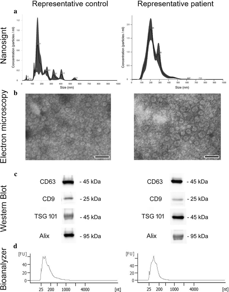
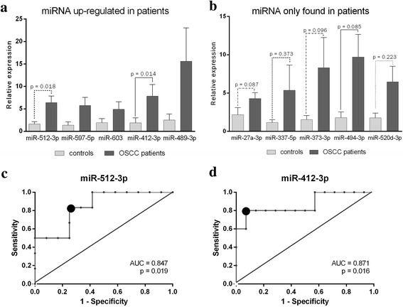
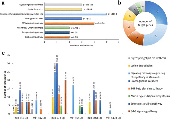
Results: A preliminary qRT-PCR array was performed on samples from 5 OSCC patients and 5 healthy controls whereby a subset of miRNAs were identified that were differentially expressed. On the basis of these results, a cohort of additional 16 patients and 6 controls were analyzed to further confirm the miRNAs that were up-regulated or selectively expressed in the previous pilot study. The following miRNAs: miR-302b-3p and miR-517b-3p were expressed only in EVs from OSCC patients and miR-512-3p and miR-412-3p were up-regulated in salivary EVs from OSCC patients compared to controls with the ROC curve showing a good discrimination power for OSCC diagnosis. The Kyoto Encyclopedia of Gene and Genomes (KEGG) pathway analysis suggested the possible involvement of the miRNAs identified in pathways activated in OSCC.
Conclusions: In this work, we suggest that salivary EVs isolated by a simple charge-based precipitation technique can be exploited as a non-invasive source of miRNAs for OSCC diagnosis. Moreover, we have identified a subset of miRNAs selectively enriched in EVs of OSCC patients that could be potential biomarkers.
Keywords: Extracellular vesicles (EVs); Oral squamous cell carcinoma (OSCC); miR-27a-3p (miR-27a); miR-302b-3p (miR-302b); miR-494; miR-517b-3p (miR-517b); miRNA-412-3p (miR-412); miRNA-512-3p (miR-512); microRNA (miRNA).
Conflict of interest statement
Ethics approval and consent to participate
The study was approved by local ethical committee “A.O.U. Città della Salute e della Scienza di Torino”, Turin, Italy (
Competing interests
GC is named as inventors in a related patent application. All other authors declare no conflict of interest.
Publisher’s Note
Springer Nature remains neutral with regard to jurisdictional claims in published maps and institutional affiliations.
Figures
References
-
- Arduino PG, Carrozzo M, Chiecchio A, Broccoletti R, Tirone F, Borra E, et al. Clinical and histopathologic independent prognostic factors in oral squamous cell carcinoma: a retrospective study of 334 cases. J Oral Maxillofac Surg. 2008;66(8):1570–1579. doi: 10.1016/j.joms.2007.12.024. - DOI - PubMed
Publication types
MeSH terms
Substances
Grants and funding
LinkOut - more resources
Full Text Sources
Other Literature Sources
Medical
Miscellaneous
