. 2018 Apr 18;6(1):29.
doi: 10.1186/s40478-018-0532-2.

Potent prion-like behaviors of pathogenic α-synuclein and evaluation of inactivation methods

Affiliations

Potent prion-like behaviors of pathogenic α-synuclein and evaluation of inactivation methods

Airi Tarutani et al. Acta Neuropathol Commun. .

Abstract

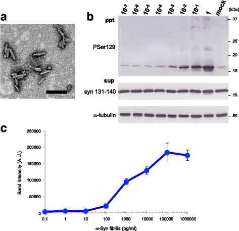
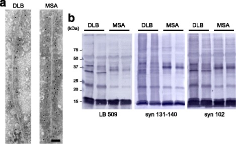
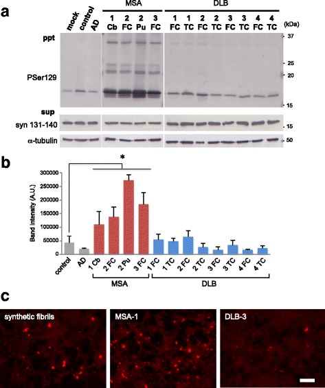
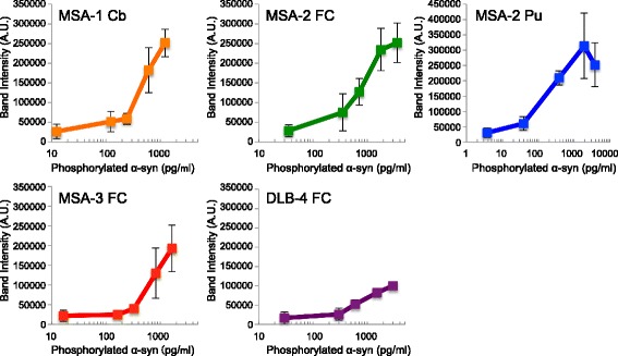
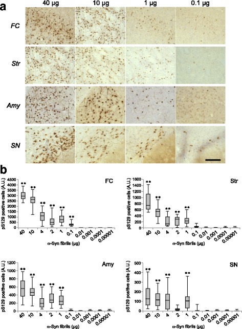
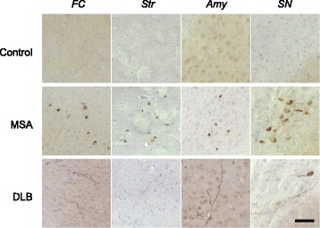
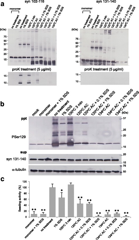
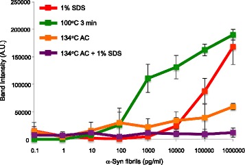
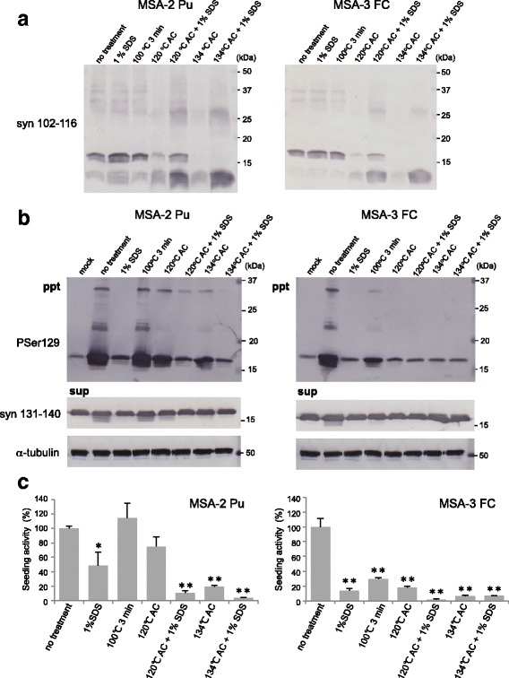
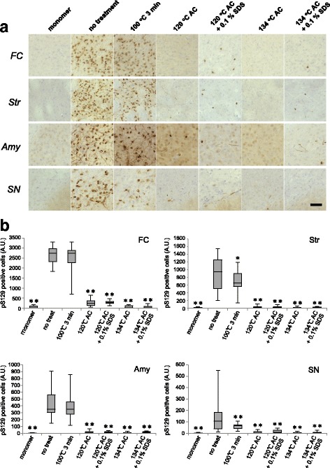
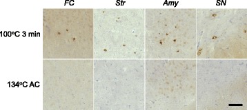
The concept that abnormal protein aggregates show prion-like propagation between cells has been considered to explain the onset and progression of many neurodegenerative diseases. Indeed, both synthetic amyloid-like fibrils and pathogenic proteins extracted from patients' brains induce self-templated amplification and cell-to-cell transmission in vitro and in vivo. However, it is unclear whether exposure to exogenous prion-like proteins can potentially cause these diseases in humans. Here, we investigated in detail the prion-like seeding activities of several kinds of pathogenic α-synuclein (α-syn), including synthetic fibrils and detergent-insoluble fractions extracted from brains of patients with α-synucleinopathies. Exposure to synthetic α-syn fibrils at concentrations above 100 pg/mL caused seeded aggregation of α-syn in SH-SY5Y cells, and seeded aggregation was also observed in C57BL/6 J mice after intracerebral inoculation of at least 0.1 μg/animal. α-Syn aggregates extracted from brains of multiple system atrophy (MSA) patients showed higher seeding activity than those extracted from patients with dementia with Lewy bodies (DLB), and their potency was similar to that of synthetic α-syn fibrils. We also examined the effects of various methods that have been reported to inactivate abnormal prion proteins (PrPSc), including autoclaving at various temperatures, exposure to sodium dodecyl sulfate (SDS), and combined treatments. The combination of autoclaving and 1% SDS substantially reduced the seeding activities of synthetic α-syn fibrils and α-syn aggregates extracted from MSA brains. However, single treatment with 1% SDS or generally used sterilization conditions proved insufficient to prevent accumulation of pathological α-syn. In conclusion, α-syn aggregates derived from MSA patients showed a potent prion-like seeding activity, which could be efficiently reduced by combined use of SDS and autoclaving.

Keywords: Inactivation; Prion-like propagation; Seeds; Strains; α-Synuclein; α-Synucleinopathy.


Conflict of interest statement

Competing interests

The authors declare that they have no competing interests.

Publisher’s Note

Springer Nature remains neutral with regard to jurisdictional claims in published maps and institutional affiliations.

Figures

Fig. 1
Fig. 1
Seed-dependent α-syn aggregation induced by serial dilutions of synthetic α-syn fibrils in SH-SY5Y cells. a Electron microscopy of human α-syn fibrils after sonication for 180 s. Negatively stained short fibrils less than 100 nm in size were observed. Scale bar, 100 nm. b Serial 10-fold dilutions of human α-syn fibrils (2 μl) were introduced into SH-SY5Y cells transiently expressing human WT α-syn in the presence of the transfection reagent. Immunoblot analysis of sarkosyl-insoluble fractions (ppt) and sarkosyl-soluble fractions (sup) extracted from mock-transfected cells or cells transfected with human α-syn fibrils in the range of 2 μg (1) to 0.2 pg (10− 7) are shown. Phosphorylated α-syn was detected with anti-phosphorylated α-syn PSer129 antibody. α-Syn was detected with anti-syn 131–140 antibody c Quantification of phosphorylated α-syn accumulated in SH-SY5Y cells exposed to serial dilutions of synthetic human α-syn fibrils. Band intensities from the immunoblot analyses shown in b were measured. The results are expressed as means ± SEM (n = 3).
Fig. 2
Fig. 2
Characterization of α-syn aggregates extracted from brains of synucleinopathy patients. a Immunoelectron microscopy of sarkosyl-insoluble fractions extracted from DLB (left) and MSA (right) patients’ brains. Electron micrographs show fibrous structures positive for PSer129 antibody EP1536Y, that were labeled with secondary antibody conjugated to 5 nm gold particles. Scale bar, 50 nm. b Immunoblot analyses of sarkosyl-insoluble fractions prepared from brains of synucleinopathy patients. Sarkosyl-insoluble α-syn (22, 29 and 37 kDa in DLB, and 22 and 32 kDa in MSA) were detected by LB509 (left), anti-syn 131–140 (center) and Syn 102 (right) antibodies
Fig. 3
Fig. 3
Prion-like properties in SH-SY5Y cells of α-syn aggregates extracted from brains of synucleinopathy patients. a Sarkosyl-insoluble fractions extracted from patients’ brains (2 μl) were introduced into SH-SY5Y cells transiently expressing human WT α-syn. Immunoblot analysis of sarkosyl-insoluble fractions (ppt) and sarkosyl-soluble fractions (sup) extracted from mock-transfected cells, and sarkosyl-insoluble fractions from cerebellum, frontal cortex and putamen of 3 MSA cases, frontal cortex and temporal cortex of 4 DLB cases, control brain and an AD case. Phosphorylated α-syn was detected with anti-phosphorylated α-syn PSer129 antibody. α-Syn was detected with anti-syn 131–140 antibody. The α-syn concentrations of Sarkosyl-insoluble fractions derived from human brains are shown in Additional file 1: Table S1A. b Quantification of immunoblot analyses shown in A. The results are expressed as means ± SEM (n = 3). *P < 0.05. c SH-SY5Y cells into which synthetic human α-syn fibrils (2 μg) or sarkosyl-insoluble fractions from cerebellum of a MSA case and temporal cortex of a DLB case (2 μl) had been introduced were fixed and immunostained with PSer129 antibody EP1536Y. Scale bar, 100 μm. Cb: cerebellum, FC: frontal cortex, Pu: putamen, TC: temporal cortex
Fig. 4
Fig. 4
Seed-dependent α-syn aggregation induced in SH-SY5Y cells by serial dilutions of insoluble fractions extracted from brains of patients with synucleinopathies. Serial dilutions of sarkosyl-insoluble fractions prepared from cerebellum, frontal cortex and putamen of 3 MSA cases and frontal cortex of a DLB case (2 μl) were introduced into SH-SY5Y cells transiently expressing human WT α-syn. Quantification of phosphorylated α-syn accumulated in SH-SY5Y cells induced by serial dilutions of pathogenic α-syn derived from brain samples. Band intensities of immunoblot analysis shown in Additional file 4: Figure S3 were measured. The results are expressed as means ± SEM (n = 3). Cb: cerebellum, FC: frontal cortex, Pu: putamen, TC: temporal cortex
Fig. 5
Fig. 5
Dose-dependent propagation following introduction of different amounts of synthetic α-syn fibrils in non-Tg mice. a Various amounts of mouse α-syn fibrils inoculated into WT mice dose-dependently induced α-syn pathology in frontal cortex, striatum, amygdala and substantia nigra. α-Syn pathology in mouse brains inoculated with 40, 10, 1, and 0.1 μg of α-syn fibrils into striatum at 3 months after inoculation is shown. The numbers of injected mice are shown in Additional file 5: Table S2A. Sections were evaluated by immunohistochemistry with PSer129 antibody EP1536Y. Scale bar, 100 μm. b Quantification of α-syn pathology in brains of mice inoculated with different amounts (40 μg to 10 pg) of mouse α-syn fibrils into striatum. The box plots show the number of pS129-positive cells in different regions. One-way ANOVA with Dunnett’s post hoc test was used for multiple comparisons to 10 pg, *P < 0.05; **P < 0.01. FC: frontal cortex, Str: striatum, Amy: amygdala, SN: substantia nigra
Fig. 6
Fig. 6
α-Syn strain-specific pathology in non-Tg mice. Inoculation of sarkosyl-insoluble fractions extracted from MSA brain (MSA-2, putamen, 5 μl) into WT mice induced PS129-positive inclusions in frontal cortex, striatum, amygdala and substantia nigra at 3 months after inoculation (middle). Sarkosyl-insoluble fractions extracted from DLB brain (5 μl) induced Lewy-neurite-like α-syn pathology at 9 months after inoculation (lower). Control brain did not induce α-syn pathology (upper). Sections were evaluated by immunohistochemistry with PSer129 antibody EP1536Y. The α-syn concentrations of sarkosyl-insoluble fractions derived from human brains are shown in Additional file 2: Table S1B. The numbers of injected mice are shown in Additional file 5: Table S2C. FC: frontal cortex, Str: striatum, Amy: amygdala, SN: substantia nigra
Fig. 7
Fig. 7
Inactivation of synthetic α-syn fibrils by SDS treatment and autoclaving. a Synthetic α-syn fibrils were subjected to various inactivation treatments and analyzed by immunoblotting with Syn 102–116 and anti-syn 131–140 antibodies (upper). Treated α-syn samples were treated with protease K (5 μg/mL) and analyzed by immunoblotting (lower). b α-Syn fibrils were subjected to various inactivation treatments (2 μl) and introduced into SH-SY5Y cells transiently expressing human WT α-syn. Immunoblot analysis of sarkosyl-insoluble fractions (ppt) and sarkosyl-soluble fractions (sup) extracted from mock-transfected cells and cells transfected with α-syn monomer and fibrils, and treated with 1% SDS for 1 h at room temperature, or boiled, or autoclaved (AC) at 120 °C with or without 0.1%, 1% SDS, or autoclaved at 134 °C with or without 0.1% or 1% SDS are shown. Phosphorylated α-syn was detected with anti-phosphorylated α-syn PSer129 antibody. α-Syn was detected with anti-syn 131–140 antibody. c Quantification of immunoblot analysis shown in b. The results are expressed as means ± SEM (n = 3). “No treatment” was taken as 100%. One-way ANOVA with Dunnett’s post hoc test were used for multiple comparisons to no treatment, *P < 0.05; **P < 0.01
Fig. 8
Fig. 8
Effects of inactivation treatments of synthetic α-syn fibrils on seeding activity in SH-SY5Y cells. Serial 10-fold dilutions of human α-syn fibrils treated with 1% SDS (red), boiling (green), autoclaving (AC) at 134 °C (orange) or autoclaving at 134 °C with 1% SDS (purple) were introduced into SH-SY5Y cells transiently expressing human WT α-syn. Phosphorylated α-syn accumulated in SH-SY5Y cells exposed to serial dilutions of synthetic human α-syn fibrils were quantified after each treatment. Band intensities from the immunoblot analyses shown in Additional file 6: Figure S4 were measured. The results are expressed as means ± SEM (n = 3)
Fig. 9
Fig. 9
Inactivation of pathogenic α-syn derived from brains of MSA patients. a Sarkosyl-insoluble fractions prepared from 2 MSA cases (MSA-2 Pu and MSA-3 FC) after various inactivation treatments were analyzed by immunoblotting with anti-syn 102–116 antibody. b Sarkosyl-insoluble fractions extracted from MSA brains after inactivation treatments (2 μl) were introduced into SH-SY5Y cells transiently expressing human WT α-syn. Immunoblot analyses of sarkosyl-insoluble fractions (ppt) and sarkosyl-soluble fractions (sup) extracted from mock-transfected cells or cells transfected with pathogenic α-syn derived from MSA brains and treated with 1% SDS for 1 h at room temperature, boiling, autoclaving (AC) at 120 °C with or without 1% SDS or AC at 134 °C with or without 1% SDS are shown. Phosphorylated α-syn was detected with anti-phosphorylated α-syn PSer129 antibody. α-Syn was detected with anti-syn 131–140 antibody. c, Quantification of immunoblot analyses shown in b. The results are expressed as means ± SEM (n = 3). “No treatment” was taken as 100%. One-way ANOVA with Dunnett’s post hoc test were used for multiple comparisons to no treatment, *P < 0.05; **P < 0.01
Fig. 10
Fig. 10
Effects of inactivation treatments of synthetic α-syn fibrils on seeding activity in non-Tg mice. a 2 mg/ml mouse α-syn fibrils were exposed to various treatments (1% SDS for 1 h at room temperature, boiling, autoclaving (AC) at 120 °C with or without 0.1% SDS or autoclaving at 134 °C with or without 0.1% SDS), and 5 μl aliquots of the resulting samples were inoculated into WT mice. α-Syn pathologies in frontal cortex, striatum, amygdala and substantia nigra at 3 months after inoculation are shown. The numbers of injected mice are shown in Additional file 5: Table S2B. Sections were evaluated by immunohistochemistry with PSer129 antibody EP1536Y. Scale bar, 100 μm. b Quantification of α-syn pathology in mouse brains inoculated into striatum with α-syn fibrils exposed to various treatments. The box plots show the numbers of pS129-positive cells in different regions. One-way ANOVA with Dunnett’s post hoc test were used for multiple comparisons to no treatment, *P < 0.05; **P < 0.01. FC: frontal cortex, Str: striatum, Amy: amygdala, SN: substantia nigra
Fig. 11
Fig. 11
Effects of inactivation treatments of pathogenic α-syn extracted from MSA patients’ brains on seeding activity in non-Tg mice, α-Syn pathology in mouse brains inoculated into striatum with sarkosyl-insoluble fractions extracted from MSA brain (MSA-2, putamen) after boiling or autoclaving (AC) at 134 °C, at 3 months after injection. The numbers of injected mice are shown in Additional file 5: Table S2C. Sections were evaluated by immunohistochemistry with PSer129 antibody EP1536Y. Scale bar, 100 μm. FC: frontal cortex, Str: striatum, Amy: amygdala, SN: substantia nigra

References

    1. Abeliovich A, Schmitz Y, Farinas I, Choi-Lundberg D, Ho WH, Castillo PE, et al. Mice lacking alpha-synuclein display functional deficits in the nigrostriatal dopamine system. Neuron. 2000;25:239–252. doi: 10.1016/S0896-6273(00)80886-7. - DOI - PubMed
    1. Anderson JP, Walker DE, Goldstein JM, de Laat R, Banducci K, Caccavello RJ, et al. Phosphorylation of Ser-129 is the dominant pathological modification of alpha-synuclein in familial and sporadic Lewy body disease. J Biol Chem. 2006;281:29739–29752. doi: 10.1074/jbc.M600933200. - DOI - PubMed
    1. Ayers JI, Brooks MM, Rutherford NJ, Howard JK, Sorrentino ZA, Riffe CJ et al (2017) Robust central nervous system pathology in transgenic mice following peripheral injection of alpha-Synuclein fibrils. J Virol 91 - PMC - PubMed
    1. Baba M, Nakajo S, Tu PH, Tomita T, Nakaya K, Lee VM, et al. Aggregation of alpha-synuclein in Lewy bodies of sporadic Parkinson's disease and dementia with Lewy bodies. Am J Pathol. 1998;152:879–884. - PMC - PubMed
    1. Bernis ME, Babila JT, Breid S, Wusten KA, Wullner U, Tamguney G. Prion-like propagation of human brain-derived alpha-synuclein in transgenic mice expressing human wild-type alpha-synuclein. Acta Neuropathol Commun. 2015;3:75. doi: 10.1186/s40478-015-0254-7. - DOI - PMC - PubMed

Publication types

MeSH terms

LinkOut - more resources