Review
. 2018 Jul 2;217(7):2261-2271.
doi: 10.1083/jcb.201802003. Epub 2018 Apr 18.

Modulation of the secretory pathway by amino-acid starvation

Affiliations
Review

Modulation of the secretory pathway by amino-acid starvation

Wessel van Leeuwen et al. J Cell Biol. .

Abstract

As a major anabolic pathway, the secretory pathway needs to adapt to the demands of the surrounding environment and responds to different exogenous signals and stimuli. In this context, the transport in the early secretory pathway from the endoplasmic reticulum (ER) to the Golgi apparatus appears particularly regulated. For instance, protein export from the ER is critically stimulated by growth factors. Conversely, nutrient starvation also modulates functions of the early secretory pathway in multiple ways. In this review, we focus on amino-acid starvation and how the function of the early secretory pathway is redirected to fuel autophagy, how the ER exit sites are remodeled into novel cytoprotective stress assemblies, and how secretion is modulated in vivo in starving organisms. With the increasingly exciting knowledge on mechanistic target of rapamycin complex 1 (mTORC1), the major nutrient sensor, it is also a good moment to establish how the modulation of the secretory pathway by amino-acid restriction intersects with this major signaling hub.


Figures

Figure 1.
Figure 1.
The dual role of COPII-coated vesicles in secretion and in autophagy. (A) Upon growing conditions, COPII-coated vesicles transport cargo from the ER (in yeast) or ERES (metazoa) to the Golgi. (1 and 1′) In yeast (1), Ypt1 is present on COPII vesicles in an inactive GDP-bound state. TRAPPIII catalyses the nucleotide exchange on Ypt1 (GTP-bound) that recruits Uso1p. In metazoa (1′), TRAPPIII has the same role on Rab1 that recruits p115. (2) In yeast, Hrr25-dependent Sec24 phosphorylation is necessary for fusion of the COPII-derived vesicle with the Golgi. (3) In mammalian cells, COPII vesicle formation is regulated by TECPR2, which protects Sec24 against proteasomal degradation and helps maintain ERES integrity. Interestingly, TECPR2 interacts with lipidated LC3C. (B) Under conditions in which autophagy is activated, ERES provide membranes for phagophore biogenesis and extension. (4) In yeast (S. cerevisiae), TRAPPIII-activated Ypt1 present on COPII-coated vesicles recruits Atg17 and the Atg1 complex. This allows the binding of Atg9-containing vesicles that in yeast derive from the Golgi. Collectively, this forms the PAS that matures into a phagophore. (5) Upon autophagy stimulation, Sec24 is specifically phosphorylated on three threonines in a Hrr25-dependent manner. This phosphorylation allows the binding to Atg9 vesicles and redirects COPII-coated vesicles to the growing phagophore. (6) Phagophores (Atg8-positive) have also been observed in close proximity to Sec13 and to Sec16. (7) In mammalian cells, autophagy induction leads to the remodeling of ERES and the relocalization of Sec12 to ERGIC in a CTAGE5- and FIB200-dependent manner. This leads to COPII vesicle budding from ERGIC, recruitment of LC3, and binding to Atg9-containing vesicles. Together, they form and/or feed the growing phagophore. (8) In mammalian cells, the TECPR2 interaction with LC3C somehow leads to the redirection of COPII vesicles to the phagophore.
Figure 2.
Figure 2.
Amino-acid starvation of Drosophila cells leads to protein transport inhibition and Sec body formation. In growing conditions, COPII-coated vesicles in Drosophila S2 cells bud from the ERES in a Sec16-dependent manner, leading to active transport through the secretory pathway. ERK7 levels are low and dPARP16 is inactive. Upon amino-acid starvation, protein transport from the ERES to the Golgi is inhibited. ERK7 level increases, leading to Sec16 release away from the ERES membrane. dPARP16 is activated and mono-ADP-ribosylates Sec16 on its SRDC, leading to the coalescence of Sec16 and COPII subunits into Sec bodies after 3–4-h starvation. MARylated, mono-ADP-ribosylated.
Figure 3.
Figure 3.
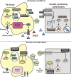
The secretion-based signaling between the Drosophila fat body and IPCs upon sensing the level of circulating amino acids. Top: When amino acids are available, they are transported into the fat body cells by Slimfast, where they activate mTORC1 (see mTORC1 activation text box). The nutrient responsive coactivator PGC-1 stimulates stunted expression, giving rise to the SunA and SunB peptides that are secreted into the hemolymph. SunA and SunB subsequently bind to their receptor Methuselah on the plasma membrane of IPCs in the brain. The IPCs transcribe the Dilp genes, leading to the accumulation of Dilps 3, 2, and 5. Activation of Methuselah triggers intracellular calcium release, which stimulates Dilps secretion to the hemolymph. In turn, Dilps activate the InR in the fat body, which increases mTORC1 activity further via the insulin-Akt pathway (see mTORC1 activation text box). Bottom: Upon amino-acid starvation, mTORC1 activation is reduced, leading to a reduction of SunA and SunB. It is also possible that their secretion is inhibited through the increase of the ERK7 level. Decreased SunA and SunB secretion leads to a decreased stimulation of Methuselah, resulting in a reduced Dilp secretion from IPCs where they accumulate. ERK7 level in the IPCs is increased, possibly contributing to secretion inhibition. Reduced Dilp secretion during amino-acid starvation effectively reduces mTORC1 activation and overall growth.

References

    1. Agrawal G., and Subramani S.. 2016. De novo peroxisome biogenesis: Evolving concepts and conundrums. Biochim. Biophys. Acta. 1863:892–901. 10.1016/j.bbamcr.2015.09.014 - DOI - PMC - PubMed
    1. Agrawal N., Delanoue R., Mauri A., Basco D., Pasco M., Thorens B., and Léopold P.. 2016. The Drosophila TNF Eiger Is an Adipokine that Acts on Insulin-Producing Cells to Mediate Nutrient Response. Cell Metab. 23:675–684. 10.1016/j.cmet.2016.03.003 - DOI - PubMed
    1. Aguilera-Gomez A., van Oorschot M.M., Veenendaal T., and Rabouille C.. 2016. In vivo vizualisation of mono-ADP-ribosylation by dPARP16 upon amino-acid starvation. eLife. 5:e21475 10.7554/eLife.21475 - DOI - PMC - PubMed
    1. Aguilera-Gomez A., Zacharogianni M., van Oorschot M.M., Genau H., Grond R., Veenendaal T., Sinsimer K.S., Gavis E.R., Behrends C., and Rabouille C.. 2017. Phospho-Rasputin Stabilization by Sec16 Is Required for Stress Granule Formation upon Amino Acid Starvation. Cell Reports. 20:935–948. 10.1016/j.celrep.2017.06.042 - DOI - PMC - PubMed
    1. Alberti S. 2017. The wisdom of crowds: regulating cell function through condensed states of living matter. J. Cell Sci. 130:2789–2796. 10.1242/jcs.200295 - DOI - PubMed

MeSH terms

Substances