

High-riding brachiocephalic (innominate) artery during surgical tracheostomy
- PMID: 29669763
- PMCID: PMC5911098
- DOI: 10.1136/bcr-2017-221802
High-riding brachiocephalic (innominate) artery during surgical tracheostomy
Abstract
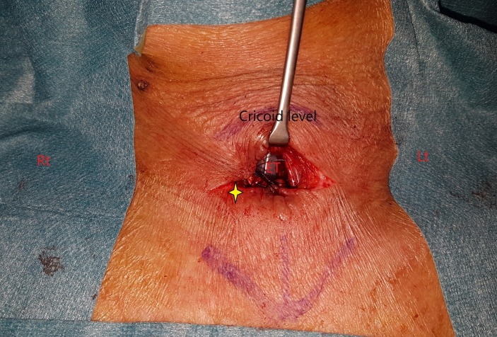
Two cases of a high-riding innominate artery, which were found during routine surgical tracheostomy. A cartilage flap was applied to cover the significant vessel to prevent the life-threatening complications. These two cases were followed up for 2 months without any adverse events. We discussed the related vascular anatomy, imaging studies and brief literature review.
Keywords: ear, nose and throat/otolaryngology; emergency medicine; head and neck surgery; intensive care; otolaryngology / ent.
© BMJ Publishing Group Ltd (unless otherwise stated in the text of the article) 2018. All rights reserved. No commercial use is permitted unless otherwise expressly granted.
Conflict of interest statement
Competing interests: None declared.
Figures


References
-
- Warwick R, Williams PL. Gray’s anatomy. 35th Br. edn Philadelphia: Saunders, 1973;1:443–5.
-
- Lang J. Klinische anatomie der halswirbelsäule: 13 tabellen. Thieme, 1991.
Publication types
MeSH terms
LinkOut - more resources
Full Text Sources
Other Literature Sources
Research Materials
Miscellaneous