. 2018 Jun 8;293(23):8861-8873.
doi: 10.1074/jbc.RA118.002215. Epub 2018 Apr 18.

Two distinct sites of client protein interaction with the chaperone cpSRP43

Affiliations

Two distinct sites of client protein interaction with the chaperone cpSRP43

Camille Z McAvoy et al. J Biol Chem. .

Abstract

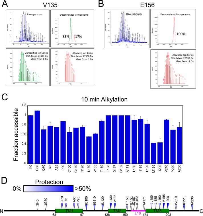
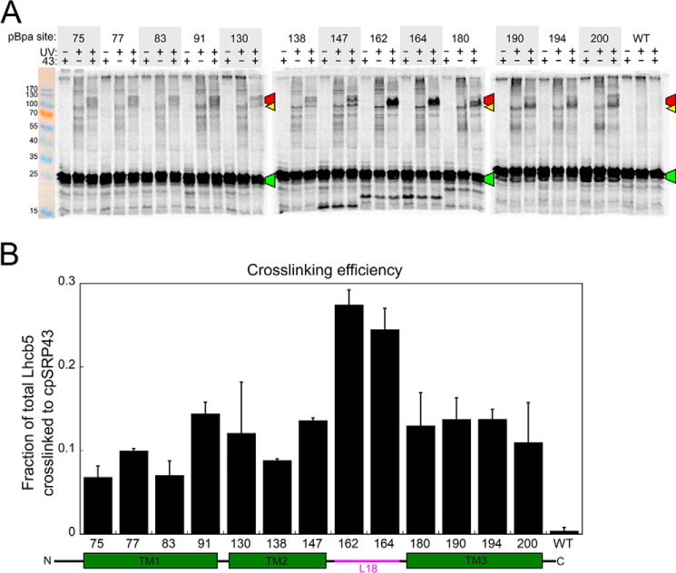
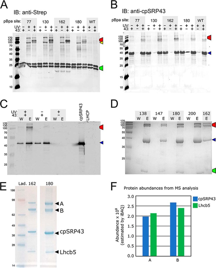
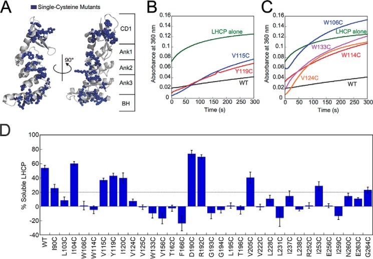
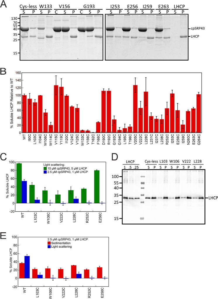
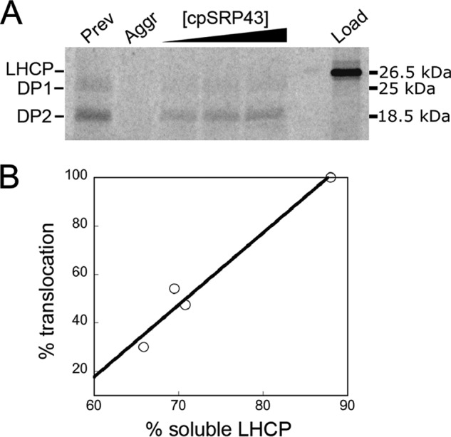
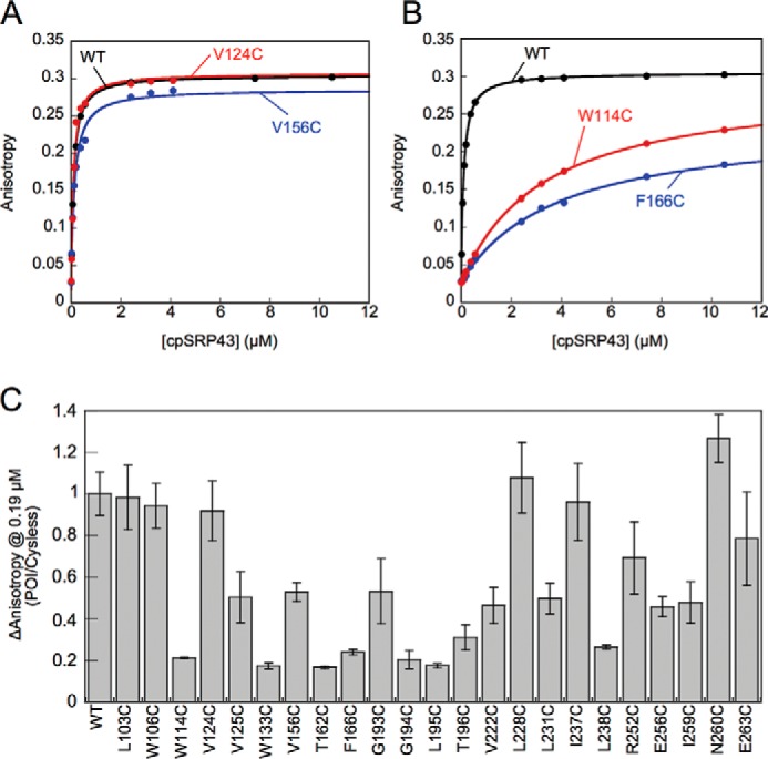
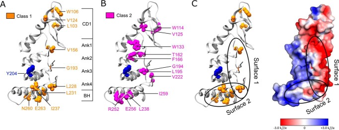
Integral membrane proteins are prone to aggregation and misfolding in aqueous environments and therefore require binding by molecular chaperones during their biogenesis. Chloroplast signal recognition particle 43 (cpSRP43) is an ATP-independent chaperone required for the biogenesis of the most abundant class of membrane proteins, the light-harvesting chlorophyll a/b-binding proteins (LHCPs). Previous work has shown that cpSRP43 specifically recognizes an L18 loop sequence conserved among LHCP paralogs. However, how cpSRP43 protects the transmembrane domains (TMDs) of LHCP from aggregation was unclear. In this work, alkylation-protection and site-specific cross-linking experiments found that cpSRP43 makes extensive contacts with all the TMDs in LHCP. Site-directed mutagenesis identified a class of cpSRP43 mutants that bind tightly to the L18 sequence but are defective in chaperoning full-length LHCP. These mutations mapped to hydrophobic surfaces on or near the bridging helix and the β-hairpins lining the ankyrin repeat motifs of cpSRP43, suggesting that these regions are potential sites for interaction with the client TMDs. Our results suggest a working model for client protein interactions in this membrane protein chaperone.

Keywords: chaperone; chloroplast; membrane protein; protein targeting; signal recognition particle (SRP).


Conflict of interest statement

The authors declare that they have no conflicts of interest with the contents of this article

Figures

Figure 1.
Figure 1.
Alkylation pattern of Lhcb5 in the cpSRP43·Lhcb5 complex show cpSRP43-induced protection on the substrate protein. A and B, mass spectrum (upper left), deconvolution (lower panels), and component analysis (upper right) for a partially alkylated Lhcb5 residue, Cys-135 (A) and a completely alkylated Lhcb5 residue, Cys-156 (B). C, summary of the NEM alkylation efficiencies at individual sites in Lhcb5. Alkylation reactions were carried out for 10 min. For each engineered cysteine, “fraction accessible” was calculated from the ratio of the fraction of alkylation in the cpSRP43·Lhcb5 complex relative to that of Lhcb5 dissolved in 6 m GdmHCl. Error bars indicate S.E., with n = 2. D, the alkylation protection pattern of Lhcb5 in complex with cpSRP43 is mapped onto the sequence of Lhcb5. Colored triangles denote the extent of protection, with white denoting the least protection (0% protection, or 100% alkylation) and blue denoting the highest observed protection (>50% protection or <50% alkylation).
Figure 2.
Figure 2.
Site-specific cross-linking suggests extensive contacts between Lhcb5 and cpSRP43. A, SDS-PAGE analysis of 35S-labeled Lhcb5 containing a photocross-linker, pBpa, at the indicated positions. Purified superactive cpSRP43 was present (+ lanes) or absent (− lanes) during translation, and samples were protected from light (− lanes) or exposed to UV light to induce pBpa cross-links (+ lanes). Marked bands indicate the cross-linked cpSRP43·Lhcb5 complex (red), free Lhcb5 (green), and Lhcb5 cross-linked to an unknown protein in the translation extract (yellow). B, summary of cross-linking efficiencies between cpSRP43 and Lhcb5 with pBpa incorporated at different sites. Cross-linking efficiency was calculated from the ratio of the cpSRP43·Lhcb5 band to total Lhcb5 (after subtraction of background from the corresponding locations in the +UV, −cpSRP43 lane) for the SDS-PAGE analysis in A and replicates (not shown). Data are reported as mean ± S.E., with n = 2.
Figure 3.
Figure 3.
Analysis of the high molecular weight cross-linked bands between cpSRP43 and Lhcb5. The color-marked bands in A–D indicate the cross-linked cpSRP43·Lhcb5 complex (red), free Strep-Lhcb5 (green), free cpSRP43 (blue), and Strep-Lhcb5 cross-linked to an unknown protein (yellow). A and B, representative Western blotting analyses of the cross-linking reactions and their controls from Fig. 2A using anti-Strep (for Strep-tagged Lhcb5; A) or anti-cpSRP43 (B) antibodies. The lower Mr band for Lhcb5 is likely a C terminally proteolyzed product of full-length Lhcb5. C, representative Western blot of affinity purification of the cross-linked cpSRP43·Lhcb5 complex based on Strep-tagged Lhcb5. The final wash (W) and elution (E) from Strep-Tactin resin were shown for reactions with (+ lanes) and without (− lanes) intein–cpSRP43 and exposure to UV light. D, representative Coomassie-stained SDS-PAGE gels for the same cross-linking reactions purified based on His6-tagged cpSRP43, with Bpa incorporated at the indicated residues of Lhcb5. E, NuPAGE gels showing the purified (as in C and D) cpSRP43·Lhcb5(162Bpa) and cpSRP43·Lhcb5(180Bpa) cross-linking reactions. The two labeled bands (A and B) were digested and sent for MS analysis. F, results for MS analysis of the abundance of cpSRP43 and Lhcb5 in bands A and B excised the gel in E for Lhcb5(162Bpa).
Figure 4.
Figure 4.
Single-cysteine mutants across the cpSRP43 SBD exhibit defects in chaperone activity in the light scattering assay. A, structure of cpSRP43 indicating all sites where cysteine mutations were made (blue). B and C, representative data showing the chaperone activity of neutral (B) and defective (C) cpSRP43 mutants. Light scattering time traces are shown for LHCP diluted into aqueous buffer (green), into a solution containing Cys-less WT (black), and into solutions containing the indicated cpSRP43 mutants. D, summary of the chaperone activity for all the single cysteine mutants of cpSRP43 measured by light scattering. Mutants exhibiting chaperone activity within 3-fold of that of Cys-less cpSRP43 are considered neutral (above dashed line), whereas mutants with lower activity are considered defective (below dashed line). Error bars indicate S.E., with n = 3–13.
Figure 5.
Figure 5.
Analysis of the chaperone activity of mutant cpSRP43 using the sedimentation assay and comparison with the results of light scattering assay. A, representative Coomassie-stained gels for analysis of the chaperone activity of cpSRP43 mutants using the sedimentation assay. C denotes lanes with cpSRP43 only; S denotes the soluble fraction; P denotes pellet. B, summary of the relative chaperone activity of all cpSRP43 mutants measured by the sedimentation assay. Values are reported relative to Cys-less cpSRP43 (WT). C, comparison of the chaperone activity of cpSRP43 mutants measured at high (green) and low (blue) protein concentrations using the light scattering assay. D, representative Western blot images for analysis of the chaperone activity of cpSRP43 mutants using the sedimentation assay at low protein concentrations. C denotes lanes with cpSRP43 only; S denotes the soluble fraction; P denotes pellet. LHCP denotes controls where indicated concentrations of purified LHCP were loaded to assess the dynamic range of Western blotting. E, comparison of the chaperone activity of cpSRP43 variants measured by the sedimentation (red; data from D and replicates) and light scattering (blue) assays at the same concentration. Data were reported as mean ± S.E., with n = 2–9.
Figure 6.
Figure 6.
Solubilization of LHCP by cpSRP43 correlates with LHCP targeting and integration. A, [35S]methionine-labeled LHCP (lane 6, Load) were preincubated under different conditions with cpSRP43 and tested for targeting and insertion into thylakoid membrane in the presence of 3 μm cpSRP43, cpSRP54, and cpFtsY. Lanes 1 and 2, 2 μl of 35S-LHCP in 8 m urea was added to 40 μl of Buffer D with (lane 1) or without (lane 2) 3 μm cpSRP43/54 and incubated for 60 min. Lanes 3–5, 2 μl of 35S-LHCP in 8 m urea was added to 33.6 μl of Buffer D and allowed to aggregate at room temperature for 60 s, followed by addition of an equimolar ratio of cpSRP43/54 to final concentrations of 5, 15, and 30 μm in a final volume of 40 μl. 20 μl of the preincubated sample was used for the LHCP integration assay. DP1 and DP2 (25 and 18.5 kDa) are the protease-protected fragments of integrated LHCP (51). The remaining 20 μl was subjected to the sedimentation assay as described under “Experimental procedures,” except that LHCP bands were quantified by autoradiography using Storm 840 (Molecular Dynamics) and ImageQuant (GE Healthcare). Details of the LHCP integration assay are described under “Experimental procedures.” B, correlation of the translocation efficiency of LHCP with the degree to which LHCP is solubilized by cpSRP43.
Figure 7.
Figure 7.
Characterization of the interaction of mutant cpSRP43s with the L18 motif. A and B, representative equilibrium titrations for the binding of WT and mutant cpSRP43s to HiLyte-Fluor 488-labeled L11. Representative data for cpSRP43 mutants that can bind L11 with high affinity are shown in A, and those for mutants exhibiting weakened L11 binding are shown in B. C, summary of the cpSRP43-induced changes in the fluorescence anisotropy of L11 at 0.19 μm, which is subsaturating for binding of Cys-less cpSRP43 to L11. The data for all mutants are normalized to that of Cys-less cpSRP43 (denoted as WT). All data are reported as mean ± S.E., with n ≥ 2.
Figure 8.
Figure 8.
Mapping two classes of cpSRP43 mutants onto the crystal structure of the cpSRP43 SBD (Protein Data Bank code 3dep). A, residues whose mutations led to defective chaperone activity for LHCP but did not disrupt L18 binding are categorized as Class I and colored in orange. B, residues whose mutations disrupted both cpSRP43's chaperone activity and its interaction with the L18 motif are categorized as Class II and colored in magenta. C, a putative model for the interaction surfaces of cpSRP43 with LHCP, with Tyr-204 (blue) interacting with the L18 sequence, and the hydrophobic surfaces formed by Ank4, BH, and the β-hairpins along the ankyrin repeat motifs involved in protection of the TMDs of LHCP. The electrostatic surface potential of the cpSRP43 SBD was generated using Adaptive Poisson-Boltzmann Solver (52) and visualized in PyMOL.

Similar articles

Cited by

References

    1. Randall L. L., and Hardy S. J. (2002) SecB, one small chaperone in the complex milieu of the cell. Cell. Mol. Life Sci. 59, 1617–1623 10.1007/PL00012488 - DOI - PMC - PubMed
    1. Walton T. A., Sandoval C. M., Fowler C. A., Pardi A., and Sousa M. C. (2009) The cavity chaperone Skp protects its substrates from aggregation but allows independent folding of substrate domains. Proc. Natl. Acad. Sci. U.S.A. 106, 1772–1777 10.1073/pnas.0809275106 - DOI - PMC - PubMed
    1. Deshaies R. J., Koch B. D., Werner-Washburne M., Craig E. A., and Schekman R. (1988) 70 kD stress protein homologues facilitate translocation of secretory and mitochondrial precursor polypeptides. Nature 332, 800–805 10.1038/332800a0 - DOI - PubMed
    1. Deshaies R. J., Koch B. D., and Schekman R. (1988) The role of stress proteins in membrane biogenesis. Trends Biochem. Sci. 13, 384–388 10.1016/0968-0004(88)90180-6 - DOI - PubMed
    1. Thoma J., Burmann B. M., Hiller S., and Müller D. J. (2015) Impact of holdase chaperones Skp and SurA on the folding of β-barrel outer-membrane proteins. Nat. Struct. Mol. Biol. 22, 795–802 10.1038/nsmb.3087 - DOI - PubMed

Publication types

MeSH terms

Substances

Associated data

LinkOut - more resources