. 2018 Jun 1;293(22):8439-8448.
doi: 10.1074/jbc.M117.818013. Epub 2018 Apr 18.

Structural insights into the mechanism of action of a biparatopic anti-HER2 antibody

Affiliations

Structural insights into the mechanism of action of a biparatopic anti-HER2 antibody

Vaheh Oganesyan et al. J Biol Chem. .

Abstract

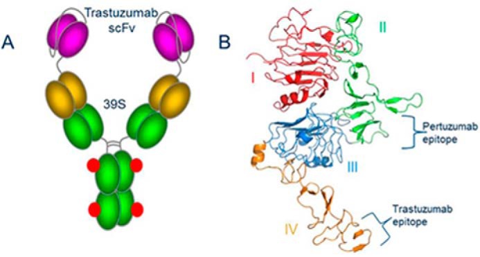
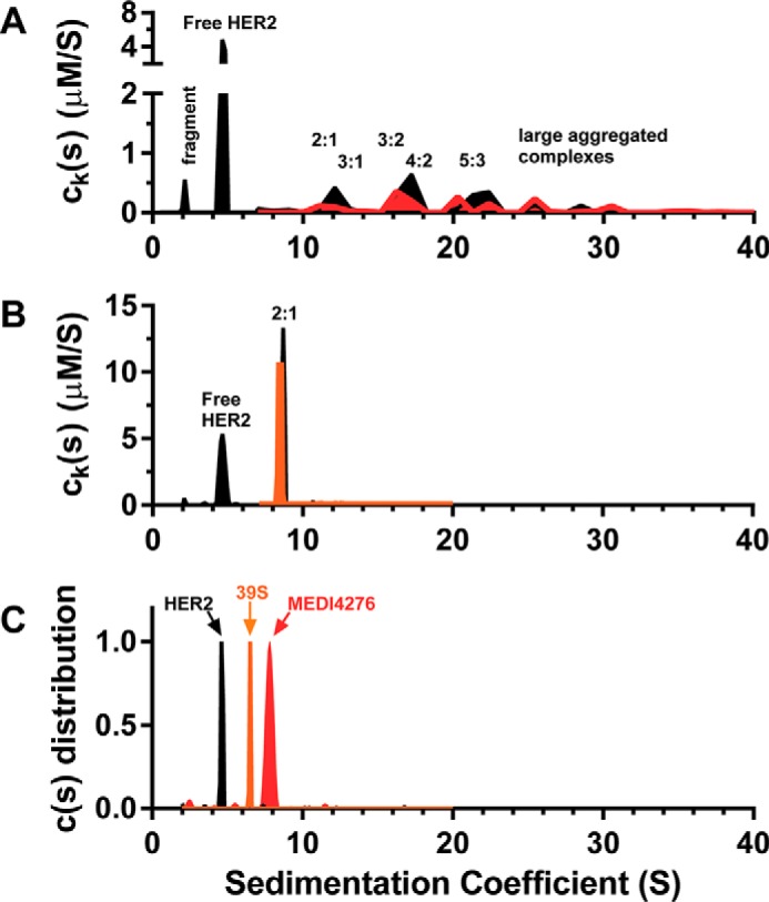
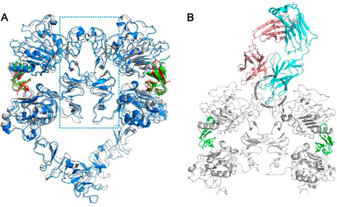
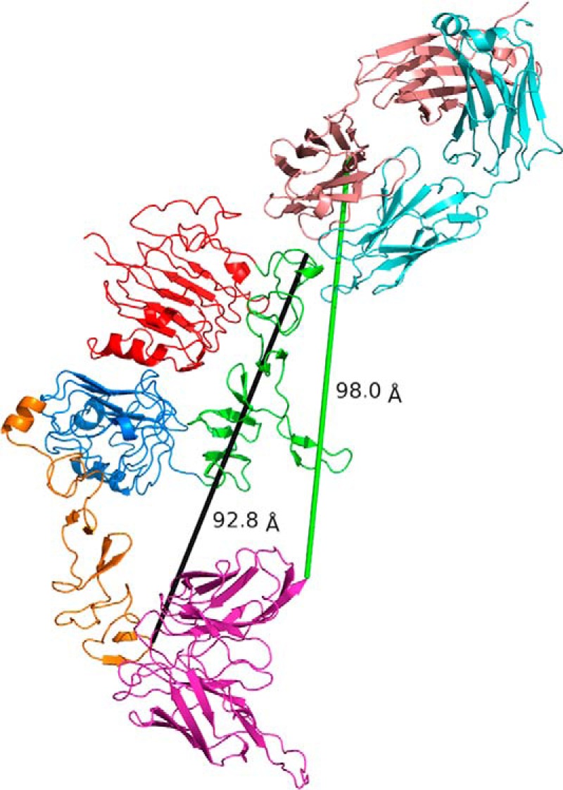
Pathways of human epidermal growth factor (EGF) receptors are activated upon ligand-dependent or -independent homo- or heterodimerization and their subsequent transphosphorylation. Overexpression of these receptors positively correlates with transphosphorylation rates and increased tumor growth rates. MEDI4276, an anti-human epidermal growth factor receptor 2 (HER2) biparatopic antibody-drug conjugate, has two paratopes within each antibody arm. One, 39S, is aiming at the HER2 site involved in receptor dimerization and the second, single chain fragment (scFv), mimicking trastuzumab. Here we present the cocrystal structure of the 39S Fab-HER2 complex and, along with biophysical and functional assays, determine the corresponding epitope of MEDI4276 and its underlying mechanism of action. Our results reveal that MEDI4276's uniqueness is based first on the ability of its 39S paratope to block HER2 homo- or heterodimerization and second on its ability to cluster the receptors on the surface of receptor-overexpressing cells.

Keywords: analytical ultracentrifugation; antibody; crystallography; epitope mapping; phosphorylation.


Conflict of interest statement

The authors declare that they have no conflicts of interest with the contents of this article

Figures

Figure 1.
Figure 1.
A, cartoon of MEDI4276 ADC. Trastuzumab scFv is shown in magenta. Linkers between variable heavy and light, scFv and 39S, and the antibody hinge region are shown as gray coils. The 39S variable region is shown in dark yellow. The 39S constant region is shown in green. Conjugation sites are shown as red dots. B, structure and domain organization of the HER2 ecd. Subdomains I through IV are colored red, green, blue, and orange, respectively. Pertuzumab and trastuzumab epitopes are shown with curly brackets. All structure-related figures were prepared using PyMOL (Schrödinger LLC).
Figure 2.
Figure 2.
A, schematic of the human/mouse chimeric variants. The individual subdomains of the human and mouse HER2 ecd were swapped as illustrated to construct LoF and GoF variants. B, binding characterization of 39S to human/mouse chimeric variants. Binding and expression were calculated as percent to WT HER2, dividing the surface plasmon resonance response of 39S or anti-His polyclonal antibody response by that of WT HER2, respectively. Replacing subdomain II or segment Leu146-Pro208 or Ser192-Pro208 of human HER2 with mouse counterparts abolished the binding of 39S (LoF II, LoF Leu146-Pro208, and LoF Ser192-Pro208). Grafting either subdomain II or the region of Ser192-Pro208 of human HER2 into the mouse molecule led to recognition of the chimera by 39S (GoF II and GoF Ser192-Pro208). Results represent the means of three independent experiments, with error bars indicating standard deviations. C, amino acid alignment of the subdomains II between human (Hu) and mouse (Mo) HER2. Identical and similar amino acids are shown in green and red, respectively. Segments denoted Leu146-Pro208, Leu159-Ile162, His171-Ser187, Ser192-Pro208, or Val250-His296 with lines are ones swapped between human and mouse HER2 to construct chimeric variants. D, mapping the 39S functional epitope on HER2 structure. The functional epitope of 39S, shown as spheres, is the most distant from the juxtamembrane part of the molecule.
Figure 3.
Figure 3.
A, content of an asymmetric unit of crystal containing 39S Fab and HER2 ecd. HER2 domains I through IV are colored red, green, blue, and orange, respectively. Fab heavy and light chains are shown in sand and cyan, respectively. B, view of interaction between 39S Fab and HER2 domain II. Electrostatic surface potential, calculated for the tip of domain II using the APBS plugin in PyMOL, shows that nature of the interaction is mostly hydrophobic. C, close-up view showing the interaction of both chains of 39S Fab with domain II of HER2. The coloring scheme is as in A and B. D, fragment of the final electron density map around glycosylated Asn237 showing two consecutive GlcNAc residues at 1.6 σ. The carving radius is 9 Å. The structure was determined to a resolution of 3.8 Å.
Figure 3.
Figure 3.
A, content of an asymmetric unit of crystal containing 39S Fab and HER2 ecd. HER2 domains I through IV are colored red, green, blue, and orange, respectively. Fab heavy and light chains are shown in sand and cyan, respectively. B, view of interaction between 39S Fab and HER2 domain II. Electrostatic surface potential, calculated for the tip of domain II using the APBS plugin in PyMOL, shows that nature of the interaction is mostly hydrophobic. C, close-up view showing the interaction of both chains of 39S Fab with domain II of HER2. The coloring scheme is as in A and B. D, fragment of the final electron density map around glycosylated Asn237 showing two consecutive GlcNAc residues at 1.6 σ. The carving radius is 9 Å. The structure was determined to a resolution of 3.8 Å.
Figure 4.
Figure 4.
A and B, MSSV results for 4:1 loading of the HER2 and MEDI4276 biparatopic antibody (A) and 4:1 loading of HER2 and the 39S mAb (B). C, control samples of HER2, 39S mAb, and MEDI4276, which were run separately and plotted together with a normalized y axis. HER2 is depicted as a black fill, 39S mAb in orange, and MEDI4276 in red.
Figure 5.
Figure 5.
A–C, inhibition of Heregulin 1-β1–dependent Akt phosphorylation was evaluated in the breast cancer T47D (A), MDA-MB-361 (B), and bladder cancer RT-112 (C) cell lines using a multiplexed Ser(P)473/total Akt sandwich immunoassay kit. D and E, the similarity of inhibition of HRG1-β1–dependent Akt phosphorylation by unconjugated MEDI4276 (Bs2–39SH) and combination of the parental antibodies. HER2 expression was evaluated in tumor xenografts by IHC test (data not shown), and the IHC intensity scoring is shown in parentheses for each cell line.
Figure 6.
Figure 6.
A, crystal structures of HER1 (blue and red for HER1 and EGF, respectively; PDB code 3NJP; 36) and HER4 (gray and green for HER4 and neuregulin, respectively; PDB code 3U7U; 22) ligand-induced dimers, superimposed, show that the dimerization interface is solely made of subdomains II (the area within the blue dotted rectangle). B, superimposition of either of those homodimers with the 39S Fab-HER2 structure shows that 39S causes steric hindrance and prevents homo- and heterodimerization of HER molecules. The area of clash is shown by the black circle.
Figure 7.
Figure 7.
39S and trastuzumab epitopes are located at opposite ends of HER2 molecule at a distance of more than 90 Å from each other (black line). The (G4S)3 linker employed in MEDI4276 can hardly stretch to even 60 Å and will not be able to cover the distance of 98 Å (green line) that would be required to allow the C-terminal residue of trastuzumab scFv and N-terminal amino acid of 39S heavy chain to bind simultaneously to the same antigen molecule. This suggests that MEDI4276 cannot bind two HER2 receptors and have all its paratopes occupied at the same time.

References

    1. Stein R. A., and Staros J. V. (2000) Evolutionary analysis of the ErbB receptor and ligand families. J. Mol. Evol. 50, 397–412 10.1007/s002390010043 - DOI - PubMed
    1. Olayioye M. A., Neve R. M., Lane H. A., and Hynes N. E. (2000) The ErbB signaling network: receptor heterodimerization in development and cancer. EMBO J. 19, 3159–3167 10.1093/emboj/19.13.3159 - DOI - PMC - PubMed
    1. Ushiro H., and Cohen S. (1980) Identification of phosphotyrosine as a product of epidermal growth factor-activated protein kinase in A-431 cell membranes. J. Biol. Chem. 255, 8363–8365 - PubMed
    1. Schreiber A. B., Libermann T. A., Lax I., Yarden Y., and Schlessinger J. (1983) Biological role of epidermal growth factor-receptor clustering: investigation with monoclonal anti-receptor antibodies. J. Biol. Chem. 258, 846–853 - PubMed
    1. Chung I., Akita R., Vandlen R., Toomre D., Schlessinger J., and Mellman I. (2010) Spatial control of EGF receptor activation by reversible dimerization on living cells. Nature 464, 783–787 10.1038/nature08827 - DOI - PubMed

Publication types