. 2018 Apr;8(4):170240.
doi: 10.1098/rsob.170240.

Single cardiac ventricular myosins are autonomous motors

Affiliations

Single cardiac ventricular myosins are autonomous motors

Yihua Wang et al. Open Biol. 2018 Apr.

Abstract

Myosin transduces ATP free energy into mechanical work in muscle. Cardiac muscle has dynamically wide-ranging power demands on the motor as the muscle changes modes in a heartbeat from relaxation, via auxotonic shortening, to isometric contraction. The cardiac power output modulation mechanism is explored in vitro by assessing single cardiac myosin step-size selection versus load. Transgenic mice express human ventricular essential light chain (ELC) in wild- type (WT), or hypertrophic cardiomyopathy-linked mutant forms, A57G or E143K, in a background of mouse α-cardiac myosin heavy chain. Ensemble motility and single myosin mechanical characteristics are consistent with an A57G that impairs ELC N-terminus actin binding and an E143K that impairs lever-arm stability, while both species down-shift average step-size with increasing load. Cardiac myosin in vivo down-shifts velocity/force ratio with increasing load by changed unitary step-size selections. Here, the loaded in vitro single myosin assay indicates quantitative complementarity with the in vivo mechanism. Both have two embedded regulatory transitions, one inhibiting ADP release and a second novel mechanism inhibiting actin detachment via strain on the actin-bound ELC N-terminus. Competing regulators filter unitary step-size selection to control force-velocity modulation without myosin integration into muscle. Cardiac myosin is muscle in a molecule.

Keywords: Qdot labelled actin under load; cardiomyopathy-linked mutants; ratcheting myosin essential light chain; single cardiac myosin mechanics; super-resolution microscopy.


Conflict of interest statement

We declare we have no competing interests.

Figures

Figure 1.
Figure 1.
SDS-PAGE of nontransgenic (NTg) and transgenic WT, A57G and E143K myosin preparations. MHC is the mouse cardiac α-myosin heavy chain. Human versus total ELC is indicated with %.
Figure 2.
Figure 2.
Actin-activated myosin ATPase versus actin concentration [A] for wild-type (WT) and mutants E143K and A57G. Error bars show standard deviation for sampling statistics given in table 1 and under experimental conditions given in Material and methods. Fitted curves use equation (1.1). Significance of ATPase rate versus [A] data in pairwise comparison for the WT and mutant species is indicated. They differ significantly with confidence level p < 0.01 as indicated by **.
Figure 3.
Figure 3.
Event versus velocity histograms (solid squares) for WT, A57G and E143K myosins and for frictional actin loading, Ff, from 0 to 465 pN. The solid red line is the baseline due to thermal/mechanical fluctuations that was subtracted from the raw data to give the data points at solid squares. Black lines are simulations conducted as described in ‘Material and methods’ and used to estimate step-size (at arrows) and step-frequency (figure 4). Natural velocity units (vu) have 1 vu = (dIt) for dI the intermediate step-size (green down arrow at approx. 5–6 nm) between the short (red up arrow at approx. 2–3 nm) and long (blue up arrow at approx. 8–9 nm) step-sizes. Step-sizes have standard deviation of approximately 0.5 nm for replicates described in ‘Statistics’. The intermediate step-size for the E143K species under 0 load differs significantly from equivalent WT species with confidence level p < 0.01 as indicated by **. Other step-sizes are not significantly different for confidence level p < 0.05.
Figure 4.
Figure 4.
The step-frequencies for the WT (top row), A57G (middle row) and E143K (bottom row) under frictional loads indicated in each panel in pN units. Curves are derived from simulation of the corresponding event–velocity histograms in figure 3 and as described in the Qdot assay event–velocity histogram simulation’ section. Columns are equivalently loaded actin for each transgenic protein. Errors are SDs for replicates described in ‘Statistics’. Intermediate step-frequency (green) for the A57G species differs significantly from equivalent WT species with confidence level p < 0.01 for no load and as indicated by **. Short (red) and long (blue) step-frequencies for the A57G and WT species also differ significantly with confidence level p < 0.01 under maximum load and as indicated by **.
Figure 5.
Figure 5.
In vitro single myosin (a) average step-size formula image 
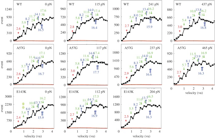
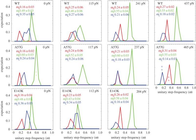
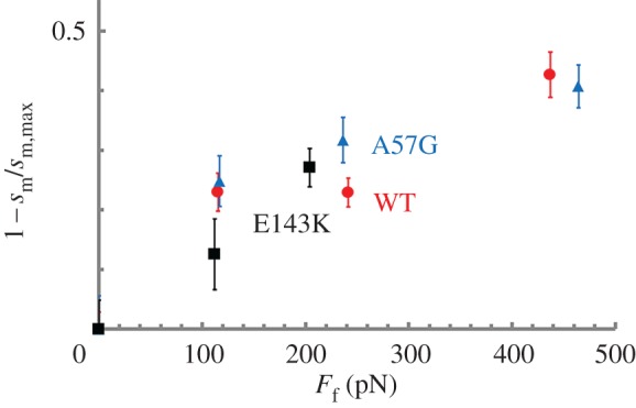
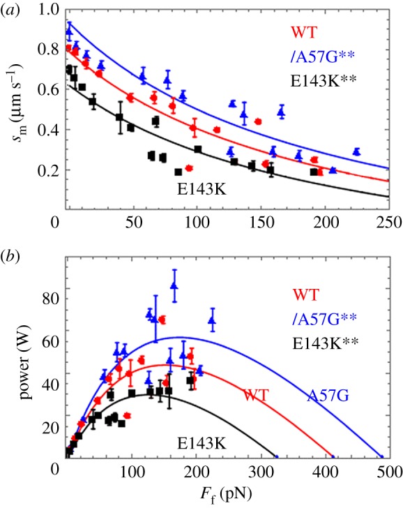
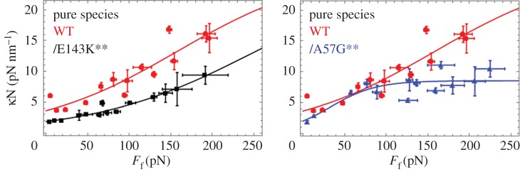
(b) average duty ratio and (c) power for WT (red), A57G (blue) and E143K (black) αcardiac myosin measured by the Qdot assay under a retarding force. Data points are derived from simulation of the corresponding event–velocity histograms in figure 3 and as described in ‘Qdot assay event–velocity histogram simulation’. Errors are standard error of the mean for the 16–55 replicates described in ‘Statistics’. Data in pairwise comparison for the WT and mutant species are indicated for each panel. They differ significantly with confidence level p < 0.05 or 0.01 indicated by * or **.
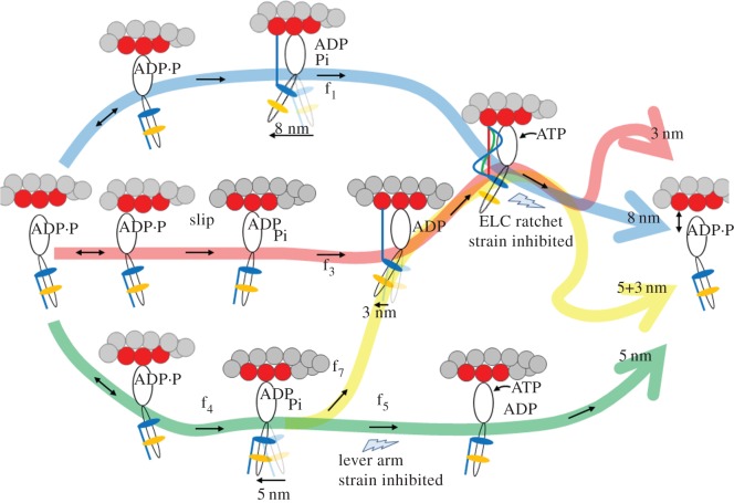
Figure 6.
Figure 6.
The four-pathway network for in vivo and in vitro cardiac myosin unitary steps. Myosin powerstroke has two sequential steps with Pi release driving the larger lever arm swing (nominal 5 nm step-size) followed by the ELC N-term binding actin and ADP release driving the smaller lever-arm swing (3 nm step-size). In the blue pathway with the 8 nm step-size, large and small steps are tightly coupled for the maximum lever-arm swing. In the bifurcated green/yellow pathway, Pi release with the 5 nm step is not immediately followed by ADP release and the 3 nm step due to the lever arm strain inhibition at the lower thunderbolt. The delay allows the powerstroke a choice to terminate with myosin detachment for a terminal 5 nm step-size (terminal half of green path) or complete the second 3 nm step (5 + 3 nm step-size via the yellow path). In vivo data provided evidence for a solo-3 nm step-size (red pathway) [12]. We propose in the figure that the myosin in this pathway slips to releases Pi without net forward movement, but then the ELC N-term binds actin permitting ADP release and completion of a solo-3 nm step. Myosin flux values through each pathway are f1 (blue pathway), f3 (red), f7 (yellow), f4 (green main channel) and f5 (green terminus) using nomenclature from [12]. Relative flux values are listed in table 2. The ELC-ratchet strain activated filter at upper thunderbolt regulates the solo-3, 8 and 5 + 3 nm pathways (red, blue and yellow, respectively). ELC ratchet strain inhibits ATP binding and ELC N-terminal detachment from actin maintaining tension at peak isometric force. The upper and lower lightning bolts indicate strain-regulated checkpoints that modulate step-frequencies for quickly responding to changing force-velocity demands.
Figure 7.
Figure 7.
The actin filament fraction under isometric loading versus loading force estimated from the motility of Qdot labelled actin and equation (2.13). Errors are standard deviation for the 16–55 replicates described in ‘Statistics’.
Figure 8.
Figure 8.
Motility velocity, sm, for myosin isoforms WT (red circles), A57G (blue triangles) and E143K (black squares) versus α-actinin concentration. Sample sizes for the SDs indicated by error bars are WT (n = 11), A57G (n = 7) and E143K (n = 7) for n the number of acquisitions corresponding to 1 in vitro motility movie per acquisition. Fitted curves are based on equation (2.2) with the friction coefficient c([α]) converted to calibrated units (pN s µm−1) using equation (2.3) and ensemble myosin driving force Fd equal to isometric force.
Figure 9.
Figure 9.
(a) Motility velocity (sm) and (b) power in aW for myosin isoforms WT (red circles), A57G (blue triangles) and E143K (black squares) versus calibrated frictional force, Ff, exerted by the α-actinin. Sample sizes for the SDs indicated by error bars are WT (n = 11), A57G (n = 7) and E143K (n = 7) for n the number of acquisitions corresponding to 1 in vitro motility movie per acquisition. Significance testing of sm versus Ff data in (a) and power versus Ff data in (b) in pairwise comparison for the WT and mutant species indicates that they differ significantly with confidence level p < 0.01 denoted by **. Fitted curves are based on equations (2.5) and (2.6) (Hill equation) for (a,b).
Figure 10.
Figure 10.
Ensemble elasticity κN versus frictional loading force Ff for each homogeneous species computed using equations (2.3) and (2.17). Two-dimensional error bars indicate standard deviation for n = 5–51 trials at each data point. Pairwise comparison for the WT and mutant species show that they differ significantly with confidence level p < 0.01 indicated by **.

References

    1. Geeves MA, Fedorov R, Manstein DJ. 2005. Molecular mechanism of actomyosin-based motility. Cell. Mol. Life Sci. 62, 1462–1477. (doi:10.1007/s00018-005-5015-5) - DOI - PMC - PubMed
    1. Huxley AF. 1957. Muscle structure and theories of contraction. Prog. Biophys. Chem. 7, 255–318. - PubMed
    1. Rayment I, Holden HM. 1994. The three-dimensional structure of a molecular motor. Trends Biochem. Sci. 19, 129–134. (doi:10.1016/0968-0004(94)90206-2) - DOI - PubMed
    1. Sherwood JJ, Waller GS, Warshaw DM, Lowey S. 2004. A point mutation in the regulatory light chain reduces the step size of skeletal muscle myosin. Proc. Natl Acad. Sci. USA 101, 10 973–10 978. (doi:10.1073/pnas.0401699101) - DOI - PMC - PubMed
    1. Steffen W, Smith D, Simmons R, Sleep J. 2001. Mapping the actin filament with myosin. Proc. Natl Acad. Sci. USA 98, 14 949–14 954. (doi:10.1073/pnas.261560698) - DOI - PMC - PubMed

Publication types

LinkOut - more resources