. 2018 May 1;115(18):4773-4778.
doi: 10.1073/pnas.1721345115. Epub 2018 Apr 18.

Origin, antigenicity, and function of a secreted form of ORF2 in hepatitis E virus infection

Affiliations

Origin, antigenicity, and function of a secreted form of ORF2 in hepatitis E virus infection

Xin Yin et al. Proc Natl Acad Sci U S A. .

Abstract

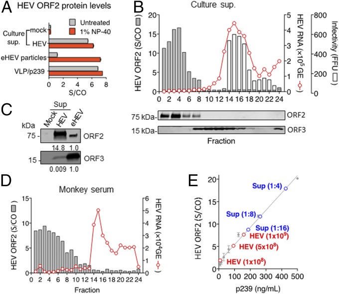
The enterically transmitted hepatitis E virus (HEV) adopts a unique strategy to exit cells by cloaking its capsid (encoded by the viral ORF2 gene) and circulating in the blood as "quasi-enveloped" particles. However, recent evidence suggests that the majority of the ORF2 protein present in the patient serum and supernatants of HEV-infected cell culture exists in a free form and is not associated with virus particles. The origin and biological functions of this secreted form of ORF2 (ORF2S) are unknown. Here we show that production of ORF2S results from translation initiated at the previously presumed AUG start codon for the capsid protein, whereas translation of the actual capsid protein (ORF2C) is initiated at a previously unrecognized internal AUG codon (15 codons downstream of the first AUG). The addition of 15 amino acids to the N terminus of the capsid protein creates a signal sequence that drives ORF2S secretion via the secretory pathway. Unlike ORF2C, ORF2S is glycosylated and exists as a dimer. Nonetheless, ORF2S exhibits substantial antigenic overlap with the capsid, but the epitopes predicted to bind the putative cell receptor are lost. Consistent with this, ORF2S does not block HEV cell entry but inhibits antibody-mediated neutralization. These results reveal a previously unrecognized aspect in HEV biology and shed new light on the immune evasion mechanisms and pathogenesis of this virus.

Keywords: antibody neutralization; hepatitis E virus; immunological decoy; leaky translation; quasi-envelopment.


Conflict of interest statement

The authors declare no conflict of interest.

Figures

Fig. 1.
Fig. 1.
HEV-infected cells release a large amount of nonvirion-associated ORF2 protein both in vitro and in vivo. (A) Detection of HEV ORF2 protein in culture supernatants (sup) (1:32 dilution) of mock or HEV (Kernow C1/p6) persistently infected Huh-7 cells, purified quasi-enveloped HEV particles (eHEV) (109 GE), and HEV VLP p239 (12.5 ng) with or without 1% Nonidet P-40 treatment. HEV ORF2 protein was measured by a commercial HEV antigen ELISA (Wantai). Signal to cut-off (S/CO) ratio was determined based on optical density values measured at 450 nm. Data represent mean ± SEM from two independent experiments each in duplicate wells. (B, Upper) Rate-zonal gradient profile of HEV RNA, ORF2 protein, and infectious virions in the culture supernatant of HEV-infected Huh-7 cells. Fractions were collected from the top of the gradient. (Lower) Western blots of HEV ORF2 and ORF3 in the same gradient fractions (each lane contained two consecutive fractions) with a chimpanzee convalescent serum (Ch1313) and a rabbit anti-ORF3 antibody, respectively. (C) Protein abundance of HEV ORF2 and ORF3 in culture supernatants of HEV persistently infected Huh-7 cells and gradient purified eHEV particles (∼109 GE). The relative protein abundance is shown below each blot. (D) Rate-zonal gradient profile of HEV RNA and ORF2 protein in a serum sample from a genotype 3 HEV (Kernow C1)-infected rhesus macaque. (E) ELISA quantitation of HEV ORF2 protein in the culture supernatant with different dilutions and Nonidet P-40-treated purified eHEV particles. Serially diluted VLP (p239) was used for creating a standard curve.
Fig. 2.
Fig. 2.
The secreted form of ORF2 (ORF2S) is glycosylated and dimerized. (A) Effects of PNGase F, O-glycosidase (O-Gly-ase), and neuraminidase (NA) treatment on ORF2 in virions and in supernatants of Huh-7 cells transfected with a WT HEV RNA or a variant carrying mutations in three putative N-linked glycosylation sites (N137/310/562Q). ORF2 was detected by a chimpanzee convalescent serum (Ch1313). (B) HEV persistently infected Huh-7 cells were treated with brefeldin A (BFA) for 24 h or left untreated (Ctrl). Cells and supernatants were collected and analyzed by immunoblotting with indicated antibodies. (C) Effects of β-mecaptomethanol (β-ME) and heat treatment (95 °C, 10 min) on the size of ORF2S in a native protein gel. (D) Determination of the size of ORF2S by size-exclusion chromatography. The majority of ORF2S was eluted as peak 1 with an estimated size of 180 kDa. The column contaminants were eluted in the second peak (peak 2). The presence of ORF2S in peak fractions was confirmed by Western blot analysis using Ch1313.
Fig. 3.
Fig. 3.
ORF2S and ORF2C are different translation products of the HEV ORF2 gene. (A) The N terminus sequence of the HEV subgenomic RNA (Kernow C1/p6) and strategies for site-directed mutagenesis. The AUG start codons for ORF3, ORF2S, and ORF2C are underlined, and the corresponding protein sequences are shown below. The first AUG and the internal AUG codons of the ORF2 gene are mutated either singly (mut1 and mut2) or in combination (mut1+2) without altering the amino acid sequence of ORF3. The putative signal peptide cleavage site in ORF2S is indicated (arrowhead). (B) qRT-PCR analysis of intracellular RNA levels in Huh-7 cells transfected with in vitro transcripts of WT or mutant HEV RNA (5-d posttransfection). Data are shown as mean ± SEM of two independent experiments each performed in duplicate wells. LOD, limit of detection. (C) Huh-7 cells transfected with WT or mutant HEV RNA for 5 d were stained with a chimpanzee convalescent serum Ch1313 (green). Nuclei were stained with DAPI (blue). (Scale bars, 100 μm.) (D) Immunoblots of intracellular and extracellular ORF2 and ORF3 proteins at 5 d after HEV RNA transfection into Huh-7 cells. β-Actin was used as a loading control. ORF2 was detected by Ch1313. ORF3 was detected with a rabbit anti-ORF3 antibody. (E) Isopycnic gradient profile of intracellular HEV RNA (as a surrogate marker of virions) in Huh-7 cells transfected with different HEV RNA (15-d posttransfection). Cells were freeze-thawed three times and treated with RNase before gradient centrifugation. Fractions were collected from the top of the gradient. (F and G) HepG2/shMAVS cells were inoculated with equal amounts of WT or mutant HEV (10,000 GE per cell) isolated from HEV RNA-transfected Huh-7 cells. Cells were stained with Ch1313 (green) and DAPI (blue) at 5-d postinoculation (F). (Scale bars, 100 μm.) Supernatant HEV RNA levels (5 d after inoculation) were measured by qRT-PCR (G). Data are shown as mean ± SEM of two independent experiments each performed in duplicate wells.
Fig. 4.
Fig. 4.
ORF2S antigenically overlaps with virions and interferes with antibody-mediated neutralization but not entry of HEV. (A) Location of six distinct antigenic clusters (C1–C6) on the protrusion domain of the HEV capsid (genotype 1). (B) Comparison of antigenicity of HEV recombinant VLP (rVLP, genotypes 1 and 4), recombinant ORF2 dimer (rDimer, genotypes 1, 3, and 4), ORF2S (OS), and virions (V) [Xinjiang-1 (genotype 1), Kernow C1/p6 (genotype 3)] in a sandwich ELISA with a rabbit anti-ORF2 antibody as a capture antibody and indicated anti-ORF2 monoclonal antibodies as the detection antibody. (C) Nonenveloped HEV or eHEV particles (1,000 GE per cell) were incubated with indicated concentrations of purified ORF2S (37 °C, 1 h) before inoculation of HepG2/shMAVS cells. Relative infectivity was calculated based on the numbers of foci determined by indirect IFAs with Ch1313 5 d later. Data represent mean ± SEM of two independent experiments each performed in duplicate wells. (D) Serially diluted monoclonal (6H8 and 9F7) or polyclonal (Ch1313) anti-HEV antibodies were mixed with indicated concentrations of purified ORF2S at 37 °C for 2 h before incubation with HEV (1 × 10⁷ GE) for another 2 h. The mixture was then inoculated to HepG2 cells (4 × 10⁴ cells per well). Percent neutralization was calculated based on the number of HEV+ foci 5 d after inoculation. Shown are representative results from two independent experiments.

References

    1. Kamar N, et al. Hepatitis E virus infection. Nat Rev Dis Primers. 2017;3:17086. - PubMed
    1. Smith DB, et al. International Committee on Taxonomy of Viruses Hepeviridae Study Group Consensus proposals for classification of the family Hepeviridae. J Gen Virol. 2014;95:2223–2232. - PMC - PubMed
    1. Pérez-Gracia MT, Suay-García B, Mateos-Lindemann ML. Hepatitis E and pregnancy: Current state. Rev Med Virol. 2017;27:e1929. - PubMed
    1. Meng XJ. Zoonotic and foodborne transmission of hepatitis E virus. Semin Liver Dis. 2013;33:41–49. - PubMed
    1. Pavio N, Meng XJ, Doceul V. Zoonotic origin of hepatitis E. Curr Opin Virol. 2015;10:34–41. - PubMed

Publication types

LinkOut - more resources