Comparative Study
. 2018 Apr 19;3(8):e97828.
doi: 10.1172/jci.insight.97828.

CX3CR1 identifies PD-1 therapy-responsive CD8+ T cells that withstand chemotherapy during cancer chemoimmunotherapy

Affiliations
Comparative Study

CX3CR1 identifies PD-1 therapy-responsive CD8+ T cells that withstand chemotherapy during cancer chemoimmunotherapy

Yiyi Yan et al. JCI Insight. .

Abstract

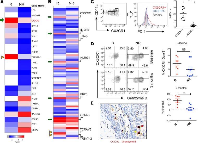
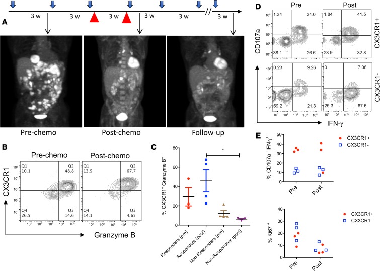
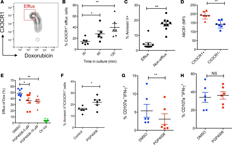
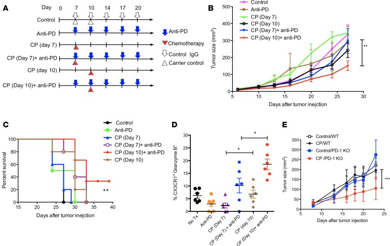
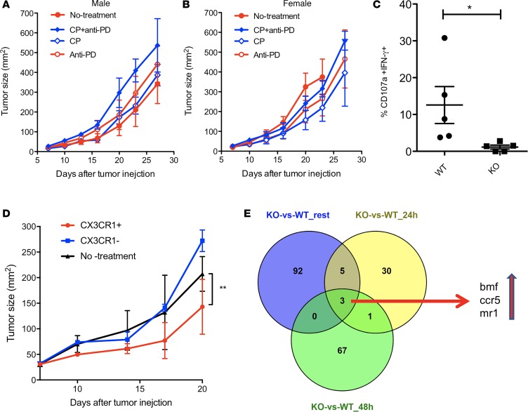
Although immune checkpoint inhibitors have resulted in durable clinical benefits in a subset of patients with advanced cancer, some patients who did not respond to initial anti-PD-1 therapy have been found to benefit from the addition of salvage chemotherapy. However, the mechanism responsible for the successful chemoimmunotherapy is not completely understood. Here we show that a subset of circulating CD8+ T cells expressing the chemokine receptor CX3CR1 are able to withstand the toxicity of chemotherapy and are increased in patients with metastatic melanoma who responded to chemoimmunotherapy (paclitaxel and carboplatin plus PD-1 blockade). These CX3CR1+CD8+ T cells have effector memory phenotypes and the ability to efflux chemotherapy drugs via the ABCB1 transporter. In line with clinical observation, our preclinical models identified an optimal sequencing of chemoimmunotherapy that resulted in an increase of CX3CR1+CD8+ T cells. Taken together, we found a subset of PD-1 therapy-responsive CD8+ T cells that were capable of withstanding chemotherapy and executing tumor rejection with their unique abilities of drug efflux (ABCB1), cytolytic activity (granzyme B and perforin), and migration to and retention (CX3CR1 and CD11a) at tumor sites. Future strategies to monitor and increase the frequency of CX3CR1+CD8+ T cells may help to design effective chemoimmunotherapy to overcome cancer resistance to immune checkpoint blockade therapy.

Keywords: Cancer immunotherapy; Cellular immune response; Immunology; Oncology; T cells.


Conflict of interest statement

Conflict of interest: The authors have declared that no conflict of interest exists.

Figures

Figure 1
Figure 1. CX3CR1+Granzyme B+CD8+ T cells in responders to PD-1 therapy.
RNA was isolated from CD11ahiCD8+ T cells in the peripheral blood of melanoma patients prior to (A) or after (B) anti–PD-1 therapy. (A) RNA-seq data show an increased transcription of CX3CR1 (arrow) and TCRVβ29-1 (arrow head) in the responders (R, n = 3) compared with the nonresponders (NR, n = 3) at baseline prior to anti–PD-1 therapy. Data represent the average levels of transcription of 3 patients (with at least 1.5-fold changes). (B) RNA-seq data show increased transcriptions of CX3CR1, CD122 (IL2RB), KLRG1, perforin (PRF1), granzyme B (GZMB) (arrows), and TCRVα5/TCRVβ4-2 (arrow heads) on week 12 after PD-1 therapy. Data represent the average levels of transcription of 3 or 2 patients (R, n = 3; NR, n = 2) with at least 2-fold changes. (C) PD-1 expression by CX3CR1+CD11ahi or CX3CR1CD11alo CD8+ T cells isolated from the peripheral blood of patients with metastatic melanoma prior to PD-1 therapy (n = 12, ***P < 0.01, paired 2-tailed t test). (D) The frequency of CX3CR1+Granzyme B+ cells among CD11ahiCD8+ T cells significantly increased in responders after anti–PD-1 therapy in melanoma patients (n = 7, **P < 0.05, Mann-Whitney U test) but not at baseline prior to PD-1 therapy. (E) Tissue staining of CX3CR1+Granzyme B+ (double-positive staining, DP) in human melanoma tissues. Original magnification ×400. One DP cell was inside the tumor bed (red arrow) and another adhered to a blood vessel, probably in a process of extravasation (yellow arrow).
Figure 2
Figure 2. Patient responses to chemoimmunotherapy with an increase of CX3CR1+Granzyme B+CD8+ T cells.
(A) Treatment schedule and clinical responses of a patient with metastatic melanoma who received pembrolizumab single-agent and chemotherapy paclitaxel and carboplatin (red arrow head) that were initiated at 175 mg/m2, and an AUC (area under curve) of 5 every 3 weeks for 2 cycles in combination with pembrolizumab. PET/CT scan results were collected at each time point (arrows) to demonstrate the disease status. Patient received a total of 12 cycles of pembrolizumab at the end of follow up, shown here. (B) Following the same schedule of treatment as in A, blood samples were collected for flow analysis of CX3CR1+Granzyme B+ among CD11ahiCD8+ T cells. (C) The frequency of CX3CR1+Granzyme B+ among CD11ahiCD8+ T cells in responders (n = 3 pre, 4 post) and nonresponders (n = 4) before and after chemotherapy as treated in A. *P < 0.05 compared between responders and nonresponders to CIT (1-way ANOVA, P = 0.024). (D) Representative flow cytometry data showing the CTL function of CX3CR1+ or CX3CR1 CD8+ T cells from one of the chemoimmunotherapy responders (n = 3) after a brief ex vivo stimulation of T cells with PMA and ionomycin. (D and E) CTL function (CD107a expression and IFN-γ production) and proliferation (Ki67 expression) of CX3CR1+ or CX3CR1 CD8+ T cells in responders (n = 3) prior to and after PD-1 therapy.
Figure 3
Figure 3. Efflux of chemotherapy drug by human CX3CR1+CD8+ T cells.
Purified human primary CD8+ T cells were loaded with Doxorubicin (1 μg/ml) for 30 minutes and then washed before further incubation for 60 minutes (A) or at indicated times (B). Gated areas in A are efflux cells (DoxloCX3CR1hi). The data was analyzed by 1-way ANOVA (*P < 0.05, **P < 0.01, n = 6). (C) CD8+ T cells were incubated with Doxorubicin (0.5 μg /ml) for 40 hours and then stained with annexin V to identify apoptotic cells. (D) Expression of ABCB1 by CX3CR1+ or CX3CR1 CD8+ T cells. (E) ABCB1 inhibitor (PGP4008) reduced the drug efflux ability of CX3CR1+CD8+ T cells. Cells incubated on ice after loading with drug were used as a negative control for drug efflux. Data was analyzed by 1-way ANOVA (*P < 0.05, **P < 0.01, n = 7). (F) ABCB1 inhibitor (PGP4008) increased the apoptosis of CX3CR1+CD8+ T cells as cultured in C. The impact of ABCB1 inhibitor on the function of human CX3CR1+CD8+ T cells incubated with (G) or without (H) chemotherapy drug (carboplatin and paclitaxel). CD8+ T cells were activated with anti-CD3/CD28 beads for 24 hours in the presence of DMSO (control) or PGP4008 (10 μM). The CTL function was measured for CD107a expression and IFN-γ production at the end of culture. The data of CD and F–H were analyzed by Mann-Whitney U test, 2-tailed (*P < 0.05; **P < 0.01, n = 5–7).
Figure 4
Figure 4. CX3CR1+Granzyme B+CD8+ T cells increased after chemoimmunotherapy.
Once B16F10 mouse melanoma tumors were palpable on day 7 after tumor injection, animals were randomly assigned to treatment groups. (A) Schedule of treatments. Mice were treated with i.p. injection of anti–PD-1 and –PD-L1 antibody (at 100 μg of each antibody) and collectively indicated as anti-PD IgG for a total of 5 doses at 3-day intervals. Carboplatin (40 μg/g) and paclitaxel (10 μg/g body weight) (collectively indicated as CP) were injected i.p. once either on day 7 or on day 10 after tumor injection. (B) Tumor growth. Data show the mean ± SEM of 5 mice per group; **P < 0.01 compared between day 7 and 10 treatment with CP plus anti-PD (2-way ANOVA). (C) The survival curve of treated animals as in B. *P < 0.05 compared between control and anti-PD plus day 10 CP (log-rank test). (D) Frequency of CX3CR1+Granzyme B+CD8+ T cells was measured in CD11ahiCD8+ cells isolated from tumor tissues on day 16 after tumor injection (*P < 0.05, n = 6, 2-way ANOVA). (E) B16F10 tumor growth in WT and PD-1–KO mice after treatment with CP) as in B on day 8 after tumor injection. One of 2 independent experiments (***P < 0.001, n = 3–5, 2-way ANOVA).
Figure 5
Figure 5. CX3CR1 is required for CD8+ CTL to reject tumors during chemoimmunotherapy.
CX3CR1-deficient (A, male mice; B, female mice) mice were injected with B16F10 tumor cells and were treated with i.p. injection of anti–PD-1 and PD-L1 antibody (at 100 μg of each antibody and collectively indicated as anti-PD) or control IgG for a total of 5 doses at 3-day intervals on day 7 after tumor injection. Carboplatin (40 μg/g) and paclitaxel (10 μg/g body weight) (collectively indicated as CP) were injected i.p. once on day 10 after tumor injection. Data show the mean ± SEM of 5 mice per group. (C) Frequency of CD107a+IFN-γ+ among CD11ahiCD8+ T cells isolated from tumor tissues decreased in CX3CR1-KO mice compared with WT mice. *P < 0.05 (Mann-Whitney U test, 2-tailed, n = 5). (D) Adoptive transfer of CX3CR1+ OT-1 CD8+ T cells, but not CX3CR1 OT-1 CD8+ T cells, suppressed the growth of B16-OVA tumors. Data show the mean ± SEM of 5 mice per group. **P < 0.01 (2-way ANOVA). One of 2 independent experiments was shown. (E) Venn diagram shows 3 genes upregulated in CX3CR1-KO CD8+ T cells compared with WT CD8+ T cells, and the upregulation of these 3 genes was shared among groups of 3 statuses (resting, 24-hour, and 48-hour activation with anti-CD3/CD28 beads in vitro).

References

    1. Coit DG, et al. Melanoma, Version 2.2016, NCCN Clinical Practice Guidelines in Oncology. J Natl Compr Canc Netw. 2016;14(4):450–473. doi: 10.6004/jnccn.2016.0051. - DOI - PubMed
    1. Rizvi NA, et al. Nivolumab in Combination With Platinum-Based Doublet Chemotherapy for First-Line Treatment of Advanced Non-Small-Cell Lung Cancer. J Clin Oncol. 2016;34(25):2969–2979. doi: 10.1200/JCO.2016.66.9861. - DOI - PMC - PubMed
    1. Langer CJ, et al. Carboplatin and pemetrexed with or without pembrolizumab for advanced, non-squamous non-small-cell lung cancer: a randomised, phase 2 cohort of the open-label KEYNOTE-021 study. Lancet Oncol. 2016;17(11):1497–1508. doi: 10.1016/S1470-2045(16)30498-3. - DOI - PMC - PubMed
    1. Yan Y, et al. The Mayo Clinic experience in patients with metastatic melanoma who have failed previous pembrolizumab treatment. J Clin Oncol. 2016;34((15_suppl);):e21014
    1. Flaherty KT, et al. Phase III trial of carboplatin and paclitaxel with or without sorafenib in metastatic melanoma. J Clin Oncol. 2013;31(3):373–379. doi: 10.1200/JCO.2012.42.1529. - DOI - PMC - PubMed

Publication types

MeSH terms