. 2018 Apr 19;3(8):e98240.
doi: 10.1172/jci.insight.98240.

Cystic fibrosis-related diabetes is caused by islet loss and inflammation

Affiliations

Cystic fibrosis-related diabetes is caused by islet loss and inflammation

Nathaniel J Hart et al. JCI Insight. .

Abstract

Cystic fibrosis-related (CF-related) diabetes (CFRD) is an increasingly common and devastating comorbidity of CF, affecting approximately 35% of adults with CF. However, the underlying causes of CFRD are unclear. Here, we examined cystic fibrosis transmembrane conductance regulator (CFTR) islet expression and whether the CFTR participates in islet endocrine cell function using murine models of β cell CFTR deletion and normal and CF human pancreas and islets. Specific deletion of CFTR from murine β cells did not affect β cell function. In human islets, CFTR mRNA was minimally expressed, and CFTR protein and electrical activity were not detected. Isolated CF/CFRD islets demonstrated appropriate insulin and glucagon secretion, with few changes in key islet-regulatory transcripts. Furthermore, approximately 65% of β cell area was lost in CF donors, compounded by pancreatic remodeling and immune infiltration of the islet. These results indicate that CFRD is caused by β cell loss and intraislet inflammation in the setting of a complex pleiotropic disease and not by intrinsic islet dysfunction from CFTR mutation.

Keywords: Cell Biology; Diabetes; Endocrinology; Genetic diseases; Islet cells.


Conflict of interest statement

Conflict of interest: The authors have declared that no conflict of interest exists.

Figures

Figure 1
Figure 1. β Cell CFTR deletion did not alter murine oral glucose tolerance or islet insulin secretion.
(A) Murine models created to excise Cftr exon 11 from β cells in an inducible fashion (β Δ11) or from the pancreas with constitutive Cre action (Panc Δ11). (B) Experimental timeline of murine models. Analyses included oral glucose tolerance testing (OGTT) on conscious animals, insulin secretion assays, and RNA sequencing of whole islets. OGTT of (C) male β cell–specific/inducible mice prior to (Pre-Tx; n = 46) and after treatment with vehicle (V; n = 17) or tamoxifen (β Δ11; n = 23) and (D) pancreatic/constitutive mice homozygous for the Cftr wt allele (Panc wt; n = 8) and mice homozygous for the Cftr FL11 allele (Panc Δ11; n = 11). Insulin secretion from isolated islets incubated in medium containing (E and H) 5.6 mM glucose (5.6 G), 16.7 mM glucose (16.7 G), or (F and I) 16.7 mM glucose and 100 μM 3-isobutyl-1-methylxanthine (16.7 G + IBMX), and (G and J) islet insulin content from β cell–specific/inducible mice (E–G: V, n = 8, 5 male, 3 female; β Δ11, n = 9, 6 male, 3 female) and pancreatic/constitutive mice (H–J: Panc wt, n = 13, 6 male, 7 female; Panc Δ11, n = 20, 10 male, 10 female). We observed slight differences in glucose-stimulated insulin secretion, cAMP-potentiated GSIS, and islet insulin content between the control animals of the β Δ11 and Panc Δ11 models. However, the control animals were individualized for each model and differ in the type of Cre recombinase expressed as well as the expression promotor (A). Red represents the β cell–specific/inducible model (β Δ11), blue the pancreatic constitutive model (Panc Δ11). Data represent mean ± SEM. No statistical significance (P < 0.05) was observed in OGTT AUC, insulin secretion, or insulin content in either model. Statistical data were calculated with 1-way ANOVA (C and D) or unpaired 2-tailed Student’s t test (E–J).
Figure 2
Figure 2. CFTR mRNA expression was minimal in human β cells, and CFTR protein was undetectable in human β cells.
Expression of CFTR and select β cell–related transcripts from published islet cell transcriptomes: (A) 270 single human β cells from 6 healthy and 4 diabetic donors (reads per kilobase of transcript per million mapped reads [RPKM], Segerstolpe and Palasantza et al., ref. 38) and (B) sorted β cells from 7 healthy adult donors (transcript per kilobase million [TPM], Blodgett et al., refs. 39). Note: Individual expression values are not presented in A, as the log2 of the mean expression value of 270 β cells was calculated to account for the approximately 85% of β cells in this data set that do not express CFTR; individual CFTR expression values are presented in Supplemental Figure 3E. Green bar, insulin; blue bars, key islet transcription factors; pink, islet hormone secretion related genes; red, CFTR. (C) Representative immunohistochemical labeling of CFTR (red), insulin (green), and glucagon (purple) in a pancreas from 3-month-old male donor. Insets depict the islet border and interior. (D) CFTR (red) channel alone (note: CFTR ductal localization and islet absence). Scale bars: 50 μm (C and D); 10 μm (insets). (E) Representative patch clamp recording of a human β cell and a INS832/13 + wtCFTR cell (n = 5 donors, 35 β cells; Supplemental Figure 5B). (F) Insulin secretion from human islets (n = 4 donors) in medium containing 1 mM glucose (1 G), 16.7 mM glucose (16.7 G), or 16.7 G plus 100 μM forskolin (16.7 G + Fsk) and no drug (white), 1 μM VX770 (blue, ivacaftor), 5 μM VX661 (green), or 5 μM VX809 (red, lumacaftor). 1 G, n = 22–24 replicates; 16.7 G, n = 11–12 replicates; 16.7 + Fsk, n = 10–12 replicates. VX770 is a selective CFTR potentiator that increases CFTR activity at the membrane and VX661, and VX809 are CFTR channel correctors that increase membrane channel density. Data represent mean ± SEM. No statistical significance (P < 0.05) was observed in in vitro human islet insulin secretion when comparing secretory responses at 1 G, 16.7 G, and 16.7 G + Fsk in the presence of absence of CFTR modulators. One-way ANOVA was used for statistical analysis.
Figure 3
Figure 3. Pancreatic processing allowed for multidisciplinary investigation of the CF pancreas.
Acquisition of intact pancreatic tissue and islets from the same pancreas allowed for integration of in situ pancreatic characteristics with in vitro islet function, the islet transcriptome, and islet immune infiltration.
Figure 4
Figure 4. CF pancreata were severely remodeled, with ectopic adipose and fibrotic tissue deposition accompanied by β cell loss.
Characteristic CF-related pancreatic pathology observed in (A) donor 1: islet aggregations (black arrowhead) with inter-islet fibrosis (blue arrowhead) and islets in adipose niches (yellow arrowhead). Pathology observed in (B) donor 2: ectopic adipose (white arrowhead) and fibrotic deposition (red arrowhead). (C) Pathology observed in donor 3: formation of fibrotic cyst-like structures with embedded dilated duct-like structures (red arrowhead) and islets in fibrotic niches (black arrowhead). The pancreata from all donors lacked discernible exocrine tissue. Scale bars: 500 μm. (D) β Cell area of CF donors (n = 7) compared with healthy pancreatic donors (n = 7). Examples of abnormal islet morphology: (E, donor 1) islet aggregations and (F, donor 2) scattering of islet cells and dilated structures within and around islets. Scale bars: 100 μm. (G) Percentage of β, α, and δ cells relative to all β, α, and δ cells in the CF pancreas (n = 7) compared with healthy donors (n = 5). Additional islet abnormalities observed in a subset of CF pancreata in (H) donor 6: β cell apoptosis (scale bar: 100 μm; 20 μm [insets]) quantified in Supplemental Figure 7 and (I) intraislet amyloid, as detected by Thioflavin S in 2 of 7 donors (scale bar: 100 μm; 15 μm [insets]). Data represent mean ± SEM. Statistical significance (P < 0.05) was observed in β cell area and α cell ratio where noted by the asterisk. Unpaired 2-tailed Student’s t test was used for statistical analysis. The squares and dots represent individual donors and are color coded according to CF donor (Table 1).
Figure 5
Figure 5. CF islet insulin content, glucose-stimulated insulin secretion, and islet gene expression were similar to healthy control islets.
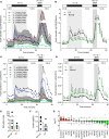
Individual donor (A) and mean (B) insulin secretion perifusion traces of CF islets (n = 5) compared with normal islets (shaded area, dotted black lines, n = 5) normalized to islet equivalents (IEQ). Individual donor (C) and mean (D) insulin secretion perifusion traces of CF islets (n = 5) compared with normal islets (shaded area, n = 5) normalized to insulin content. Entire perifusion traces are in Supplemental Figure 8. Stimulation index (E) comparing the ratio of 16.7 G/5.6 G insulin secretory rates. (F) Islet insulin content and (G) whole-islet fold changes of key islet regulatory transcripts in CF islets (n = 5) relative to whole healthy islets (n = 5). Data represent mean ± SEM. No statistical difference (P < 0.05) was detected using an unpaired 2-tailed Student’s t test in E and F. Ordinary 2-way ANOVA was used to compare islet perifusions and is discussed in the text. The dots represent individual donors and are color coded according to CF donor (Table 1).
Figure 6
Figure 6. CF pancreas and islets showed marked immune infiltration.
CD45 (red) and insulin (green) labeling of CF pancreata from (A) donor 1 and (B) donor 2. Magnification of boxed areas shows populations of CD45+ cells at the periphery and within islets (scale bars: 100 μm; 50 μm [insets]). (C) Percentage contribution of CD45+ cells to CF and normal pancreata. (D) Number of CD45+ cells identified within 20 μm of islets (n = 25 islets/donor) from CF donors (n = 6). The dots represent individual islets and are color-coded according to CF donor (Table 1). Each concentric circle represents the number of immune cells within 20 μm of the islet, and each arrow indicates the number of immune cells in that concentric circle. The number increases outward. (E) Selected immune-related genes from whole-islet RNA sequencing of CF donors (n = 5) versus healthy donors (n = 5). T cell lines grown from donor 5 islets were stimulated with and without soluble anti-CD3 overnight and soluble anti-CD28, GolgiPlug, and CD107a staining antibody were then added to all wells for 6 hours. Intracellular staining of a representative CD8+ T cell line is shown for (F) CD107a, (G) IFN-γ, and (H) TNF-α. Data represent mean ± SEM. Statistical significance (P < 0.05) was observed in the percentage of pancreatic CD45+ cells indicated by the asterisk. Unpaired 2-tailed Student’s t test was used for statistical analysis.
Figure 7
Figure 7. Integrated model of CFRD pathogenesis.
Pancreatic autolysis and remodeling results in destruction of the islet niche and environment, β cell loss, and immune infiltration. Islet loss and inflammation combined with the numerous physiological derangements observed in CF, particularly those responsible for nutrient assimilation, lead to insulin insufficiency and CFRD.

References

    1. Cystic Fibrosis Foundation Patient Registry 2016 Annual Data Report. Cystic Fibrosis Foundation. https://www.cff.org/Research/Researcher-Resources/Patient-Registry/2016-... Accessed April 10, 2018.
    1. Moran A, et al. Clinical care guidelines for cystic fibrosis-related diabetes: a position statement of the American Diabetes Association and a clinical practice guideline of the Cystic Fibrosis Foundation, endorsed by the Pediatric Endocrine Society. Diabetes Care. 2010;33(12):2697–2708. doi: 10.2337/dc10-1768. - DOI - PMC - PubMed
    1. Marshall BC, Butler SM, Stoddard M, Moran AM, Liou TG, Morgan WJ. Epidemiology of cystic fibrosis-related diabetes. J Pediatr. 2005;146(5):681–687. doi: 10.1016/j.jpeds.2004.12.039. - DOI - PubMed
    1. Gudipaty L, Rickels MR. Pancreatogenic (Type 3c) Diabetes. The Pancreapedia: Exocrine Pancreas Knowledge Base. https://doi.org/10.3998/panc.2015.35.
    1. O’Riordan SM, Robinson PD, Donaghue KC, Moran A, ISPAD Clinical Practice Consensus Management of cystic fibrosis-related diabetes. Pediatr Diabetes. 2008;9(4 Pt 1):338–344. - PubMed

Publication types

MeSH terms