. 2018 May 1;9(5):446.
doi: 10.1038/s41419-018-0474-4.

Inhibition of cyclin-dependent kinase 4 as a potential therapeutic strategy for treatment of synovial sarcoma

Affiliations

Inhibition of cyclin-dependent kinase 4 as a potential therapeutic strategy for treatment of synovial sarcoma

Xiaoyang Li et al. Cell Death Dis. .

Abstract

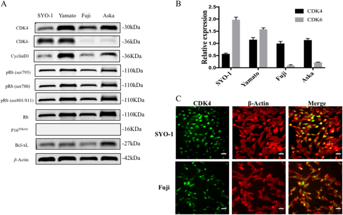
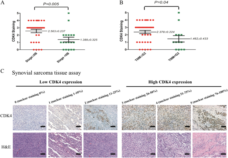
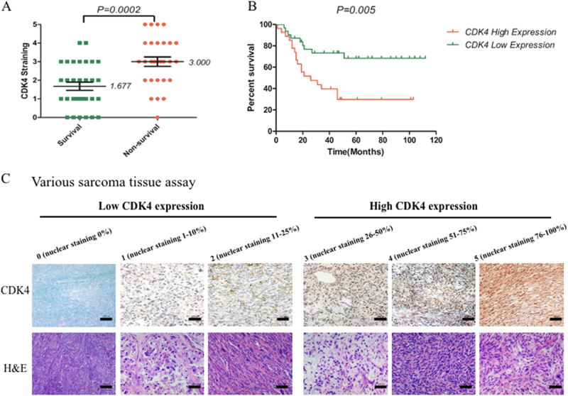
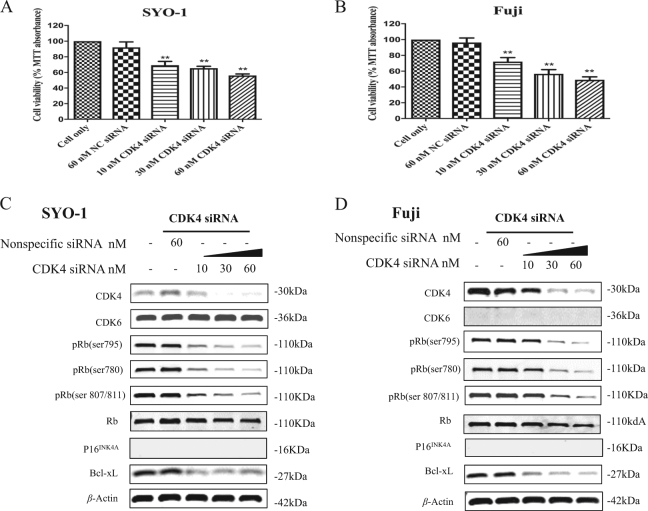
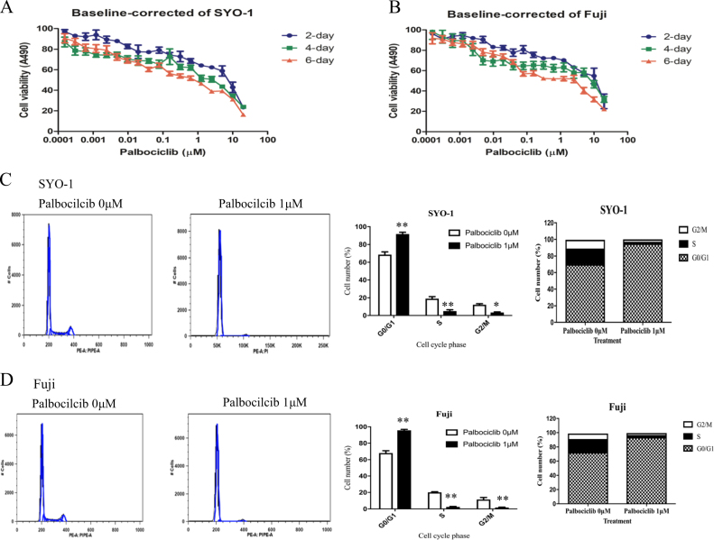
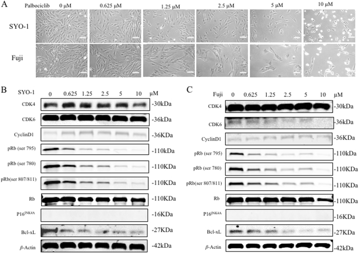
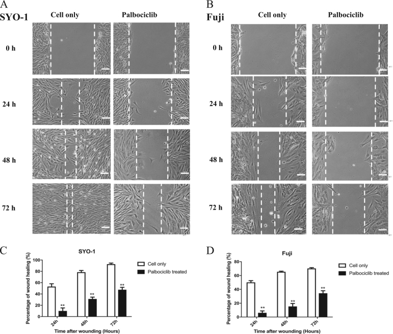
Synovial sarcoma is a highly aggressive but rare form of soft tissue malignancy that primarily affects the extremities of the arms or legs, for which current chemotherapeutic agents have not been proven to be very effective. The cyclin-dependent kinase 4/6-retinoblastoma protein (CDK4/6-Rb) pathway of cell cycle control is known to be aberrant in a large proportion of cancers. Recently, CDK4 inhibitors have successfully been used pre-clinically for the treatment of many human cancers, and in 2015, following the success of clinical trials, the FDA approved the first selective CDK4/6 inhibitor, palbociclib, for the treatment of endocrine therapy resistant breast cancers. However, the expression and therapeutic potential of targeting CDK4 in synovial sarcoma remains unclear. In the present study, we report that CDK4 is highly expressed in human synovial sarcoma, and high CDK4 expressions are associated with poor prognosis in sarcomas patients and the clinical stage and the TNM grade in synovial sarcoma patients. Knockdown of CDK4 with specific small interference RNAs inhibits cell proliferation and enhances apoptotic effects in synovial sarcoma cells. CDK4 inhibitor palbociclib suppresses synovial sarcoma cell proliferation and growth in a dose and time-dependent manner. Palbociclib also inhibits the CDK4/6-Rb signaling pathway and promotes cell apoptosis without changing CDK4/6 protein levels, suggesting that palbociclib only represses the hyper-activation, not the expression of CDK4/6. Flow cytometry analysis reveals that palbociclib induces G1 cell-cycle arrest and apoptotic effects by targeting the CDK4/6-Rb pathway in synovial sarcoma cells. Furthermore, wound healing assays demonstrate that inhibition of the CDK4/6-Rb pathway by palbociclib significantly decreases synovial sarcoma cell migration in vitro. Our study highlights the importance of the CDK4/6-Rb pathway in human synovial sarcoma pathogenesis, and the role of the current selective CDK4/6 inhibitor, palbociclib, as a potential promising targeted therapeutic agent in the treatment of human synovial sarcoma.


Conflict of interest statement

The authors declare that they have no conflict of interest.

Figures

Fig. 1
Fig. 1. CDK4 is highly expressed in human synovial sarcoma cell lines.
a Expression levels of CDK4, CDK6, cyclin D1, p16INK4A, Rb, pRb (Ser795, 780 and 807/811) and Bcl-xL in four synovial sarcoma cell lines SYO-1, Yamato, Fuji, and Aska were detected using Western blotting. b Relative expressions of CDK4 and CDK6 compared with β-actin in different synovial sarcoma cell lines were figured. c Expression of CDK4 in SYO-1 and Fuji cells was assessed by immunofluorescence with antibodies to CDK4 and β-Actin. Cells were visualized under a fluorescence microscope after incubation with Alexa Fluor 488 goat anti-rabbit IgG (green) or Alexa Fluor 594 goat anti-mouse IgG (red), and the images were merged (scale bar, 50 μm)
Fig. 2
Fig. 2. CDK4 expression levels correlate to clinicopathological characteristics of synovial sarcoma patients.
CDK4 levels in human synovial sarcoma tissue microarray were determined by immunohistochemistry, and the correlation of CDK4 expression with clinicopathological characteristics of synovial sarcoma patients was evaluated. a Distribution of CDK4 staining scores among clinical stage≥IIB and clinical stage<IIB synovial sarcoma tissues. b Distribution of CDK4 staining scores in the synovial sarcoma tissues from TNM grade≥G2 patients and TNM grade<G2 patients. c Representative images of different immunohistochemical staining intensities of CDK4 ( × 200; scale bar=100 µm). On the basis of the percentage of cells with positive nuclear staining, CDK4 staining patterns were categorized into 6 groups: 0, no nuclear staining; 1+:<10% of positive cells; 2+, 10–25% of positive cells; 3+, 26–50% of positive cells; 4+, 51–75% of positive cells; 5+,>75% of positive cells
Fig. 3
Fig. 3. CDK4 expression levels associate with clinicopathological characteristics of various sarcomas patients.
CDK4 levels in human various sarcomas tissue microarray were determined by immunohistochemistry, and the relationship between CDK4 expressions and clinicopathological characteristics of various sarcomas patients was analyzed. a Distribution of CDK4 staining scores in the synovial sarcoma tissue samples from survival and non-survival patients. b Kaplan–Meier survival curve of sarcoma patients with CDK4 high staining (≥3) or low staining (<3). c Representative images of different immunohistochemical staining intensities of CDK4 (×200; scale bar:100 µm). On the basis of the percentage of cells with positive nuclear staining, CDK4 staining patterns were categorized into 6 groups: 0, no nuclear staining; 1+:<10% of positive cells; 2+, 10–25% of positive cells; 3+, 26–50% of positive cells; 4+, 51–75% of positive cells; 5+, >75% of positive cells
Fig. 4
Fig. 4. CDK4/6-Rb pathway inhibition induced by CDK4 specific siRNA (#SASI_Hs01_00122488) decreases cell proliferation and promotes apoptosis in synovial sarcoma cells.
Human synovial sarcoma SYO-1 and Fuji cells were transfected with increasing concentrations of CDK4 specific siRNA (#SASI_Hs01_00122488) or nonspecific siRNA, and cell proliferation and growth was determined subsequently. a, b Cell viability was determined by MTT assay after siRNA transfection for 5 days. c, d The respective proteins of CDK4/6-Rb-apoptosis pathway in cells were examined by Western blotting after 48 h of siRNA transfection. **P< 0.01 compared with the cell only group
Fig. 5
Fig. 5. Inhibition of CDK4/6-Rb pathway by palbociclib decreases cell proliferation and induces cell cycle arrest in human synovial sarcomas.
Human synovial sarcoma SYO-1 and Fuji cells were treated with increasing concentrations of palbociclib for the indicated time, and cell proliferation and growth was determined subsequently. a, b Cell viability was determined by MTT assays after palbociclib exposure for 2, 4, and 6 days. The cell cycle of SYO-1 and Fuji cells was assessed after exposure to palbociclib (1 µM) for 24 h by flow cytometry analysis. Representative images of cell cycle distribution in SYO-1 (c) and Fuji (d) cells with or without palbociclib treatment. Cell numbers in different cell cycle phases were counted. Different cell cycle phase rates were also analyzed. *P< 0.05, **P< 0.01 compared with the cell only group
Fig. 6
Fig. 6. Inhibition of CDK4/6-Rb pathway by palbociclib alters phosphorylation of Rb and promotes apoptosis in human synovial sarcomas.
a Human synovial sarcoma SYO-1 and Fuji cells were treated with increasing concentrations of palbociclib for the indicated time, and the morphologic changes of cells were observed by microscopy after 2 days of palbociclib treatment (scale bar, 50 μm). The expression of respective proteins in CDK4/6-Rb pathway and apoptosis in SYO-1 (b) and Fuji (c) cells was examined by western blotting after 3 days of palbociclib treatment
Fig. 7
Fig. 7. Inhibition of the CDK4/6-Rb pathway by palbociclib reduced human synovial sarcoma cell migration in vitro.
After exposure to 1 µM of palbociclib for the indicated time, the cell migration of SYO-1 and Fuji cells was determined by wound healing assays. a Representative images of SYO-1 cell migration after palbociclib treatment for 24, 48, and 72 h (scale bar, 50 μm). b Representative images of Fuji cell migration after palbociclib treatment for 24, 48, and 72 h (scale bar, 50 μm). c Cell migration distance of SYO-1 cells was measured after palbociclib treatment. d Cell migration distance of Fuji cells was measured after palbociclib treatment. ***P< 0.001 compared with the cell only group

Similar articles

Cited by

References

    1. Choong PF, Rydholm A, Gustafson P. Patients with soft-tissue sarcomas. Ann. Surg. 1995;221:203–205. - PMC - PubMed
    1. Clark J, et al. Identification of novel genes, SYT and SSX, involved in the t(X;18)(p11.2; q11.2) translocation found in human synovial sarcoma. Nat. Genet. 1994;7:502–508. doi: 10.1038/ng0894-502. - DOI - PubMed
    1. Crew AJ, et al. Fusion of SYT to two genes, SSX1 and SSX2, encoding proteins with homology to the Kruppel-associated box in human synovial sarcoma. Embo. J. 1995;14:2333–2340. - PMC - PubMed
    1. Spillane AJ, A’Hern R, Judson IR, Fisher C, Thomas JM. Synovial sarcoma: a clinicopathologic, staging, and prognostic assessment. J. Clin. Oncol.: Off. J. Am. Soc. Clin. Oncol. 2000;18:3794–3803. doi: 10.1200/JCO.2000.18.22.3794. - DOI - PubMed
    1. Lewis JJ, et al. Synovial sarcoma: a multivariate analysis of prognostic factors in 112 patients with primary localized tumors of the extremity. J. Clin. Oncol.: Off. J. Am. Soc. Clin. Oncol. 2000;18:2087–2094. doi: 10.1200/JCO.2000.18.10.2087. - DOI - PubMed

Publication types

MeSH terms