. 2018 Jan 24:2018:7934362.
doi: 10.1155/2018/7934362. eCollection 2018.

TRAIL-Dependent Resolution of Pulmonary Fibrosis

Affiliations

TRAIL-Dependent Resolution of Pulmonary Fibrosis

David M Habiel et al. Mediators Inflamm. .

Abstract

Idiopathic pulmonary fibrosis (IPF) is the most common form of interstitial lung disease characterized by the persistence of activated myofibroblasts resulting in excessive deposition of extracellular matrix proteins and profound tissue remodeling. In the present study, the expression of tumor necrosis factor- (TNF-) related apoptosis-inducing ligand (TRAIL) was key to the resolution of bleomycin-induced pulmonary fibrosis. Both in vivo and in vitro studies demonstrated that Gr-1+TRAIL+ bone marrow-derived myeloid cells blocked the activation of lung myofibroblasts. Although soluble TRAIL was increased in plasma from IPF patients, the presence of TRAIL+ myeloid cells was markedly reduced in IPF lung biopsies, and primary lung fibroblasts from this patient group expressed little of the TRAIL receptor-2 (DR5) when compared with appropriate normal samples. IL-13 was a potent inhibitor of DR5 expression in normal fibroblasts. Together, these results identified TRAIL+ myeloid cells as a critical mechanism in the resolution of pulmonary fibrosis, and strategies directed at promoting its function might have therapeutic potential in IPF.


Figures

Figure 1
Figure 1
Bone marrow mobilization using systemically administered clodronate liposomes increased the accumulation of Gr-1+ myeloid cells in the lung and is associated with reduced pulmonary fibrosis. (a) Mice either received an intraperitoneal injection of liposomes containing clodronate (Clod Lipo) or empty liposomes (Lipo) at 1 day prior to (D−1) an intrapulmonary bleomycin challenge. All mice were subsequently sacrificed at day 5 after bleomycin challenge, and lungs from D−1 Clod Lipo- or Lipo-treated mice were digested to generate a cellular suspension, and cells were stained with monoclonal antibodies directed against Gr-1, CD11b, CD11c, F4/80, CD3, CD19, γδ TCR, or NK 1.1 and analyzed by flow cytometry. (b) Mice either received an intraperitoneal injection of liposomes containing clodronate (Clod Lipo) either 1 day prior to (D−1) or at 14 days after (D+14) or empty liposomes (Lipo) one day prior to intrapulmonary bleomycin challenge. All mice were subsequently sacrificed at day 21 after bleomycin challenge and their lungs were isolated for analysis. Whole lungs were fixed, paraffin-embedded, sectioned, and Masson's trichrome-stained to visualize ECM. Shown are representative images taken at 200x magnification. (c) Quantitative PCR analysis of procollagen 3 and fibronectin 1 in whole lung samples. (d) ELISA analysis of whole lung levels of IFN-γ, IL-4, IL-13, TGF-β, CCL22, and CCL17. Data are mean ± SEM, n = 5–10/group, P ≤ 0.05 compared with Lipo-alone group.
Figure 2
Figure 2
Gr-1+ myeloid cells were recruited in a temporal manner to the lung and their depletion exacerbated bleomycin-induced fibrosis. Bleomycin-challenged mice were killed at days 14, 21, and 42 and their lungs were isolated for analysis. (a) Lungs were digested to generate a cellular suspension; the cells were stained with the following antibodies for flow cytometry: Gr-1, CD11b, CD11c, F4/80, and CD80. (b) Mice were unchallenged (i.e., naïve) or received bleomycin 14 or 21 days previously. Other groups of mice (n = 5–10/group) were treated with control IgG or anti-Gr-1 mAb at day 21 and were subsequently analyzed at day 42 after bleomycin. Whole lungs were fixed, paraffin-embedded, sectioned, and Masson's trichrome-stained to visualize ECM. Shown are representative images taken at 200x magnification (naïve, day 14, and day 21) or 40x magnification (day 42 and day 42 + αGr-1). (c) Quantitative PCR analysis of procollagen 3 and fibronectin 1 in whole lung samples. Data are mean ± SEM, n = 5–10/group, P ≤ 0.05 compared with IgG control group.
Figure 3
Figure 3
Adoptive transfer of Gr-1+ myeloid cells reduced lung fibrosis. Gr-1+ cells were purified from the lungs of bleomycin-challenged D−1 Clod Lipo-treated mice at day 5 after bleomycin. (a) Purified Gr-1+ myeloid cells from lung were spun onto a slide using a cytospin and stained, and a morphologic analysis was performed using light microscopy (left panel; 1000x magnification). Both lung- and bone marrow-purified CD11b+Gr-1+ and CD11b+Gr-1 myeloid cells were stained for CD80, CD86, MHCII, CD62L, and F4/80 for flow cytometry analysis. (b) Quantitative PCR analysis of RNA isolated from Gr-1+ myeloid cells was used to determine transcript levels of arginase, NOS2, IDO, and IL-10. (c) Purified Gr-1+ myeloid cells were adoptively transferred into the lungs of bleomycin-challenged mice at days 5 (D+5) or 14 (D+14) after bleomycin. Lung samples were removed at day 21 after bleomycin. Lungs were fixed, paraffin-embedded, sectioned, and stained with Masson's trichrome. Shown are representative images taken at 100x magnification. (d) Procollagen 3 and fibronectin 1 expression were determined by quantitative PCR using RNA isolated from whole lung samples. (e) ELISA was used to determine whole lung levels of IFN-γ, IL-4, IL-13, TGF-β, CCL22, and CCL17. Data are mean ± SEM, n = 5–10 mice/group; P ≤ 0.05 and ∗∗ P ≤ 0.01 compared with the appropriate control group.
Figure 4
Figure 4
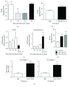
Gr-1+ myeloid cells decreased collagen, fibronectin, and TGF-β expression in cultured primary lung fibroblasts. Mice were treated with Clod Lipo or Lipo one day prior to bleomycin challenge. Five days after bleomycin administration, Gr-1+ myeloid cells were MACS-purified from the lungs of challenged mice, and 1 × 105 Gr-1+ cells were cocultured with 5 × 105 primary mouse lung fibroblasts for 24 h. Lung fibroblasts were either untreated or exposed to IL-13 at 10 ng/ml for 24 h and washed prior to coculture. After coculture, the Gr-1+ myeloid cells were subsequently washed away prior to analysis of the fibroblasts. (a) Fibroblasts were fixed, permeabilized, and stained with anticollagen mAb prior to confocal microscopy. Shown are representative images taken at 1000x magnification. (b) The expression of fibronectin 1, procollagen 1, procollagen 3, and TGF-β was determined using quantitative PCR analysis of adherent primary lung fibroblasts. (c) Quantitative PCR analysis of RNA was used to determine the expression of granzyme, perforin, and TRAIL in purified Gr-1+ myeloid cells. (d) Purified Gr-1+ myeloid cells were stained for CD11b, and TRAIL and analyzed by flow cytometry. (e) Purified mouse lung fibroblasts were treated with increasing concentrations of sTRAIL for 24 h, washed, and quantitative PCR analysis was used to determine the expression of TGF-β, procollagen 1, and procollagen 3. Data are mean ± SEM, n = 3–5 independent experiments. All statistics were performed using unpaired parametric t-tests; P ≤ 0.05.
Figure 5
Figure 5
TRAIL expression is required for the optimal resolution of lung fibrosis in vivo. (a) Mice received bleomycin and were killed at days 14, 21, and 42 after challenge and ELISA was used to determine the concentration of TRAIL in whole lung samples. As a control, whole lung samples from naïve mice were subjected to the same analysis. (b) Mice were treated with Clod Lipo one day (D−1) prior to bleomycin, and ELISA was used to determine the whole lung levels of TRAIL. (c) Bleomycin-challenged mice were analyzed at days 14, 21, and 42 after challenge. Another group of mice were injected with IgG control or anti-Gr-1 mAb on day 21 and analyzed at day 42. Quantitative PCR analysis was used to determine TRAIL and peroxiredoxin 6 transcript expression in whole lung RNA samples. (d) Purified Gr-1+ myeloid cells were adoptively transferred into bleomycin-challenged mice at day 5 or 14 after bleomycin. At day 21, ELISA was used to determine TRAIL levels in all groups of mice. (e) Mice were injected by intraperitoneal injection with anti-TRAIL mAb from days 14 to 21 after bleomycin, and quantitative PCR was used to determine whole lung transcript expression of procollagen 1 and procollagen 3. Data are mean ± SEM, n = 5–10 mice/group, P ≤ 0.05 compared with the appropriate control group. All statistics were performed using unpaired parametric t-tests; ∗∗ P ≤ 0.01 and ∗∗∗ P ≤ 0.001.
Figure 6
Figure 6
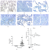
Altered TRAIL and DR5 expression in plasma, whole lung tissues, and primary fibroblasts from IPF patients. (a–f) Lung biopsies were obtained from nonfibrotic disease or IPF patients, fixed, paraffin-embedded, sectioned, and stained for TRAIL, DR5, or CD33. (a, c, e) Normal lungs stained for TRAIL, DR5, and CD33, respectively. (b, d, f) IPF lungs stained for TRAIL, DR5, and CD33, respectively. Statistical analysis were performed using ordinary one-way ANOVA test; P ≤ 0.05. (g) Plasma was collected from normal volunteers and IPF patients and ELISA was used to determine soluble TRAIL concentrations. (h) Lung fibroblasts were purified from nonfibrotic or IPF lung biopsies. Fibroblasts were left untreated or treated 10 ng/ml of IL-13 for 24 h. Quantitative PCR was used to determine the expression of TRAIL and DR5. Data are mean ± SEM, n = 3–5 independent experiments.

References

    1. du Bois R. M. Strategies for treating idiopathic pulmonary fibrosis. Nature Reviews Drug Discovery. 2010;9(2):129–140. doi: 10.1038/nrd2958. - DOI - PubMed
    1. Gan Y., Herzog E. L., Gomer R. H. Pirfenidone treatment of idiopathic pulmonary fibrosis. Therapeutics and Clinical Risk Management. 2011;7:39–47. doi: 10.2147/TCRM.S12209. - DOI - PMC - PubMed
    1. Noble P. W., Albera C., Bradford W. Z., et al. Pirfenidone in patients with idiopathic pulmonary fibrosis (CAPACITY): two randomised trials. The Lancet. 2011;377(9779):1760–1769. doi: 10.1016/S0140-6736(11)60405-4. - DOI - PubMed
    1. Drakopanagiotakis F., Xifteri A., Polychronopoulos V., Bouros D. Apoptosis in lung injury and fibrosis. European Respiratory Journal. 2008;32(6):1631–1638. doi: 10.1183/09031936.00176807. - DOI - PubMed
    1. Wynn T. A. Integrating mechanisms of pulmonary fibrosis. The Journal of Experimental Medicine. 2011;208(7):1339–1350. doi: 10.1084/jem.20110551. - DOI - PMC - PubMed

MeSH terms

Substances

LinkOut - more resources