Case Reports
. 2018 Apr 4:12:207.
doi: 10.3389/fnins.2018.00207. eCollection 2018.

Selective Fiber Degeneration in the Peripheral Nerve of a Patient With Severe Complex Regional Pain Syndrome

Affiliations
Case Reports

Selective Fiber Degeneration in the Peripheral Nerve of a Patient With Severe Complex Regional Pain Syndrome

Adrien Yvon et al. Front Neurosci. .

Abstract

Aims: Complex regional pain syndrome (CRPS) is characterized by chronic debilitating pain disproportional to the inciting event and accompanied by motor, sensory, and autonomic disturbances. The pathophysiology of CRPS remains elusive. An exceptional case of severe CRPS leading to forearm amputation provided the opportunity to examine nerve histopathological features of the peripheral nerves. Methods: A 35-year-old female developed CRPS secondary to low voltage electrical injury. The CRPS was refractory to medical therapy and led to functional loss of the forelimb, repeated cutaneous wound infections leading to hospitalization. Specifically, the patient had exhausted a targeted conservative pain management programme prior to forearm amputation. Radial, median, and ulnar nerve specimens were obtained from the amputated limb and analyzed by light and transmission electron microscopy (TEM). Results: All samples showed features of selective myelinated nerve fiber degeneration (47-58% of fibers) on electron microscopy. Degenerating myelinated fibers were significantly larger than healthy fibers (p < 0.05), and corresponded to the larger Aα fibers (motor/proprioception) whilst smaller Aδ (pain/temperature) fibers were spared. Groups of small unmyelinated C fibers (Remak bundles) also showed evidence of degeneration in all samples. Conclusions: We are the first to show large fiber degeneration in CRPS using TEM. Degeneration of Aα fibers may lead to an imbalance in nerve signaling, inappropriately triggering the smaller healthy Aδ fibers, which transmit pain and temperature. These findings suggest peripheral nerve degeneration may play a key role in CRPS. Improved knowledge of pathogenesis will help develop more targeted treatments.

Keywords: complex regional pain syndrome (CRPS); nerve histology; peripheral nerve; remak bundle; transmission electron microscopy.


Figures

Figure 1
Figure 1
Diagram showing the level of forearm amputation as well as the levels at which the proximal and distal nerve samples were taken. Original biopsies were 3 cm long, which were further dissected during sample processing, fixation, and embedding for electron microscopy.
Figure 2
Figure 2
Light microscopy of a nerve fascicle from the distal ulnar nerve sample (bar represents 50 μm). The perineurium and endoneurium are visualized. The circle indicates a cluster of healthy myelinated axons. Arrows indicate examples of degenerating fibers. Degenerative features included myelin breakdown and collapsed nerve fibers. Stain: toluidine blue.
Figure 3
Figure 3
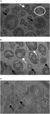
Transmission electron microscopy (TEM) images of proximal ulnar (A), distal radial (B), and proximal radial (C) nerve samples. Bar represents 5 μm. White arrow in (A) indicates healthy nerve fiber with Schwann cell, black arrows indicate entire fiber degeneration, and the white circle shows healthy Remak bundles (groups of unmyelinated fibers). In (B) white arrows show degenerating myelin while black arrows show degenerating Remak bundles. In (C) black arrow indicate denervated Schwann cell bands (unmyelinated axon loss).
Figure 4
Figure 4
Histogram showing the percentage of healthy and degenerative myelinated nerve fibers in our study. Forty-seven to fifty-four percent of myelinated nerve fibers showed evidence of degeneration.
Figure 5
Figure 5
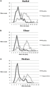
Nerve fiber distribution according to size (μm). n = 796. Gray: healthy; black: degenerative. (A) Radial nerve; smaller fibers were largely spared from degeneration, whilst the larger fibers are affected (B) Ulnar nerve; similar distribution to radial nerve, with two clear peaks of healthy and degenerative fibers (C) Median nerve; similar distribution to both radial and median nerve. The peak of healthy fibers count corresponds mainly to Aδ and Aβ fibers whilst the degeneration peak corresponds to Aα fibers.

Comment in

References

    1. Albrecht P. J., Hines S., Eisenberg E., Pud D., Finlay D. R., Connolly M. K., et al. . (2006). Pathologic alterations of cutaneous innervation and vasculature in affected limbs from patients with complex regional pain syndrome. Pain 120, 244–266. 10.1016/j.pain.2005.10.035 - DOI - PubMed
    1. Allen G., Galer B. S., Schwartz L. (1999). Epidemiology of complex regional pain syndrome: a retrospective chart review of 134 patients. Pain 80, 539–544. 10.1016/S0304-3959(98)00246-2 - DOI - PubMed
    1. Baron R., Fields H. L., Janig W., Kitt C., Levine J. D. (2002). National Institutes of Health Workshop: reflex sympathetic dystrophy/complex regional pain syndromes–state-of-the-science. Anesth. Analg. 95, 1812–1816. 10.1097/00000539-200212000-00064 - DOI - PubMed
    1. Birklein F., O'Neill D., Schlereth T. (2015). Complex regional pain syndrome: an optimistic perspective. Neurology 84, 89–96. 10.1212/WNL.0000000000001095 - DOI - PubMed
    1. Birklein F., Schmelz M., Schifter S., Weber M. (2001). The important role of neuropeptides in complex regional pain syndrome. Neurology 57, 2179–2184. 10.1212/WNL.57.12.2179 - DOI - PubMed

Publication types

LinkOut - more resources