

The Evaluation of Root Fracture with Cone Beam Computed Tomography (CBCT): An Epidemiological Study
- PMID: 29670714
- PMCID: PMC5899817
- DOI: 10.4317/jced.54009
The Evaluation of Root Fracture with Cone Beam Computed Tomography (CBCT): An Epidemiological Study
Abstract
Background: The aim of this study was evaluation of the cone-beam computed tomography (CBCT) image of 50 patients at the ages of 8-15 suspecting root fracture and root fracture occurred, exposed to dental traumatic. In additionally, this study was showed effect of crown fracture on root fracture healing.
Material and methods: All of the individuals included in the study were obtained images with the cone-beam computed tomography range of 0,3 voxel and 8.9 seconds.(i-CAT®, Model 17-19, Imaging SciencesInternational, Hatfield, Pa USA).The information obtained from the history and CBCT images of patients were evaluated using chi-square test statistical method the mean and the distribution of the independent variables.
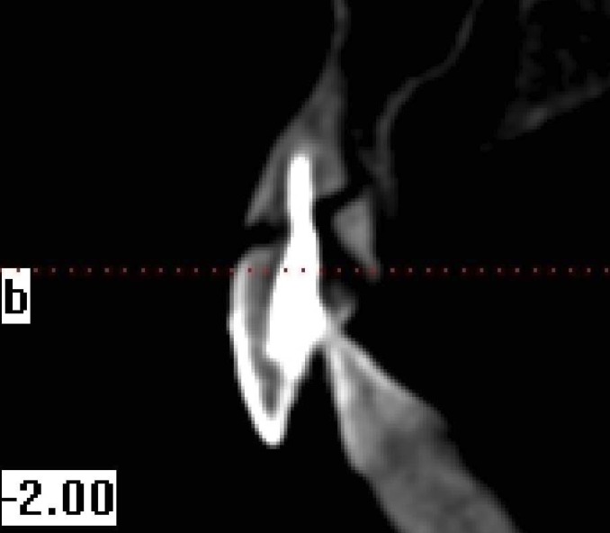
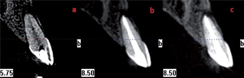
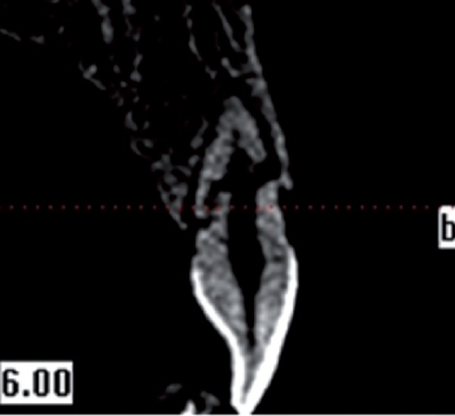
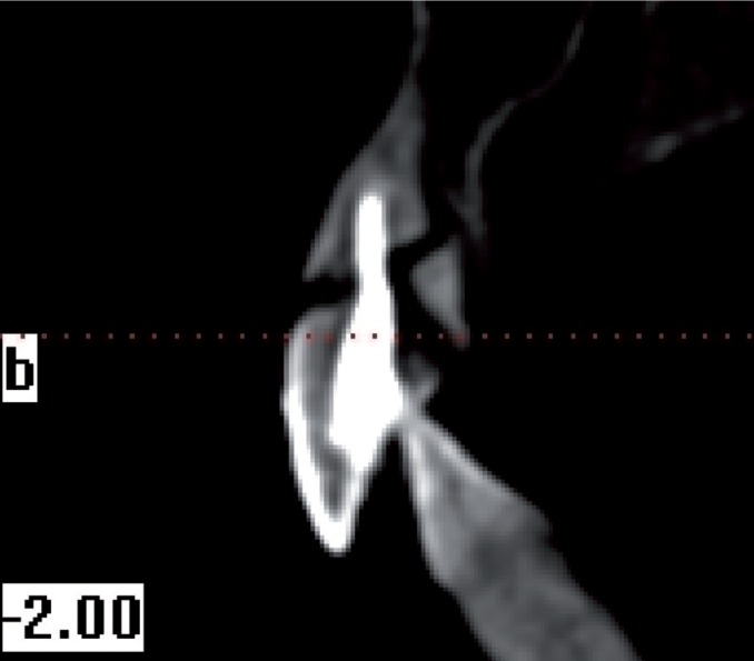
Results: 50 children, have been exposed to trauma, was detected root fracture injury in 97 teeth. Horizontal root fracture 63.9% of the 97 tooth, the oblique in 31.9%, both the horizontal and oblique in 1.03%, partial fracture in 2.06% ,and both horizontally and vertical in 1.03% was observed.The most affected teeth, respectively of, are the maxillary central incisor (41.23% left, right, 37.11%), maxillary left lateral incisor (9.27%), maxillary right lateral incisor (11.34%), and mandibular central incisor (1.03%).
Conclusions: Crown fractures have negative effects on spontaneous healing of root fractures. CBCT are used selected as an alternative to with conventional radiography for diagnosis of root fractures. In particular, ıt's cross-sectional image is quite useful and has been provided more conveniences seeing the results of diagnosis and treatment for clinician. Key words:Root fracture, CBCT, Epidemiolog.
Conflict of interest statement
Conflict of interest statement: The authors declare that they have no conflict of interest.
Figures
References
-
- Caldas AF, Burgos MEA. A retrospective study of traumatic dental injuries in a Brazilian dental trauma clinic. Dent Traumatol. 2001;17:250–253. - PubMed
-
- Lauridsen E, Hermann NV, Gerds TA, Kreiborg S, Andreasen JO. Pattern of traumatic dental injuries in the permanent dentition among children, adolescents, and adults. Dent Traumatol. 2012;28:358–363. - PubMed
-
- Malmgren B, Andreasen JO, Flores MT, Robertson A, DiAngelis AJ, Andersson L. International Association of Dental Traumatology guidelines for the management of traumatic dental injuries: 3. Injuries in the primary dentition. Dent Traumatol. 2012;28:174–182. - PubMed
-
- Andreasen JO, Andreasen FM, Meja`re I, Cvek M. Healing of 400 intra-alveolar root fractures. 2. Effect of treatment factors such as treatment delay, repositioning, splinting type and period and antibiotics. Dent Traumatol. 2004;20:203–211. - PubMed
-
- Heithersay GS, Kahler B. Healing responses following transverse root fracture: a historical review and case reports showing healing with (a) calcified tissue and (b) dense fibrous connective tissue. Dent Traumatol. 2013;29:253–265. - PubMed
LinkOut - more resources
Full Text Sources
Other Literature Sources
Miscellaneous