Review
. 2018 Apr 4:6:79.
doi: 10.3389/fped.2018.00079. eCollection 2018.

Echocardiographic Evaluation of Ventricular Function-For the Neonatologist and Pediatric Intensivist

Affiliations
Review

Echocardiographic Evaluation of Ventricular Function-For the Neonatologist and Pediatric Intensivist

Cécile Tissot et al. Front Pediatr. .

Abstract

In the neonatal and pediatric intensive care setting, bedside cardiac ultrasound is often used to assess ventricular dimensions and function. Depending upon the underlying disease process, it is necessary to be able to evaluate the systolic and diastolic function of left and or right ventricles. The systolic function of left ventricle is mostly assessed qualitatively on visual inspection "eye-balling" and quantitatively by measuring circumferential fraction shortening or calculating the ejection fraction by Simpson's planimetry. The assessment of left ventricular diastolic function relies essentially on the mitral valve and pulmonary venous Doppler tracings or tissue Doppler evaluation. The right ventricular particular shape and anatomical position does not permit to use the same parameters for measuring systolic function as is used for the LV. Tricuspid annular plane systolic excursion (TAPSE) and S' velocity on tissue Doppler imaging are more often used for quantitative assessment of right ventricle systolic function. Several parameters proposed to assess right ventricle systolic function such as fractional area change, 3D echocardiography, speckle tracking, and strain rate are being researched and normal values for children are being established. Diastolic function of right ventricle is evaluated by tricuspid valve and hepatic venous Doppler tracings or on tissue Doppler evaluation. The normal values for children are pretty similar to adults while normal values for the neonates, especially preterm infants, may differ significantly from adult population. The normal values for most of the parameters used to assess cardiac function in term neonates and children have now been established.

Keywords: bedside cardiac ultrasound; echocardiography; functional echocardiography; intensive care; neonatology; pediatric; point-of-care; ventricular function.


Figures

Figure 1
Figure 1
M-mode echocardiography from a parasternal short-axis view showing the left ventricular cavity over the cardiac cycle (see ECG tracing) during systole and diastole. Abbreviations: IVS, interventricular septum; LV, left ventricle; LVEDD, left ventricle end-diastolic dimension; LVESD, left ventricle end-systolic dimension; LVPW, left ventricle posterior wall.
Figure 2
Figure 2
M-mode echocardiography obtained from a patient (left image) with normal left ventricular systolic function (FS = 34% and EF = 65%) and in a patient (right image) with dilated cardiomyopathy with severe left ventricular systolic dysfunction (FS = 8% and EF = 16%), LVEDD with green arrow and LVESD with blue arrow. Abbreviations: EF, ejection fraction; FS, fractional shortening; LVEDD, left ventricle end-diastolic dimension; LVESD, left ventricle end-systolic dimension.
Figure 3
Figure 3
Assessment of left ventricular systolic function by planimetry using the modified Simpson’s method and allowing estimation of the ejection fraction (EF) by tracing the endocardial left ventricular border in end-diastole (LVEDV) and in end-systole (LVESV), obtained from a patient with moderate left ventricular systolic dysfunction (EF = 30%). Abbreviations: LVEF, left ventricular ejection fraction; LVEDV, left ventricle end-diastolic volume; LVESV, left ventricle end-systolic volume; SV, stroke volume.
Figure 4
Figure 4
Doppler tracing from mitral regurgitation (MR) allowing measurement of the MR dP/dt max obtained between 1 and 3 m/s, on a normal patient (left image) and on a patient with left ventricular dysfunction (right image). An MR dP/dt max <500 mmHg/s is indicative of left ventricular systolic dysfunction. The same can be applied to tricuspid regurgitation (TR dP/dt max) but has to be measured between 1 and 2 m/s.
Figure 5
Figure 5
Tissue Doppler imaging tracing obtained at the septal annulus showing S′ (systolic) wave, E′ (early diastolic) wave, and A′ (late diastolic related to atrial contraction) wave.
Figure 6
Figure 6
Myocardial performance index (=MPI or Tei index) obtained from tissue Doppler (above image) and from Doppler (below image). The MPI is calculated as the (IVCT + IVRT)/ET or (B − A)/A. The MPI allows evaluation of systolic and diastolic ventricular function. Abbreviations: ET, ejection time; IVCT, isovolumetric contraction time; IVRT, isovolumetric relaxation time.
Figure 7
Figure 7
Evaluation of diastolic function in a patient with severe diastolic dysfunction: (1) mitral inflow Doppler pattern with E and A wave reversal (above left image); (2) mitral inflow Doppler pattern with sharp E wave DT (above right image); (3) pulmonary venous Doppler with prominent diastolic flow, S/D reversal and increased velocity of AR wave (below left image); (4) TDI at the mitral annulus with E′/A′ reversal and decreased E′ wave velocity <8 cm/s (below right image).
Figure 8
Figure 8
M-mode obtained from the tricuspid annulus allowing measurement of the tricuspid annular plane systolic excursion (TAPSE) in a patient with pulmonary hypertension and right ventricular (RV) dysfunction. A TAPSE value <16 cm is indicative of RV systolic dysfunction. The same can be obtained from the mitral annulus to obtain the mitral annular plane systolic excursion.
Figure 9
Figure 9
2D echocardiography from an apical four-chamber in a patient with pulmonary hypertension view allowing measurement of the right ventricular fractional area change (FAC) by tracing the endocardial border to obtain the RVEDA and RVESA. A FAC value <35% is indicative of right ventricular systolic dysfunction. Abbreviations: RVEDA, right ventricular end-diastolic area; RVESA, right ventricular end-systolic area.
Figure 10
Figure 10
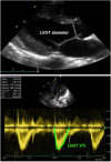
Echocardiographic assessment of cardiac output: 2D echocardiography long-axis view with measurement of LVOT diameter (above image), allowing for calculation of the LVOT CSA and Doppler tracing from an apical five-chamber view (below image) with tracing of the LVOT velocity to obtain the LVOT VTI. Abbreviations: CSA, cross-sectional area (calculated as π × r2 and r, radius, diameter/2): LVOT, left ventricular outflow tract; VTI, velocity time integral (or mean velocity).

References

    1. Al-Aweel I, Pursley DM, Rubin LP, Shah B, Weisberger S, Richardson DK. Variations in prevalence of hypotension, hypertension, and vasopressor use in NICUs. J Perinatol (2001) 21:272–8.10.1038/sj.jp.7210563 - DOI - PubMed
    1. Sankar J, Das RR, Jain A, Dewangan S, Khilnani P, Yadav D, et al. Prevalence and outcome of diastolic dysfunction in children with fluid refractory septic shock – a prospective observational study. Pediatr Crit Care Med (2014) 15(9):e370–8.10.1097/PCC.0000000000000249 - DOI - PubMed
    1. Noori S, Seri I. Evidence-based versus pathophysiology-based approach to diagnosis and treatment of neonatal cardiovascular compromise. Semin Fetal Neonatal Med (2015) 20(4):238–45.10.1016/j.siny.2015.03.005 - DOI - PubMed
    1. Azhibekov T, Soleymani S, Lee BH, Noori S, Seri I. Hemodynamic monitoring of the critically ill neonate: an eye on the future. Semin Fetal Neonatal Med (2015) 20(4):246–54.10.1016/j.siny.2015.03.003 - DOI - PubMed
    1. Giesinger RE, McNamara PJ. Hemodynamic instability in the critically ill neonate: an approach to cardiovascular support based on disease pathophysiology. Semin Perinatol (2016) 40(3):174–88.10.1053/j.semperi.2015.12.005 - DOI - PubMed

LinkOut - more resources