

T-614 Promotes Osteoblastic Cell Differentiation by Increasing Dlx5 Expression and Regulating the Activation of p38 and NF- κ B
- PMID: 29670900
- PMCID: PMC5836304
- DOI: 10.1155/2018/4901591
T-614 Promotes Osteoblastic Cell Differentiation by Increasing Dlx5 Expression and Regulating the Activation of p38 and NF- κ B
Abstract
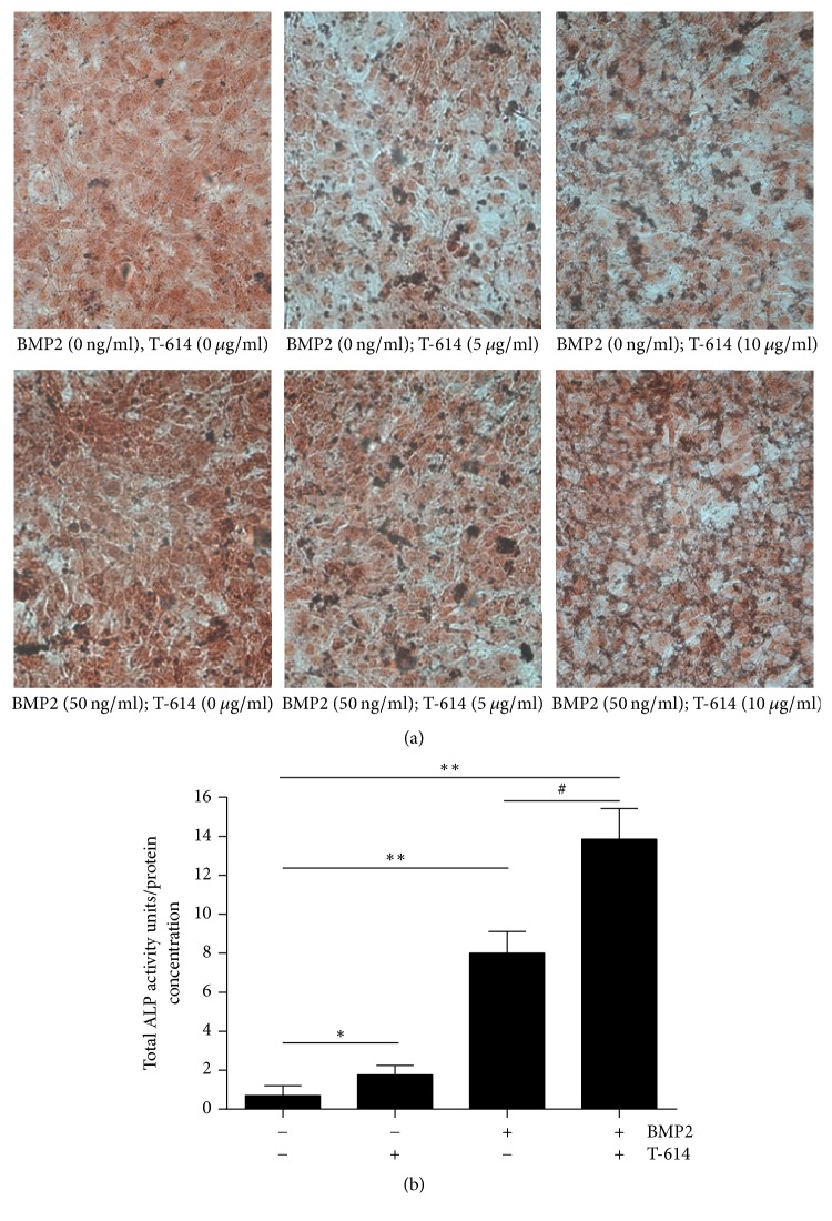
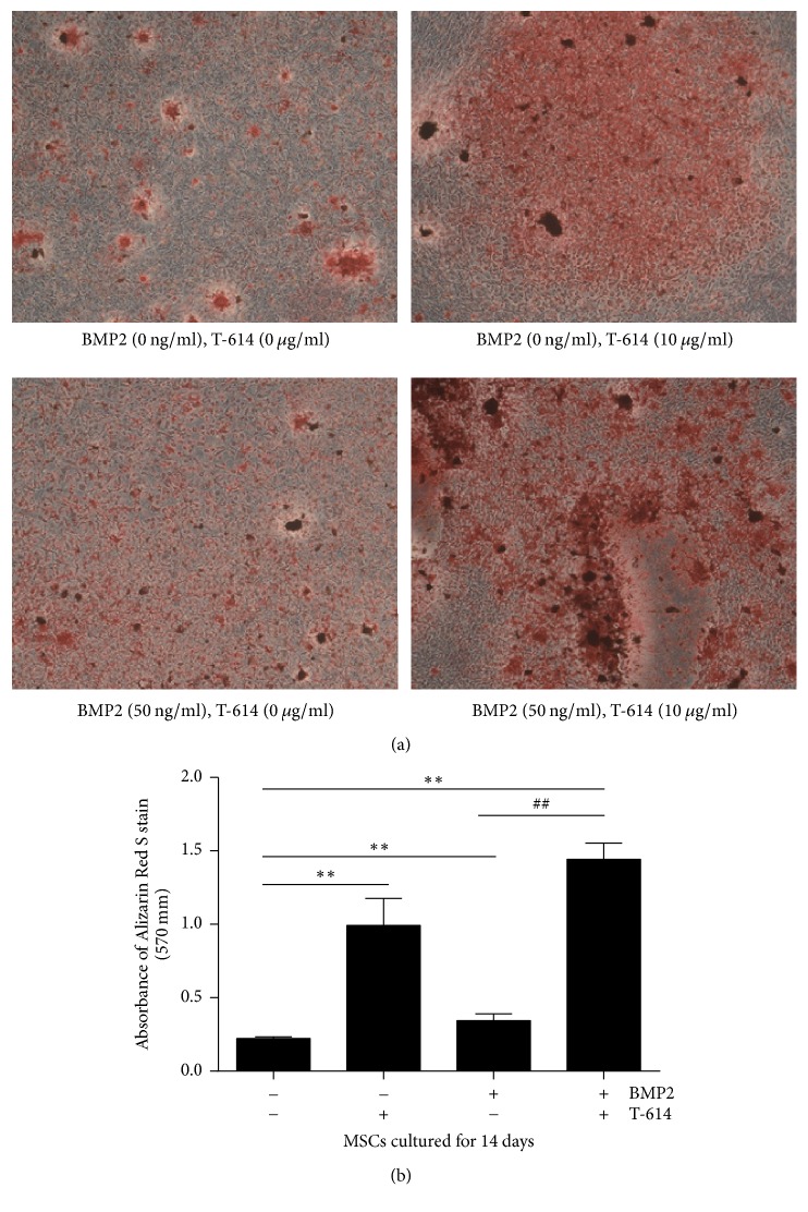
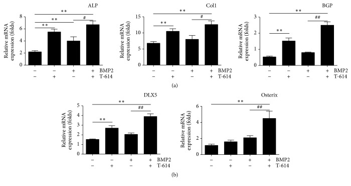
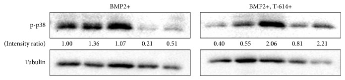
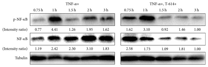
Rheumatoid arthritis (RA) is an autoimmune inflammatory disease characterized by bone loss. Degree of inflammation has been identified as an important initiator of skeletal damage in RA. Iguratimod (T-614) is an anti-inflammatory agent which has been reported to show the inhibitory effect of bone destruction in RA. However, the role of T-614 in osteoblast differentiation is still not clear. In this study, we intended to find the effect of T-614 on the osteogenesis process. We detected osteogenesis markers and transcription factors associated with osteoblastic lineage and bone formation in the culture of mesenchymal stem cells which differentiate osteoblast. The contents and activity of alkaline phosphatase, levels of collagen type I and bone gla protein, and calcium nodule formation were increased significantly after T-614 treated. Meanwhile, the mRNAs expressions of Osterix and Dlx5 were also found to be increased significantly by real-time PCR. The changes of levels of phosphorylation of p38 and NF-κB were also detected by Western blot. The results showed that T-614 promotes osteoblastic differentiation by increasing the expression of Osterix and Dlx5 and increasing the activation of P38. T-614 could advance the ectopic expression of NF-κB to suppress inflammation, which indirectly inhibits the damage of the osteoblasts.
Figures
Similar articles
-
Gliotoxin potentiates osteoblast differentiation by inhibiting nuclear factor-κB signaling.Mol Med Rep. 2015 Jul;12(1):877-84. doi: 10.3892/mmr.2015.3524. Epub 2015 Mar 20. Mol Med Rep. 2015. PMID: 25816130 Free PMC article.
-
Ugonin K promotes osteoblastic differentiation and mineralization by activation of p38 MAPK- and ERK-mediated expression of Runx2 and osterix.Eur J Pharmacol. 2011 Oct 15;668(3):383-9. doi: 10.1016/j.ejphar.2011.06.059. Epub 2011 Jul 26. Eur J Pharmacol. 2011. PMID: 21806985
-
Upregulated osterix expression elicited by Runx2 and Dlx5 is required for the accelerated osteoblast differentiation in PP2A Cα-knockdown cells.Cell Biol Int. 2018 Apr;42(4):403-410. doi: 10.1002/cbin.10902. Epub 2017 Nov 15. Cell Biol Int. 2018. PMID: 29068100
-
Sika pilose antler type I collagen promotes BMSC differentiation via the ERK1/2 and p38-MAPK signal pathways.Pharm Biol. 2017 Dec;55(1):2196-2204. doi: 10.1080/13880209.2017.1397177. Pharm Biol. 2017. PMID: 29115171 Free PMC article.
-
CD200 expression in human cultured bone marrow mesenchymal stem cells is induced by pro-osteogenic and pro-inflammatory cues.J Cell Mol Med. 2016 Apr;20(4):655-65. doi: 10.1111/jcmm.12752. Epub 2016 Jan 16. J Cell Mol Med. 2016. PMID: 26773707 Free PMC article.
Cited by
-
Research progress on the clinical application and mechanism of iguratimod in the treatment of autoimmune diseases and rheumatic diseases.Front Immunol. 2023 Sep 21;14:1150661. doi: 10.3389/fimmu.2023.1150661. eCollection 2023. Front Immunol. 2023. PMID: 37809072 Free PMC article. Review.
-
Melatonin attenuates degenerative disc degression by downregulating DLX5 via the TGF/Smad2/3 pathway in nucleus pulposus cells.JOR Spine. 2024 Nov 13;7(4):e70014. doi: 10.1002/jsp2.70014. eCollection 2024 Dec. JOR Spine. 2024. PMID: 39539538 Free PMC article.
-
Iguratimod as a New Drug for Rheumatoid Arthritis: Current Landscape.Front Pharmacol. 2020 Feb 26;11:73. doi: 10.3389/fphar.2020.00073. eCollection 2020. Front Pharmacol. 2020. PMID: 32174824 Free PMC article. Review.
-
Clinical effectiveness of iguratimod based on real-world data of patients with rheumatoid arthritis.Clin Rheumatol. 2021 Jan;40(1):123-132. doi: 10.1007/s10067-020-05208-y. Epub 2020 Jun 6. Clin Rheumatol. 2021. PMID: 32506311
-
Iguratimod: Novel Molecular Insights and a New csDMARD for Rheumatoid Arthritis, from Japan to the World.Life (Basel). 2021 May 20;11(5):457. doi: 10.3390/life11050457. Life (Basel). 2021. PMID: 34065413 Free PMC article. Review.
References
-
- Wei Y., Sun X., Hua M., Tan W., Wang F., Zhang M. Inhibitory effect of a novel antirheumatic drug T-614 on the IL-6-induced RANKL/OPG, IL-17, and MMP-3 expression in synovial fibroblasts from rheumatoid arthritis patients. BioMed Research International. 2015;2015 doi: 10.1155/2015/214683.214683 - DOI - PMC - PubMed
MeSH terms
Substances
LinkOut - more resources
Full Text Sources
Other Literature Sources
