Inflammatory Human Umbilical Cord-Derived Mesenchymal Stem Cells Promote Stem Cell-Like Characteristics of Cancer Cells in an IL-1 β-Dependent Manner
- PMID: 29670904
- PMCID: PMC5835289
- DOI: 10.1155/2018/7096707
Inflammatory Human Umbilical Cord-Derived Mesenchymal Stem Cells Promote Stem Cell-Like Characteristics of Cancer Cells in an IL-1 β-Dependent Manner
Abstract
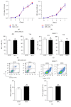
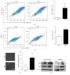
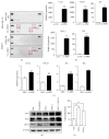
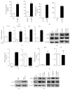
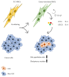
To ensure the safety of clinical applications of MSCs, thorough understanding of their impacts on tumor initiation and progression is essential. Here, to further explore the complex dialog between MSCs and tumor cells, umbilical cord-derived mesenchymal stem cells (UC-MSCs) were employed to be cocultured with either breast or ovarian cancer cells. Though having no obvious influence on proliferation or apoptosis, UC-MSCs exerted intense stem cell-like properties promoting effects on both cancer models. Cocultured cancer cells showed enriched side population, enhanced sphere formation ability, and upregulated pluripotency-associated stem cell markers. Human cytokine array and real-time PCR revealed a panel of MSC-derived prostemness cytokines CCL2, CXCL1, IL-8, and IL-6 which were induced upon coculturing. We further revealed IL-1β, a well-characterized proinflammatory cytokine, to be the inducer of these prostemness cytokines, which was generated from inflammatory UC-MSCs in an autocrine manner. Additionally, with introduction of IL-1RA (an IL-1 receptor antagonist) into the coculturing system, the stem cell-like characteristics promoting effects of inflammatory UC-MSCs were partially blocked. Taken together, these findings suggest that transduced inflammatory MSCs work as a major source of IL-1β in tumor microenvironment and initiate the formation of prostemness niche via regulating their secretome in an IL-1β-dependent manner.
Figures

Similar articles
-
Human Umbilical Cord Mesenchymal Stem Cells Promote Breast Cancer Metastasis by Interleukin-8- and Interleukin-6-Dependent Induction of CD44(+)/CD24(-) Cells.Cell Transplant. 2015;24(12):2585-99. doi: 10.3727/096368915X687462. Epub 2015 Feb 18. Cell Transplant. 2015. PMID: 25695620
-
Assessment of tumor promoting effects of amniotic and umbilical cord mesenchymal stem cells in vitro and in vivo.J Cancer Res Clin Oncol. 2019 May;145(5):1133-1146. doi: 10.1007/s00432-019-02859-6. Epub 2019 Feb 25. J Cancer Res Clin Oncol. 2019. PMID: 30805774 Free PMC article.
-
Interleukin-1β induces CXCR3-mediated chemotaxis to promote umbilical cord mesenchymal stem cell transendothelial migration.Stem Cell Res Ther. 2018 Oct 25;9(1):281. doi: 10.1186/s13287-018-1032-9. Stem Cell Res Ther. 2018. PMID: 30359318 Free PMC article.
-
Interleukin-1 receptor antagonist (IL-1Ra) and IL-1Ra producing mesenchymal stem cells as modulators of diabetogenesis.Autoimmunity. 2010 Jun;43(4):255-63. doi: 10.3109/08916930903305641. Autoimmunity. 2010. PMID: 19845478 Review.
-
The role of Interleukin 1 receptor antagonist in mesenchymal stem cell-based tissue repair and regeneration.Biofactors. 2020 Mar;46(2):263-275. doi: 10.1002/biof.1587. Epub 2019 Nov 22. Biofactors. 2020. PMID: 31755595 Review.
Cited by
-
A Novel Aurora Kinase Inhibitor Attenuates Leukemic Cell Proliferation Induced by Mesenchymal Stem Cells.Mol Ther Oncolytics. 2020 Aug 5;18:491-503. doi: 10.1016/j.omto.2020.08.001. eCollection 2020 Sep 25. Mol Ther Oncolytics. 2020. PMID: 32953983 Free PMC article.
-
IL7-IL12 Engineered Mesenchymal Stem Cells (MSCs) Improve A CAR T Cell Attack Against Colorectal Cancer Cells.Cells. 2020 Apr 3;9(4):873. doi: 10.3390/cells9040873. Cells. 2020. PMID: 32260097 Free PMC article.
-
Key Factor Regulating Inflammatory Microenvironment, Metastasis, and Resistance in Breast Cancer: Interleukin-1 Signaling.Mediators Inflamm. 2021 Sep 23;2021:7785890. doi: 10.1155/2021/7785890. eCollection 2021. Mediators Inflamm. 2021. PMID: 34602858 Free PMC article. Review.
-
CAR-T Cells/-NK Cells in Cancer Immunotherapy and the Potential of MSC to Enhance Its Efficacy: A Review.Biomedicines. 2022 Mar 30;10(4):804. doi: 10.3390/biomedicines10040804. Biomedicines. 2022. PMID: 35453554 Free PMC article. Review.
References
-
- de Windt T. S., Vonk L. A., Slaper-Cortenbach I. C. M., et al. Allogeneic Mesenchymal Stem Cells Stimulate Cartilage Regeneration and Are Safe for Single-Stage Cartilage Repair in Humans upon Mixture with Recycled Autologous Chondrons. Stem Cells. 2017;35(1):256–264. doi: 10.1002/stem.2475. - DOI - PubMed
-
- Mishra S. K., Rana P., Khushu S., Gangenahalli G. Therapeutic prospective of infused allogenic cultured mesenchymal stem cells in traumatic brain injury mice: A longitudinal proton magnetic resonance spectroscopy assessment. Stem Cells Translational Medicine. 2017;6(1):316–329. doi: 10.5966/sctm.2016-0087. - DOI - PMC - PubMed
MeSH terms
Substances
LinkOut - more resources
Full Text Sources
Other Literature Sources

