

Characterisation of DOG-1 Expression in Salivary Gland Tumours and Comparison with Myoepithelial Markers
- PMID: 29671211
- PMCID: PMC6513803
- DOI: 10.1007/s12105-018-0917-3
Characterisation of DOG-1 Expression in Salivary Gland Tumours and Comparison with Myoepithelial Markers
Abstract
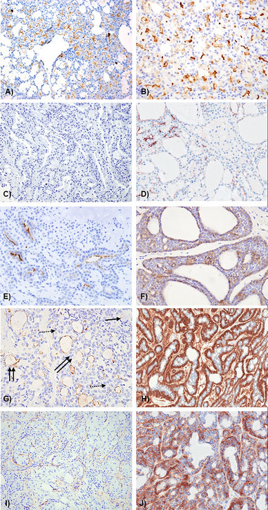
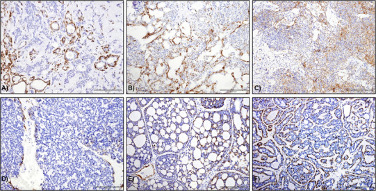
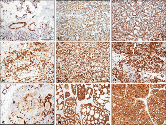
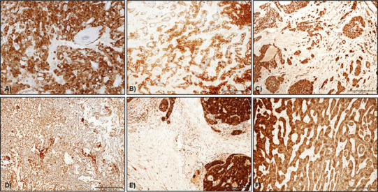
DOG1 is an established diagnostic marker for gastrointestinal stromal tumours (GIST), but has been reported in salivary gland tumours (SGT) as an acinar and intercalated duct marker. However, its specificity and distribution is not well established. The aim of this study was to evaluate the diagnostic utility of DOG-1 expression in SGT in addition to comparing it with myoepithelial markers. Normal salivary tissue and SGT (n = 184) were examined for expression of DOG1 and a range of myoepithelial markers. SGT included: acinic cell carcinoma (ACC, n = 15), secretory carcinoma (SC, n = 9), pleomorphic adenoma (PA, n = 49), carcinoma ex-PA (Ca ex-PA, n = 11), adenoid cystic carcinoma (AdCC, n = 20), polymorphous adenocarcinoma (PAC, n = 6), myoepithelioma (n = 6), myoepithelial carcinoma (MC, n = 2), basal cell adenoma (BCA, n = 14), canalicular adenoma (CA, n = 19), mucoepidermoid carcinoma (MEC, n = 11), oncocytoma (n = 2), adenocarcinoma NOS (AdNOS, n = 4), basal cell adenocarcinoma (BCAC, n = 2), salivary duct carcinoma (SDC, n = 3) and papillary cystadenocarcinoma (PCAC, n = 1). Normal acini and ACC (14/15) showed strong luminal DOG1 staining; SC were largely negative with only focal expression in 3/9 cases. Luminal staining was seen in PA (14/49), PAC (4/6), Ca ex-PA (4/11) and AdCC (6/20). 8/11 MEC showed luminal and/or mucous cell staining. No staining was seen in myoepithelioma, MC, CA, adNOS and BCAC. BCA showed strong staining of myoepithelial cells in some cases (5/14). Variable myoepithelial DOG1 staining was seen in PA, Ca ex PA, BCA, SDC and PCAC which was not as consistent as myoepithelial markers such as calponin, p63 and αSMA. Absence of DOG1 can differentiate ACC from SC, but staining is variable in PA, PLGA and Ca ex-PA. Myoepithelial staining in some tumours but not in normal gland suggests a wider distribution in SGT than originally envisaged.
Keywords: Acinic cell carcinoma; DOG-1; Luminal; Myoepithelial; Salivary gland tumours; Secretory carcinoma.
Conflict of interest statement
Conflict of interest
No conflict of interest to disclose.
Ethical Approval
All procedures performed in studies involving human participants were in accordance with the ethical standards of the institutional and/or national research committee and with the 1964 Helsinki declaration and its later amendments or comparable ethical standards. NRES Committee Yorkshire and Humber—Sheffield (South Sheffied REC)—Reference 05/Q2305/127.
Figures
References
-
- Espinosa I, Lee CH, Kim MK, Rouse BT, Subramanian S, Montgomery K, Varma S, Corless CL, Heinrich MC, Smith KS, Wang Z, Rubin B, Nielsen TO, Seitz RS, Ross DT, West RB, Cleary ML, van de Rijn M. A novel monoclonal antibody against DOG1 is a sensitive and specific marker for gastrointestinal stromal tumors. Am J Surg Pathol. 2008;32(2):210–218. doi: 10.1097/PAS.0b013e3181238cec. - DOI - PubMed
-
- Simon S, Grabellus F, Ferrera L, Galietta L, Schwindenhammer B, Mühlenberg T, Taeger G, Eilers G, Treckmann J, Breitenbuecher F, Schuler M, Taguchi T, Fletcher JA, Bauer S. DOG1 regulates growth and IGFBP5 in gastrointestinal stromal tumors. Cancer Res. 2013;73(12):3661–3670. doi: 10.1158/0008-5472.CAN-12-3839. - DOI - PMC - PubMed
Publication types
MeSH terms
Substances
LinkOut - more resources
Full Text Sources
Other Literature Sources
Medical
