Review
. 2018 Aug;9(4):499-510.
doi: 10.1007/s13244-018-0604-7. Epub 2018 Apr 18.

Neuroimaging of Meckel's cave in normal and disease conditions

Affiliations
Review

Neuroimaging of Meckel's cave in normal and disease conditions

Ajay Malhotra et al. Insights Imaging. 2018 Aug.

Abstract

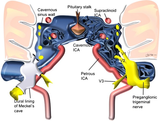
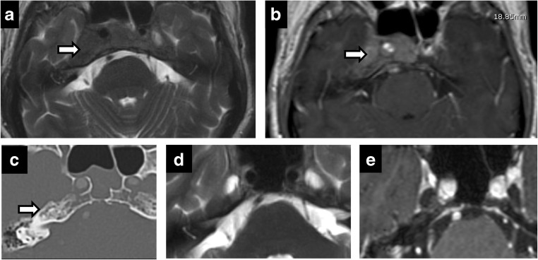
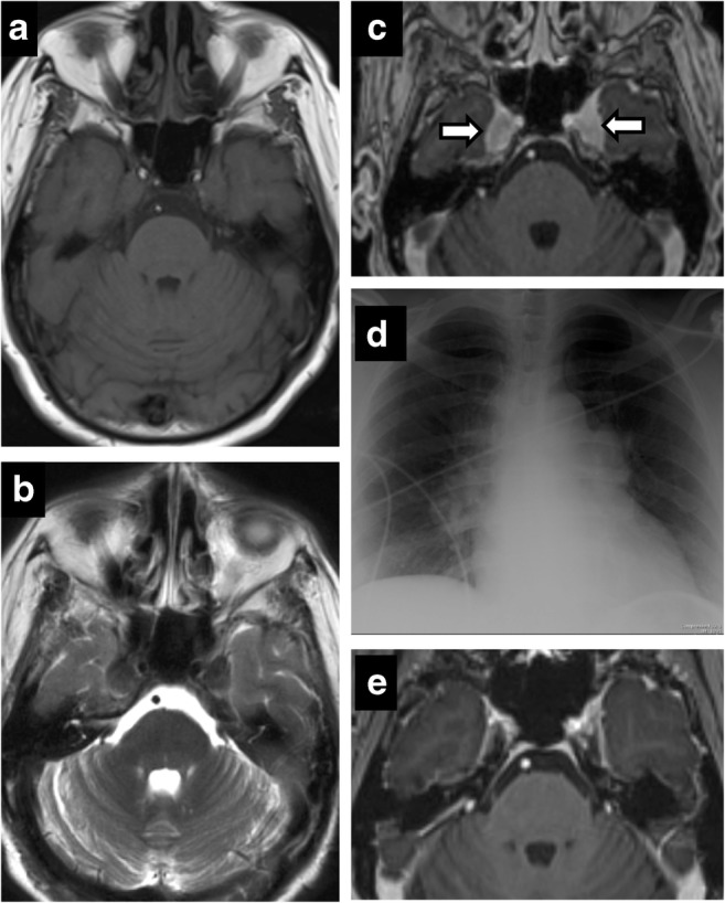
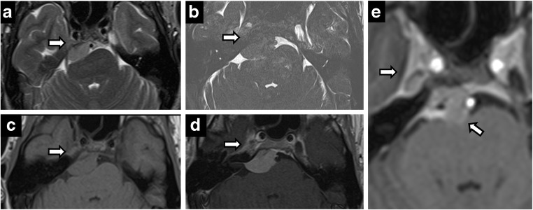
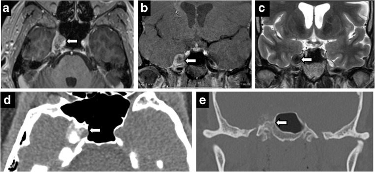
Meckel's cave is a dural recess in the posteromedial portion of the middle cranial fossa that acts as a conduit for the trigeminal nerve between the prepontine cistern and the cavernous sinus, and houses the Gasserian ganglion and proximal rootlets of the trigeminal nerve. It serves as a major pathway in perineural spread of pathologies such as head and neck neoplasms, automatically upstaging tumours, and is a key structure to assess in cases of trigeminal neuralgia. The purpose of this pictorial review is threefold: (1) to review the normal anatomy of Meckel's cave; (2) to describe imaging findings that identify disease involving Meckel's cave; (3) to present case examples of trigeminal and non-trigeminal processes affecting Meckel's cave.

Teaching points: • Meckel's cave contains the trigeminal nerve between prepontine cistern and cavernous sinus. • Assessment is essential for perineural spread of disease and trigeminal neuralgia. • Key imaging: neural enhancement, enlargement, perineural fat/CSF effacement, skull base foraminal changes.

Keywords: Meckel’s cave; Neuralgia; Perineural; Skull base; Trigeminal.


Figures

Fig. 1
Fig. 1
Schematic representation of Meckel’s cave and relationship with adjacent structures
Fig. 2
Fig. 2
Perineural spread of tumour. Coronal T2 (a), axial C+ (b), coronal C+ (c), coronal C+ (d). A 60-year-old man with right CN V symptoms. Expansile T2 hypointense, enhancing lesion filling the right Meckel’s cave extending through the foramen ovale and along V2 in the foramen rotundum
Fig. 3
Fig. 3
Perineural spread of tumour. A 45-year-old man with left facial pain and numbness. CT showing enhancing soft tissue along the course of left CN V, expanding into the foramen rotundum (a), infraorbital foramen (b) and foramen ovale (c). Posteriorly, the enhancing tumour extends to the left Meckel’s cave. Rapid expansion on follow-up MRI (d) with intense uptake on positron emission tomography (PET) (e) in left CN V including foramen ovale (arrow)
Fig. 4
Fig. 4
Diffuse infantile pontine glioma (a) with significant increase in size on the 3-month study (b) with perineural spread along left CN V into Meckel’s cave
Fig. 5
Fig. 5
Herpes zoster reactivation. A 62-year-old woman with right perioral numbness for 2 weeks and vesicular rash. Postcontrast axial (a) and coronal (b, c) images showing tubular enhancement along the right CN V from the origin, through the cisternal segment and extending to Meckel’s cave
Fig. 6
Fig. 6
Persistent trigeminal artery. Axial T2-weighted imaging (a) and CT angiography (b). Vascular channel connecting the left internal carotid artery and the basilar artery, and running through the medial aspect of Meckel’s cave
Fig. 7
Fig. 7
Trigeminal AVM. A 58-year-old man with right facial neuralgia. Axial T2 (a) and axial C+ (b) showing abnormal serpentine vasculature along cisternal segment of right CN V extending into Meckel’s cave on DSA (c)—arteriovenous shunting from right anterior inferior cerebellar artery (AICA) with prominent draining vein
Fig. 8
Fig. 8
Trigeminal schwannoma. Coronal C+ (a), axial T2 (b), axial T1 (c), axial C+ (d): expansile enhancing mass in the right Meckel’s cave with a large, lobulated cystic component along the cisternal segment of the right CN V
Fig. 9
Fig. 9
Schwannoma of Meckel’s cave in a patient with neurofibromatosis type 2. Axial T2 (a), axial C+ (b), coronal C+ (c) showing an enhancing lesion within the right Meckel’s cave. Note additional bilateral vestibular schwannomas
Fig. 10
Fig. 10
Dural ectasia in neurofibromatosis type 1. Axial T2 (a), axial C+ (b), CT (c). Bilateral enlargement of Meckel’s cave—CSF isointense and no abnormal enhancement. Smooth scalloping and remodelling of petrous apex on CT (c)
Fig. 11
Fig. 11
Lymphoma. Axial T2 (a), axial C+ (b), CT (c). Bilateral CN VI palsy and right facial pain. Mottled appearance of clivus and petrous apices on CT—caused by T2 hypointense, enhancing infiltrating lesion. Post treatment (d, e)—complete resolution
Fig. 12
Fig. 12
Sarcoidosis. Axial T1 (a), axial T2 (b), axial T1 C+ (c). A 53-year-old woman with facial pain and numbness. Enhancing, T2 hypointense lesions symmetrically involving the bilateral Meckel’s cave. Chest X-ray (d)—bilateral hilar and mediastinal lymphadenopathy. Follow-up (e)—complete resolution
Fig. 13
Fig. 13
Charcot Marie Tooth (CMT) disease. Fusiform thickened bilateral CN V (a-d)—including V3 segment extending through Foramen ovale (arrows in b, c) and V2 segment in the foramen rotundum (arrow in d). Diffusely thickened spinal nerves (e)
Fig. 14
Fig. 14
Meningioma (extrinsic). Axial T2 (a), T2 SPACE (b), axial T1 (c), axial C+ (d). Right petroclival T2 hypointense, enhancing mass invading the right Meckel’s cave. Postoperative residual enhancing lesion in the right Meckel’s cave and abutting basilar artery (e)
Fig. 15
Fig. 15
Meningioma (intrinsic). Axial C+ (a), coronal C+ (b), coronal T2 (c), axial CT (d), coronal CT (e). Circumscribed enhancing right Meckel’s cave mass—hypointense on T2. CT shows calcifications with hyperostosis of adjacent bone
Fig. 16
Fig. 16
Tolosa-Hunt syndrome. Axial T1 C+ (a, b), axial T2 (c), coronal C+ (d). A 23-year-old woman with acute onset of painful left diplopia. Asymmetric enhancing tissue in left cavernous sinus extending to Meckel’s cave and through the foramen ovale (b, d). Follow-up—complete resolution (e)
Fig. 17
Fig. 17
Petrous apex cephalocele. Coronal (a, d) and axial (b, e) T2-weighted images of a cystic petrous apex lesion that communicates with the posterolateral portion of Meckel’s cave. No abnormal enhancement on axial post-contrast images (c, f)
Fig. 18
Fig. 18
Epidermoid. Expansion of left Meckel’s cave by a left cerebellopontine angle mass on axial T2 (a), showing no enhancement (b) and restricted diffusion (c). Postoperative residual tissue in left Meckel’s cave (d, e)
Fig. 19
Fig. 19
Chondrosarcoma. Axial T2 (a), axial T1 (b), axial T1 C+ (c), CT (d). A 61-year-old woman with effacement of the right Meckel’s cave by an expansile petrous apex mass that is hyperintense on T2, hypointense on T1 and shows avid enhancement on post contrast image. CT shows features of slow growing lesion. Note preserved Meckel’s cave on the left
Fig. 20
Fig. 20
Bilateral cavernous ICA aneurysms. Axial C+ (a), coronal T2 (b), CT (c) and (d). A 73-year-old woman with left facial pain. Bilateral cavernous enhancing, partially calcified lesions encroaching the Meckel’s cave—left greater than right

References

    1. Aaron GP, Illing E, Lambertsen Z, et al. Enlargement of Meckel's cave in patients with spontaneous cerebrospinal fluid leaks. Int Forum Allergy Rhinol. 2017;7:421–424. doi: 10.1002/alr.21891. - DOI - PubMed
    1. Abdel Aziz KM, Van Loveren HR. Primary lymphoma of Meckel's cave mimicking trigeminal schwannoma: case report. Neurosurgery. 1999;44:859–862. doi: 10.1097/00006123-199904000-00096. - DOI - PubMed
    1. Aho TR, Wallace RC, Pitt AM, et al. Charcot-Marie-tooth disease: extensive cranial nerve involvement on CT and MR imaging. AJNR Am J Neuroradiol. 2004;25:494–497. - PMC - PubMed
    1. Borges A. Skull base tumours part I: imaging technique, anatomy and anterior skull base tumours. Eur J Radiol. 2008;66:338–347. doi: 10.1016/j.ejrad.2008.01.017. - DOI - PubMed
    1. Borges A, Casselman J. Imaging the cranial nerves: part I: methodology, infectious and inflammatory, traumatic and congenital lesions. Eur Radiol. 2007;17:2112–2125. doi: 10.1007/s00330-006-0575-9. - DOI - PubMed

LinkOut - more resources