. 2018 Jul;29(7):1505-1511.
doi: 10.1007/s13361-018-1946-6. Epub 2018 Apr 18.

An Internal Standard for Assessing Phosphopeptide Recovery from Metal Ion/Oxide Enrichment Strategies

Affiliations

An Internal Standard for Assessing Phosphopeptide Recovery from Metal Ion/Oxide Enrichment Strategies

Joao A Paulo et al. J Am Soc Mass Spectrom. 2018 Jul.

Abstract

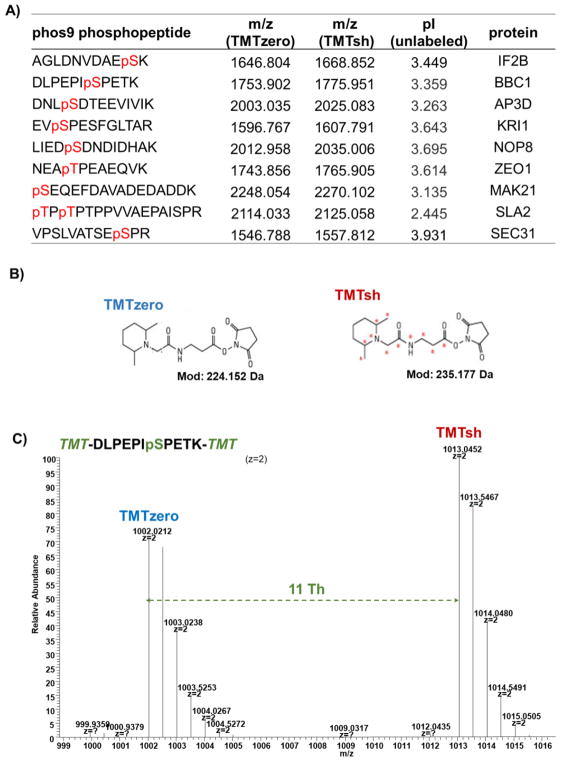
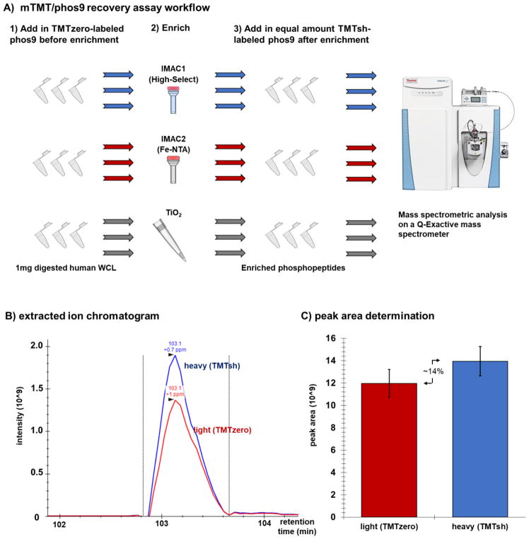
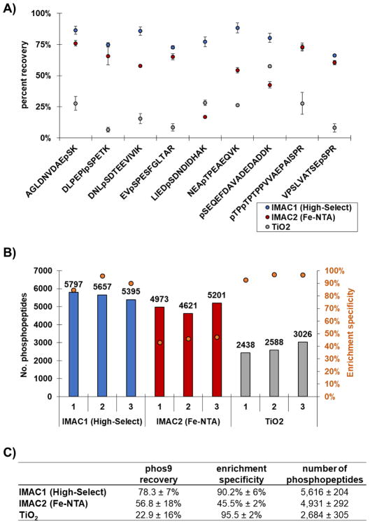
Phosphorylation-mediated signaling pathways have major implications in cellular regulation and disease. However, proteins with roles in these pathways are frequently less abundant and phosphorylation is often sub-stoichiometric. As such, the efficient enrichment, and subsequent recovery of phosphorylated peptides, is vital. Mass spectrometry-based proteomics is a well-established approach for quantifying thousands of phosphorylation events in a single experiment. We designed a peptide internal standard-based assay directed toward sample preparation strategies for mass spectrometry analysis to understand better phosphopeptide recovery from enrichment strategies. We coupled mass-differential tandem mass tag (mTMT) reagents (specifically, TMTzero and TMTsuper-heavy), nine mass spectrometry-amenable phosphopeptides (phos9), and peak area measurements from extracted ion chromatograms to determine phosphopeptide recovery. We showcase this mTMT/phos9 recovery assay by evaluating three phosphopeptide enrichment workflows. Our assay provides data on the recovery of phosphopeptides, which complement other metrics, namely the number of identified phosphopeptides and enrichment specificity. Our mTMT/phos9 assay is applicable to any enrichment protocol in a typical experimental workflow irrespective of sample origin or labeling strategy. Graphical Abstract ᅟ.

Keywords: Lumos; Phosphorylation; TMT super-heavy; TMT0; TMTsh; TMTzero; mTRAQ.


Conflict of interest statement

Competing interest

The authors acknowledge no competing interests.

Figures

Figure 1
Figure 1. TMT reagents and peptides used in this experiment
A) The table lists the phos9 phosphopeptide sequences, m/z for TMTzero- and TMTsh-labeled phosphopeptides, isoelectric point for unlabeled phosphopeptide, and associated protein. B) Chemical structures of TMTzero and TMTsh. C) Spectrum highlighting the mass difference between a doubly charged, lysine-terminating peptide (DLPEPIpSPETK) labeled with TMTzero and TMTsh.
Figure 2
Figure 2. Experimental overview of phosphopeptide enrichment strategy
A) TMTzero-labeled phos9 was added to 1mg aliquots of trypsinized human whole cell lysate (WCL). Phosphopeptides were enriched using one of three methods (High-Select, Fe-NTA, TiO2) with three technical replicates. TMTsh-labeled phos9 was spiked into the enriched sample. The sample was desalted and analyzed on a QExactive mass spectrometer. B) Example of an extracted ion chromatogram, as displayed in Skyline. C) Peak area comparison as determined in Skyline.
Figure 3
Figure 3. Evaluation of phosphorylated peptides identified by each enrichment method
A) The dot plot illustrates the efficiency of recovery (peak area ratio of TMTzero to TMTsh) for phos9 phosphopeptides. Error bars represent standard error of the mean (s.e.m.) for three independent replicate samples. B) The bar plot illustrates the total number of phosphorylated peptides identified by each method. The overlaid orange circles represent the enrichment specificity (percentage of peptides that are phosphorylated) for each method. C) The table summarizes the findings for the phos9 peptide cocktail (mean ± SD).

References

    1. Paulo JA, Gaun A, Gygi SP. Global Analysis of Protein Expression and Phosphorylation Levels in Nicotine-Treated Pancreatic Stellate Cells. Journal of proteome research. 2015;14:4246–4256. - PMC - PubMed
    1. Dephoure N, Gould KL, Gygi SP, Kellogg DR. Mapping and analysis of phosphorylation sites: a quick guide for cell biologists. Molecular biology of the cell. 2013;24:535–542. - PMC - PubMed
    1. Wu R, Haas W, Dephoure N, Huttlin EL, Zhai B, Sowa ME, Gygi SP. A large-scale method to measure absolute protein phosphorylation stoichiometries. Nature methods. 2011;8:677–683. - PMC - PubMed
    1. Wu R, Dephoure N, Haas W, Huttlin EL, Zhai B, Sowa ME, Gygi SP. Correct interpretation of comprehensive phosphorylation dynamics requires normalization by protein expression changes. Mol Cell Proteomics. 2011;10:M111 009654. - PMC - PubMed
    1. Huttlin EL, Jedrychowski MP, Elias JE, Goswami T, Rad R, Beausoleil SA, Villen J, Haas W, Sowa ME, Gygi SP. A tissue-specific atlas of mouse protein phosphorylation and expression. Cell. 2010;143:1174–1189. - PMC - PubMed

MeSH terms