. 2019 Oct;10(4):467-479.
doi: 10.1177/1947603518769714. Epub 2018 Apr 19.

Human Articular Chondrocytes Retain Their Phenotype in Sustained Hypoxia While Normoxia Promotes Their Immunomodulatory Potential

Affiliations

Human Articular Chondrocytes Retain Their Phenotype in Sustained Hypoxia While Normoxia Promotes Their Immunomodulatory Potential

Claire Mennan et al. Cartilage. 2019 Oct.

Abstract

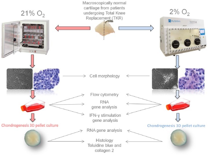
Objective: To assess the phenotype of human articular chondrocytes cultured in normoxia (21% O2) or continuous hypoxia (2% O2).

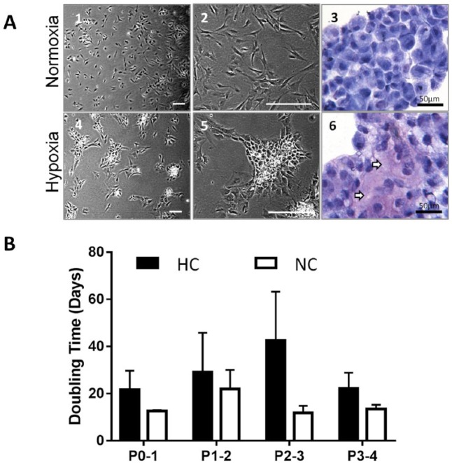
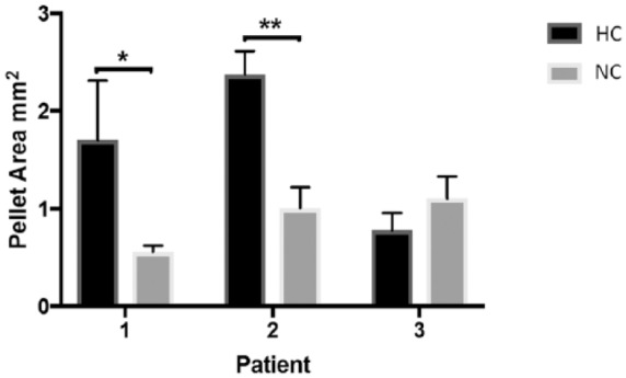
Design: Chondrocytes were extracted from patients undergoing total knee replacement (n = 5) and cultured in ~21% (normoxic chondrocytes, NC) and 2% (hypoxic chondrocytes, HC) oxygen in both monolayer and 3-dimensional (3D) pellet culture and compared with freshly isolated chondrocytes (FC). Cells were assessed by flow cytometry for markers indicative of mesenchymal stromal cells (MSCs), chondrogenic-potency and dedifferentiation. Chondrogenic potency and immunomodulatory gene expression was assessed in NC and HC by reverse transcription quantitative polymerase chain reaction. Immunohistochemistry was used to assess collagen II production following 3D pellet culture.

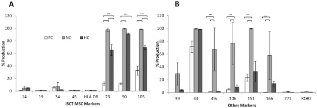
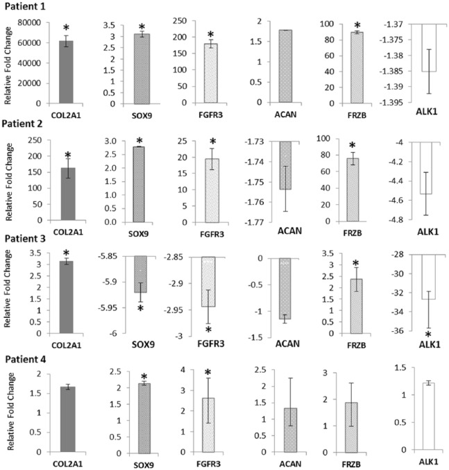
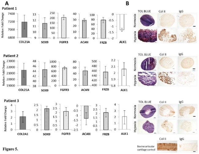
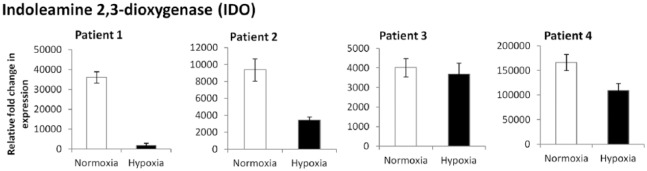
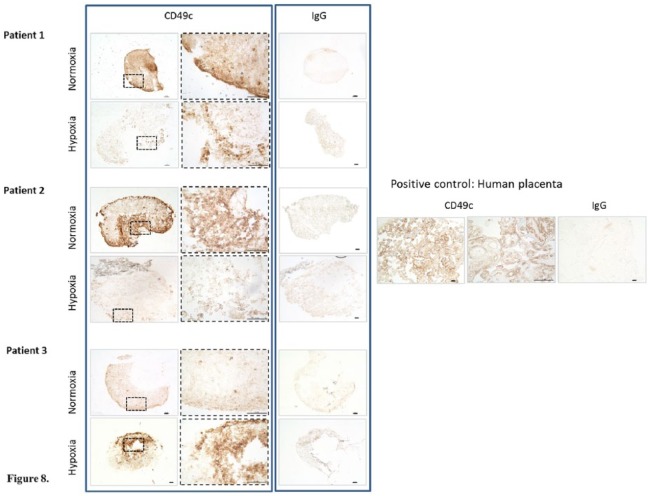
Results: NC were positive (>97%, n = 5) for MSC markers, CD73, CD90, and CD105, while HC demonstrated <90% positivity (n = 4) and FC (n = 5) less again (CD73 and CD90 <20%; CD105 <40%). The markers CD166 and CD151, indicative of chondrogenic de-differentiation, were significantly higher on NC compared with HC and lowest on FC. NC also produced the highest levels of CD106 and showed the greatest levels of IDO expression, following interferon-γ stimulation, indicating immunomodulatory potential. NC produced the highest levels of CD49c (>60%) compared with HC and FC in which production was <2%. Hypoxic conditions upregulated expression of SOX9, frizzled-related protein (FRZB), fibroblast growth factor receptor 3 (FGFR3), and collagen type II (COL2A1) and downregulated activin receptor-like kinase 1 (ALK1) in 3 out of 4 patients compared with normoxic conditions for monolayer cells.

Conclusions: Hypoxic conditions encourage retention of a chondrogenic phenotype with some immunomodulatory potential, whereas normoxia promotes dedifferentiation of chondrocytes toward an MSC phenotype with loss of chondrogenic potency but enhanced immunomodulatory capacity.

Keywords: Sustained hypoxia; cartilage repair; chondrogenic; hypoxic workstation; immunomodulation.


Conflict of interest statement

Declaration of Conflicting Interests: The author(s) declared no potential conflicts of interest with respect to the research, authorship, and/or publication of this article.

Figures

Figure 1.
Figure 1.
Experimental workflow. Macroscopically normal human cartilage was collected in media conditioned to 2% O2.
Figure 2.
Figure 2.
(A) Representative images from cells at passage 3 (P3) in normoxia (NC) (21% O2) and hypoxia (HC) (2% O2). (1 and 2) NC. (3) Cytospin stained with toluidine blue. (4 and 5) HC. (6) Cytospin stained with toluidine blue (arrows indicate glycosaminoglycan staining, purple metachromasia). (B) Doubling time (days) of NC (n = 4) and HC (n = 4). Bars represent the mean ± SD. Scale bars represent 200 µm unless otherwise stated.
Figure 3.
Figure 3.
Flow cytometry data showing the presence of cell markers indicative of (A) mesenchymal stem cell (MSC; using the ISCT criteria). (B) Other markers indicative of chondrogenic dedifferentiation, potency, and immunomodulation on freshly isolated chondrocytes (FC) (n = 5), normoxic chondrocytes (NC) at P3-4 (n = 5), and hypoxic chondrocytes (HC) at P3-4 (n = 4). Error bars indicate the mean ± SD.
Figure 4.
Figure 4.
Reverse transcription quantitative polymerase chain reaction (RT-qPCR) showing the expression of genes associated with chondrogenic potency, from monolayer chondrocytes cultured in normoxia (NC) and hypoxia (HC) at passage 3-4. Gene expression for HC and NC were normalized to the reference gene HPRT1. Data for HC are expressed relative to NC. Stars indicate genes that are significantly up- or downregulated.
Figure 5.
Figure 5.
(A) Reverse transcription quantitative polymerase chain reaction (RT qPCR) showing the expression of genes associated with chondrogenic potency, from 3-dimensional chondrogenic pellets cultured in normoxia (NC) and hypoxia (HC) at passage 3-4. Gene expression for HC and NC were normalized to the reference gene HPRT1. Data for HC are expressed relative to NC. (B) Chondrogenesis was assessed after 28 days in pellet culture by staining sections for glycosaminoglycan (GAG) using toluidine blue and type II collagen by immunohistochemistry. Stars indicate genes that are significantly up- or downregulated. Scale bars represent 100 µm.
Figure 6.
Figure 6.
The mean area of chondrogenic pellets grown in normoxia (NC) (21% O2) and hypoxia (HC) (2% O2). Data are taken from triplicate pellets from 3 different patients. Bars indicate the mean ± SD.
Figure 7.
Figure 7.
Reverse transcription quantitative polymerase chain reaction (RT-qPCR) showing the expression of the IDO gene in P3-4 monolayer chondrocytes cultured in normoxia (NC) and hypoxia (HC) following stimulation with 25 ng/mL interferon-γ (IFN-γ) for 24 hours. Gene expression was normalized to HPRT1. Gene expression for IFN-γ stimulated chondrocytes is expressed relative to those grown in normal media without inflammatory stimulus.
Figure 8.
Figure 8.
Immunohistochemistry showing CD49c staining of chondrogenic pellets cultured in normoxia (NC) and hypoxia (HC). Scale bars represent 100 μm.

References

    1. Mobasheri A, Rayman MP, Gualillo O, Sellam J, van der Kraan P, Fearon U. The role of metabolism in the pathogenesis of osteoarthritis. Nat Rev Rheumatol. 2017;13(5):302-11. doi:10.1038/nrrheum.2017.50. - DOI - PubMed
    1. Silver IA. Measurement of pH and ionic composition of pericellular sites. Philos Trans R Soc Lond B Biol Sci. 1975;271(912):261-72. - PubMed
    1. Caron MMJ, Emans PJ, Coolsen MME, Voss L, Surtel DA, Cremers A, et al. Redifferentiation of dedifferentiated human articular chondrocytes: comparison of 2D and 3D cultures. Osteoarthritis Cartilage. 2012;20(10):1170-8. doi:10.1016/j.joca.2012.06.016. - DOI - PubMed
    1. Duan L, Ma B, Liang Y, Chen J, Zhu W, Li M, et al. Cytokine networking of chondrocyte dedifferentiation in vitro and its implications for cell-based cartilage therapy. Am J Transl Res. 2015;7(2):194-208. - PMC - PubMed
    1. Duval E, Leclercq S, Elissalde JM, Demoor M, Galéra P, Boumédiene K. Hypoxia-inducible factor 1α inhibits the fibroblast-like markers type I and type III collagen during hypoxia-induced chondrocyte redifferentiation: hypoxia not only induces type II collagen and aggrecan, but it also inhibits type I and type III collagen in the hypoxia-inducible factor 1α-dependent redifferentiation of chondrocytes. Arthritis Rheum. 2009;60(10):3038-48. doi:10.1002/art.24851. - DOI - PubMed

Publication types

MeSH terms