. 2018 May 8;57(18):2590-2596.
doi: 10.1021/acs.biochem.8b00131. Epub 2018 Apr 19.

Optochemical Control of Protein Localization and Activity within Cell-like Compartments

Affiliations

Optochemical Control of Protein Localization and Activity within Cell-like Compartments

Reese M Caldwell et al. Biochemistry. .

Abstract

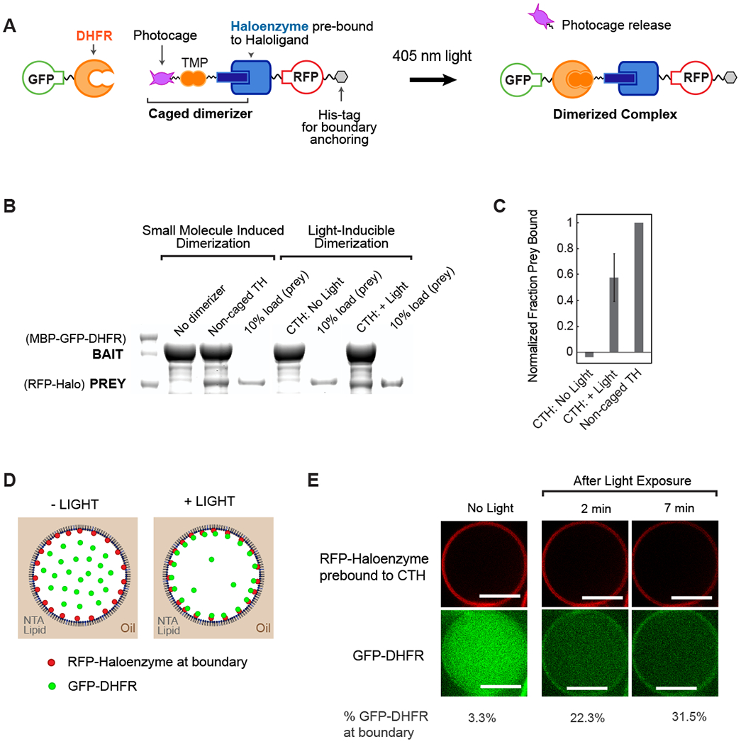
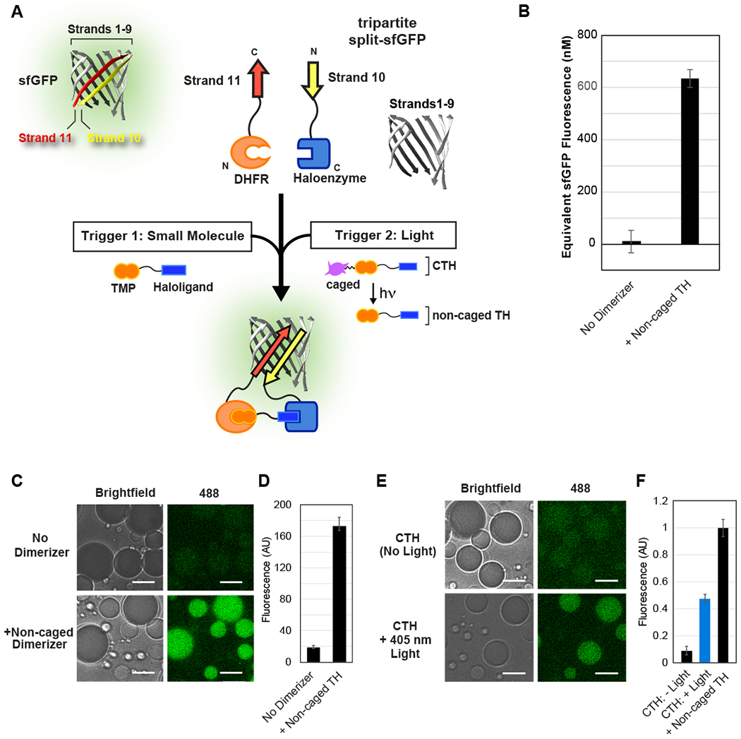
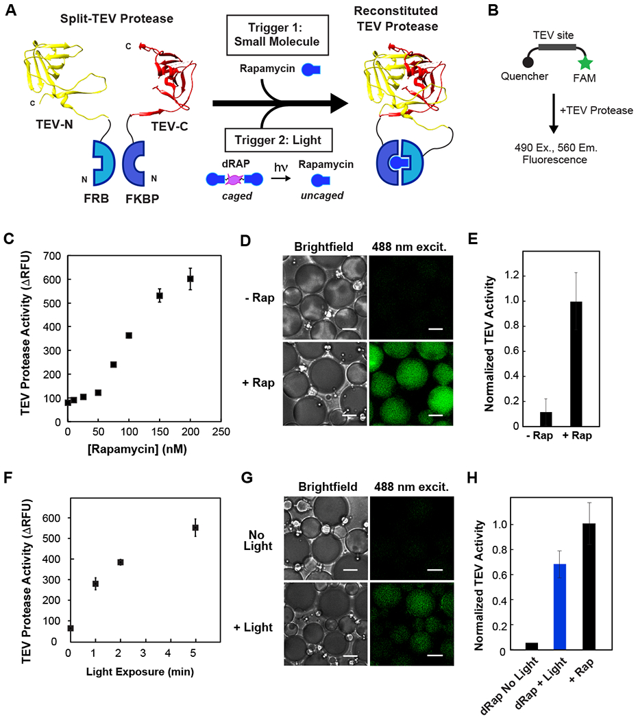
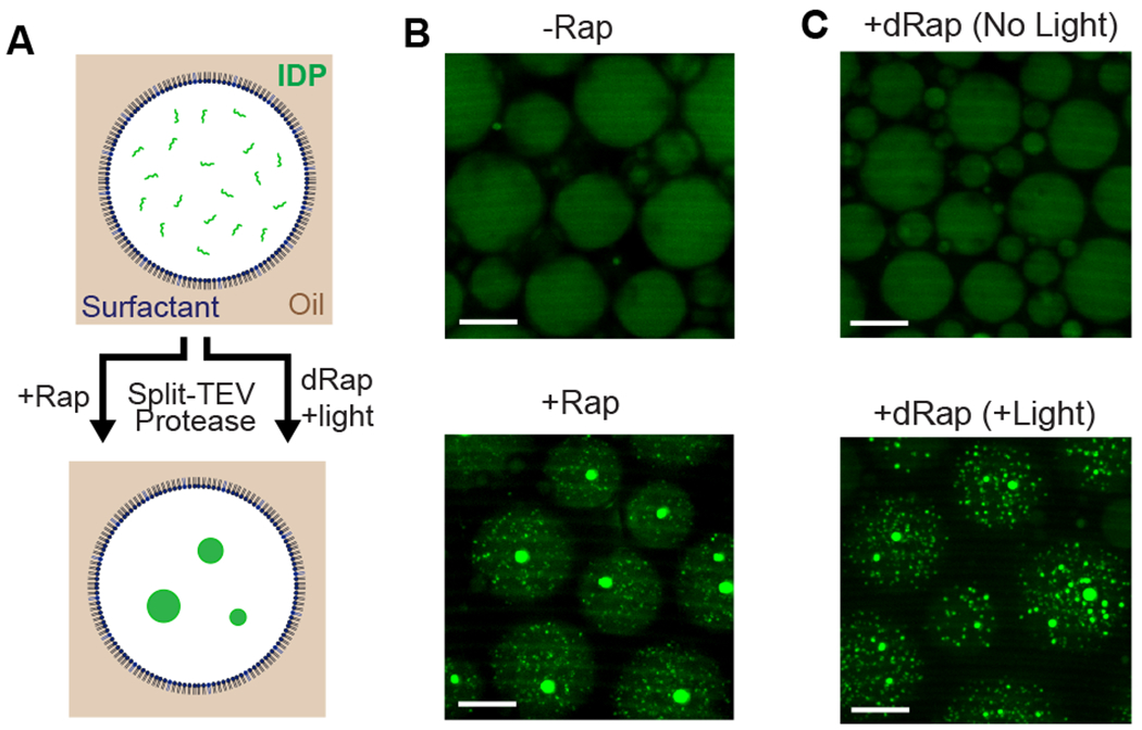
We report inducible dimerization strategies for controlling protein positioning, enzymatic activity, and organelle assembly inside synthetic cell-like compartments upon photostimulation. Using a photocaged TMP-Haloligand compound, we demonstrate small molecule and light-induced dimerization of DHFR and Haloenzyme to localize proteins to a compartment boundary and reconstitute tripartite sfGFP assembly. Using photocaged rapamycin and fragments of split TEV protease fused to FRB and FKBP, we establish optical triggering of protease activity inside cell-size compartments. We apply light-inducible protease activation to initiate assembly of membraneless organelles, demonstrating the applicability of these tools for characterizing cell biological processes in vitro. This modular toolkit, which affords spatial and temporal control of protein function in a minimal cell-like system, represents a critical step toward the reconstitution of a tunable synthetic cell, built from the bottom up.


Figures

Fig. 1.
Fig. 1.. Optochemical control of protein spatial localization in cell-like compartments.
(A) Schematic overview of photocaged TMP-Halo inducible dimerization system. (B) Haloenzyme and DHFR bind to one another in the presence of non-caged TMP-Halo dimerizer, but not in absence of dimerizer, in a pulldown assay. Photouncaging of caged TMP-Halo (CTH) leads to comparable binding to TH; there is undetectable binding in the dark. Caged and non-caged compounds were prebound to Haloenzyme protein. (C) Quantification of CTH and non-caged TH pulldown binding, normalized to positive control, non-caged TH binding. Error bar shows standard deviation from the mean, n = 3 (D) Schematic of light-inducible protein recruitment to the boundary inside synthetic cell-like compartments. 5% DGS-NTA(Ni) lipid and 95% POPC in decane phase. 1 μM His10-RFP-Halo, 0.1 μM GST-GFP-DHFR in aqueous phase. (E) His10-RFP-Halo binds to DGS-NTA(Ni) lipid in the droplet boundary. Recruitment of GST-GfP-DHFR to the boundary is triggered by 405 nM illumination, which uncages CTH. Scalebar 10 μm.
Fig. 2.
Fig. 2.. Inducible reconstitution of tripartite sfGFP assembly and activity.
(A) Schematic of tripartite sfGFP system, and fusion to DHFR and Halo domains to enable chemical or optical control of sfGFP reconstitution. Non-caged and caged versions of TMP-Halo compound are prebound to the sfGFP.Strand10-Haloenzyme protein. (B) Non-caged dimerizer promotes reconstitution of sfGFP fluorescence in a plate reader assay. The increase in fluorescence over 12 hours was interpolated to a dilution series of full sfGFP (Fig. S3C), yielding concentrations of sfGFP reconstitution. (C-D) Small molecule induced assembly of tripartite sfGFP inside cell-like compartments depends on the presence of non-caged TH dimerizer. Fluorescence was quantified 18 hours after addition of dimerizer and encapsulation, to allow for sufficient chromophore maturation. (E-F) Light-inducible reconstitution of sfGFP inside water-in-oil emulsions, normalized to non-caged TH positive control. Approximately 50% uncaging achieved using 1 s exposure to 405 nm laser light. All experiments used 3 μM DHFR-sfGFP.Strand11,3 μM sfGFP.Strand10-Haloenzyme, and 24 μM sfGFP.Strands1-9. Scale bar: 10 μm.
Fig. 3.
Fig. 3.. Activation of split TEV protease in cell-like compartments using small molecule and light-inducible dimerization.
(A) Schematic of photocaged rapamycin, dRap, and split TEV fragments fused to FRB and FKBP. (B) Schematic of TEV activity assay: upon substrate cleavage, a FAM fluorophore is released from quencher. (C) Dose-dependence of rapamycin mediated TEV reconstitution. Assay uses 125 nM of split TEV proteins and varying concentrations of rapamycin. (D-E) Chemically induced reconstitution of TEV activity inside celllike compartments. Equimolar concentration of rapamycin promotes TEV protease activity; there is low background activity in the absence of rapamycin. (F) Optical uncaging of dRap at various exposure times, promotes TEV reconstitution in a plate reader assay. 125 nM split TEV and 73 nM dRap. (G-H) Temporal triggering of TEV activation within cell-like compartments using light. 10 min exposure to 365 nm UV light to uncage dRap within emulsions. Minimal background activity in non-illuminated samples. For 3D-E and 3G-H, 500 nM split TEV proteins with equimolar rapamycin or equivalent dRap. For 3G-H, activity from a control without dimerizer was subtracted from conditions with dimerizer present. Scale bar: 5 μm.
Fig. 4.
Fig. 4.. Triggering formation of membraneless organelles in cell-like compartments using small molecule and light-inducible protease activity.
(A) Schematic of fluorescently-tagged IDP formation of membraneless organelles. MBP solubilization domain is cleaved from the IDP, resulting in phase separation and formation of membraneless organelles. (B) Rapamycin-dependent formation of protein droplets within emulsions. In absence of rapamycin, IDP remains soluble and well mixed. 1 μM split TEV was +/− equimolar dimerizer and 30 μM IDP, in the presence of 25% Xenopus egg extract. (C) Light-induced TEV activation and formation of membraneless organelles in cell-like compartments. Compartments encapsulated with 1 μM split TEV, 500 nM dRap, 30 μM IDP, 25% egg extract; either kept in dark or exposed to 365 nm UV light for 10 minutes. For 4B-C, emulsions were imaged 12 hours post-induction. Scalebar: 20 μm.

References

    1. Ankenbruck N, Courtney T, Naro Y, Deiters A Angew Chem Int Ed Engl 57, 2768–2798.(2018) - PMC - PubMed
    1. Levskaya A, Weiner OD, Lim WA, Voigt CA Nature 461, 997–1001. (2009) - PMC - PubMed
    1. Buckley CE, Moore RE, Reade A, Goldberg AR, Weiner OD, Clarke JDW Dev Cell 36, 117–126. (2016) - PMC - PubMed
    1. Strickland D, Lin Y, Wagner E, Hope CM, Zayner J, Antoniou C, Sosnick TR, Weiss EL, Glotzer M Nat Methods 9, 379–384. (2012) - PMC - PubMed
    1. Hallett RA, Zimmerman SP, Yumerefendi H, Bear JE, Kuhlman B ACS Synth Biol 5, 53–64. (2016) - PMC - PubMed

Publication types

MeSH terms