. 2018 Aug 1;315(2):G302-G317.
doi: 10.1152/ajpgi.00005.2018. Epub 2018 Apr 19.

P21-activated kinase 4 in pancreatic acinar cells is activated by numerous gastrointestinal hormones/neurotransmitters and growth factors by novel signaling, and its activation stimulates secretory/growth cascades

Affiliations

P21-activated kinase 4 in pancreatic acinar cells is activated by numerous gastrointestinal hormones/neurotransmitters and growth factors by novel signaling, and its activation stimulates secretory/growth cascades

Irene Ramos-Alvarez et al. Am J Physiol Gastrointest Liver Physiol. .

Abstract

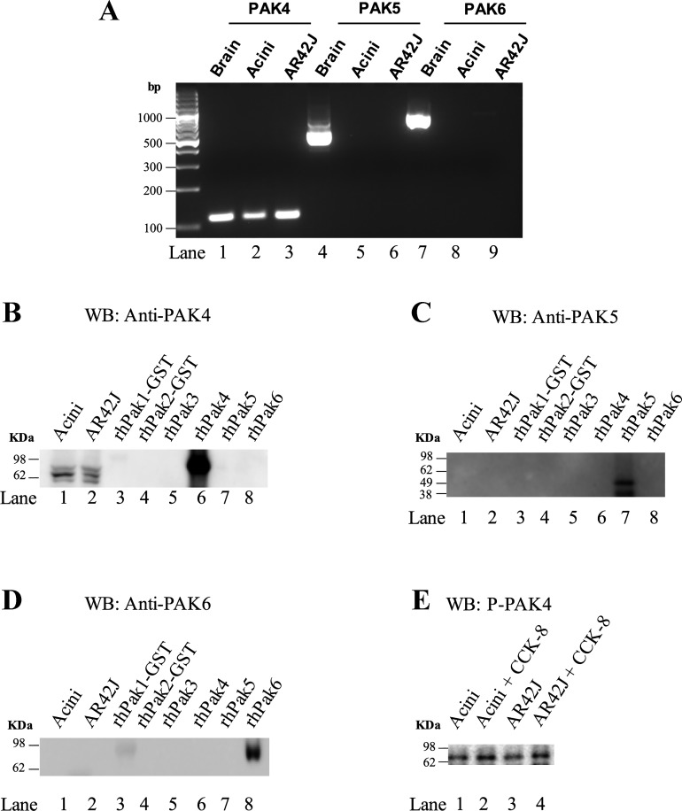
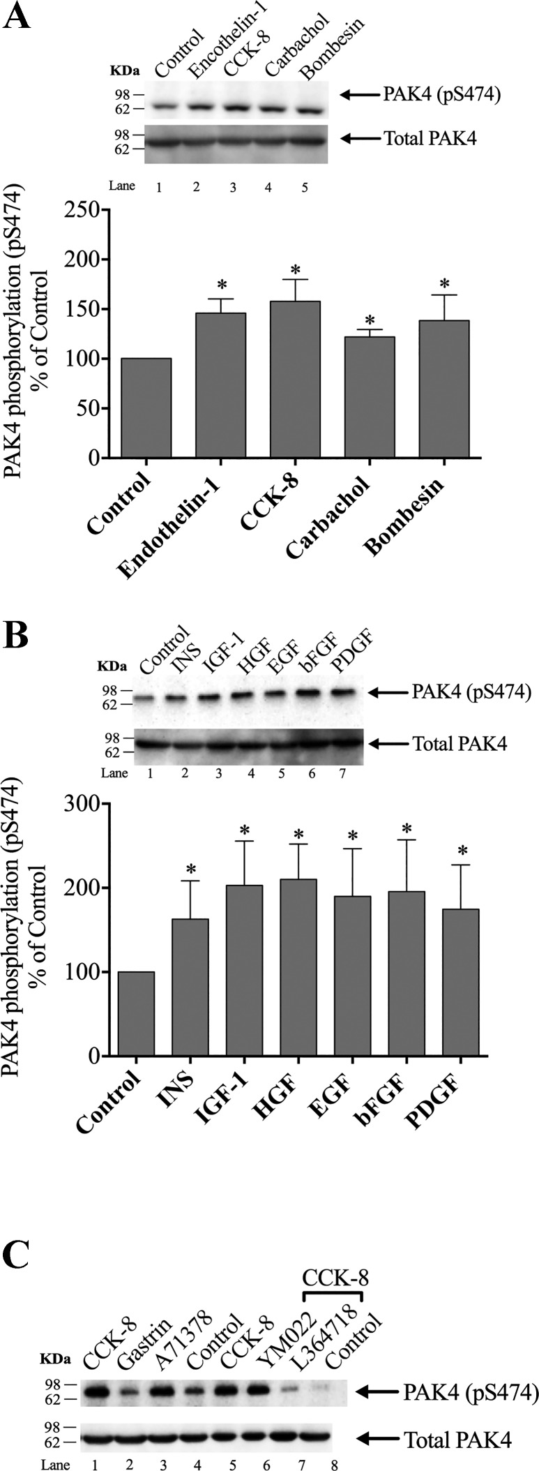
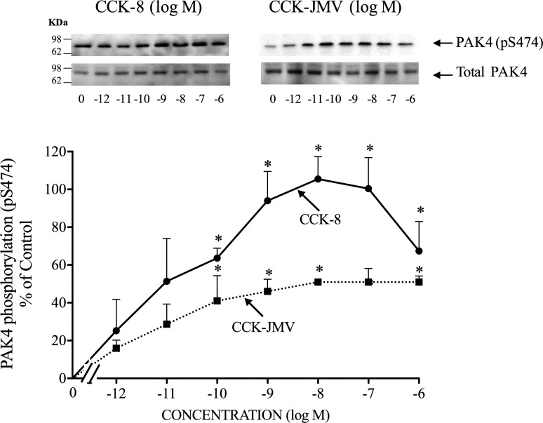
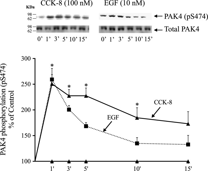
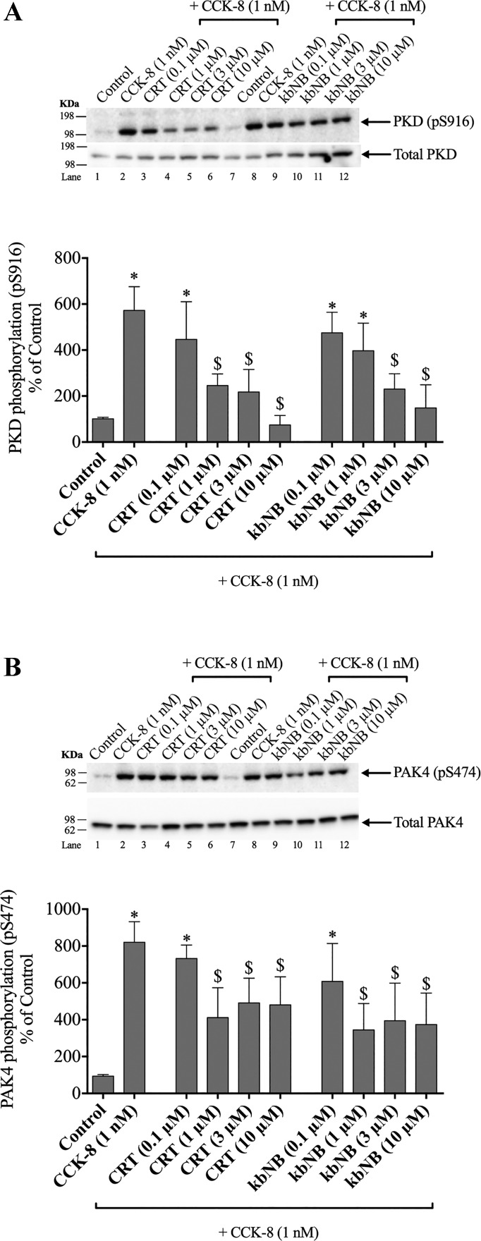
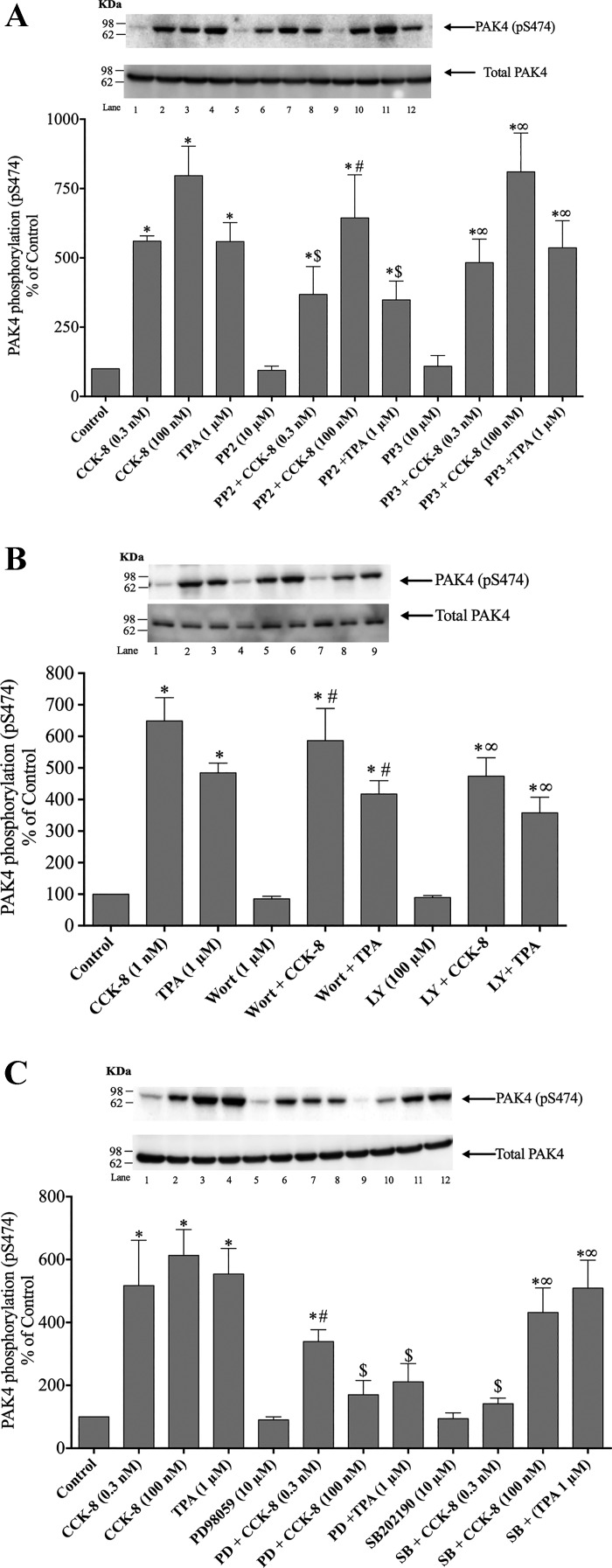
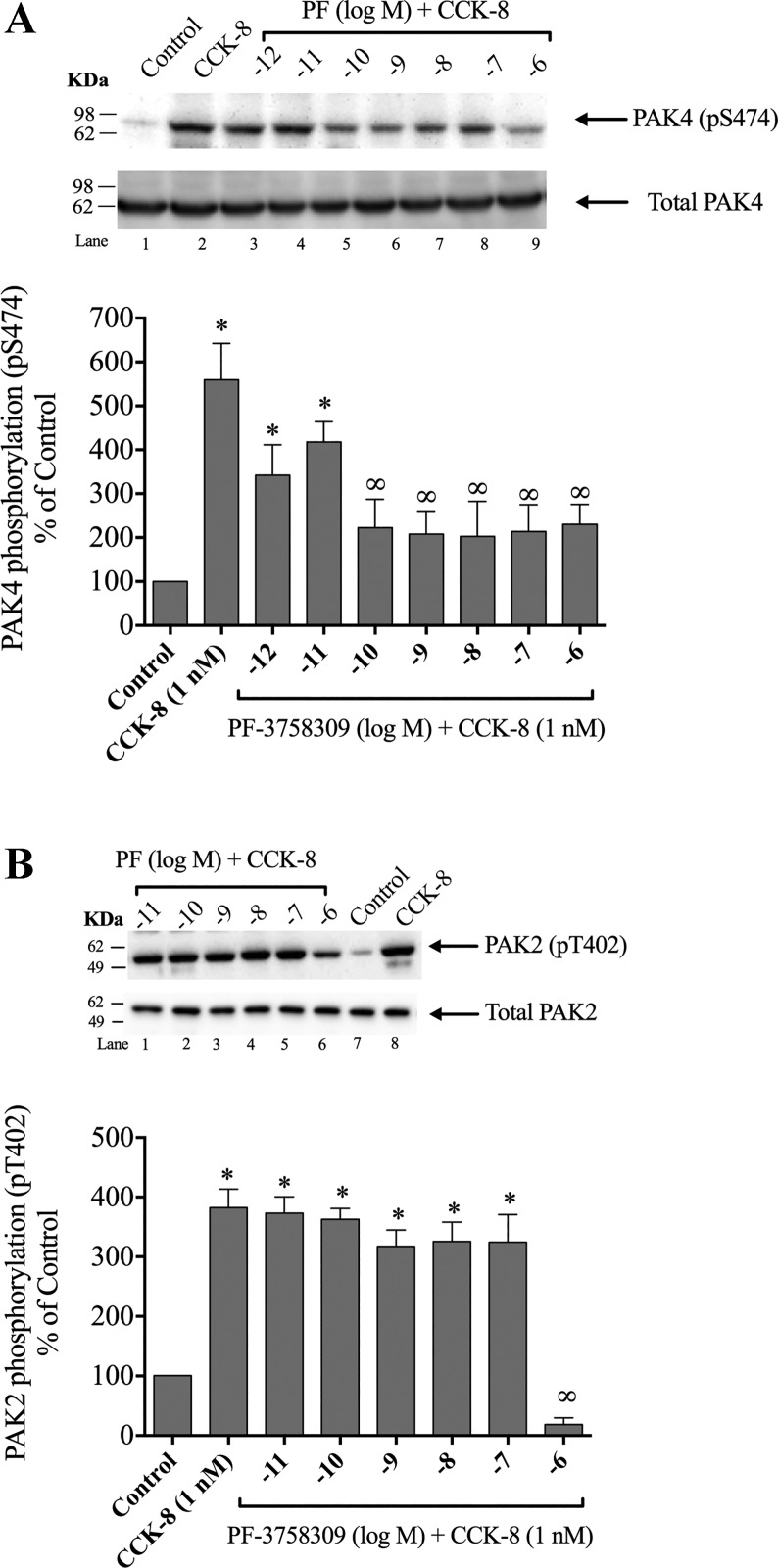
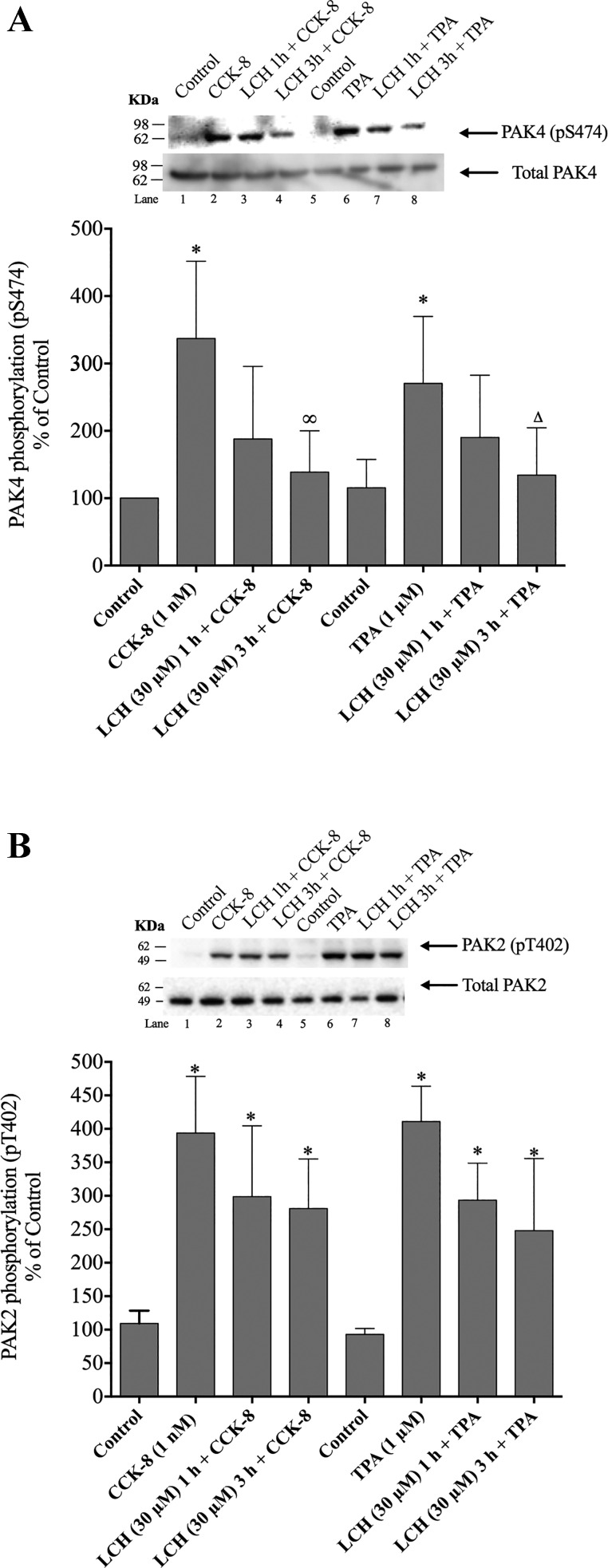
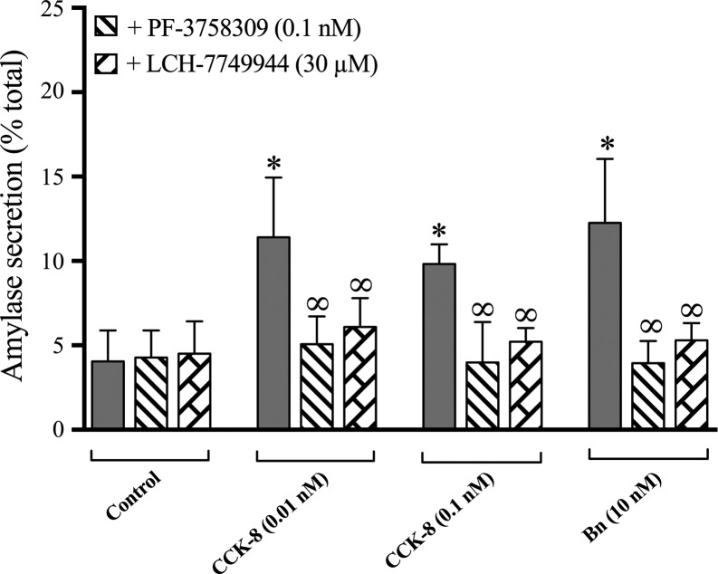
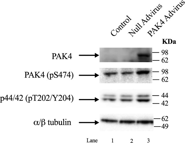
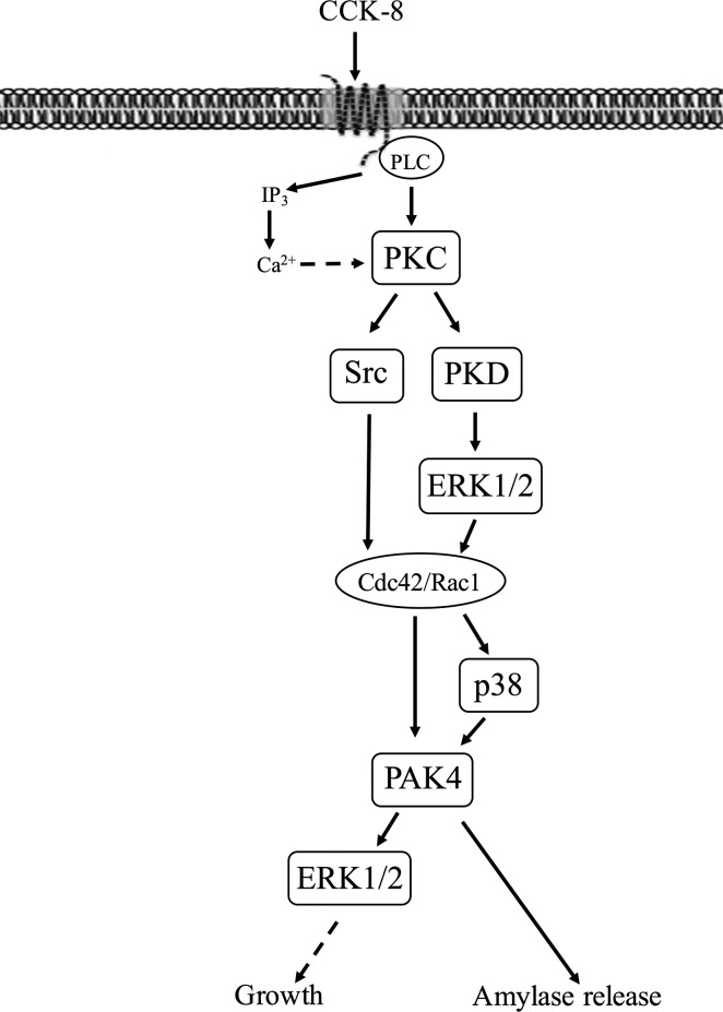
p21-activated kinases (PAKs) are highly conserved serine/threonine protein kinases, which are divided into two groups: group-I (PAKs1-3) and group-II (PAKs4-6). In various tissues, Group-II PAKs play important roles in cytoskeletal dynamics and cell growth as well as neoplastic development/progression. However, little is known about Group-II PAK's role in a number of physiological events, including their ability to be activated by gastrointestinal (GI) hormones/neurotransmitters/growth factors (GFs). We used rat pancreatic acini to explore the ability of GI hormones/neurotransmitters/GFs to activate Group-II-PAKs and the signaling cascades involved. Only PAK4 was detected in pancreatic acini. PAK4 was activated by endothelin, secretagogues-stimulating phospholipase C (bombesin, CCK-8, and carbachol), by pancreatic GFs (insulin, insulin-like growth factor 1, hepatocyte growth factor, epidermal growth factor, basic fibroblast growth factor, and platelet-derived growth factor), and by postreceptor stimulants (12-O-tetradecanoylphobol-13-acetate and A23187 ). CCK-8 activation of PAK4 required both high- and low-affinity CCK1-receptor state activation. It was reduced by PKC-, Src-, p44/42-, or p38-inhibition but not with phosphatidylinositol 3-kinase-inhibitors and only minimally by thapsigargin. A protein kinase D (PKD)-inhibitor completely inhibited CCK-8-stimulated PKD-activation; however, stimulated PAK4 phosphorylation was only inhibited by 60%, demonstrating that it is both PKD-dependent and PKD-independent. PF-3758309 and LCH-7749944, inhibitors of PAK4, decreased CCK-8-stimulated PAK4 activation but not PAK2 activation. Each inhibited ERK1/2 activation and amylase release induced by CCK-8 or bombesin. These results show that PAK4 has an important role in modulating signal cascades activated by a number of GI hormones/neurotransmitters/GFs that have been shown to mediate both physiological/pathological responses in acinar cells. Therefore, in addition to the extensive studies on PAK4 in pancreatic cancer, PAK4 should also be considered an important signaling molecule for pancreatic acinar physiological responses and, in the future, should be investigated for a possible role in pancreatic acinar pathophysiological responses, such as in pancreatitis. NEW & NOTEWORTHY This study demonstrates that the only Group-II p21-activated kinase (PAK) in rat pancreatic acinar cells is PAK4, and thus differs from islets/pancreatic cancer. Both gastrointestinal hormones/neurotransmitters stimulating PLC and pancreatic growth factors activate PAK4. With cholecystokinin (CCK), activation is PKC-dependent/-independent, requires both CCK1-R affinity states, Src, p42/44, and p38 activation. PAK4 activation is required for CCK-mediated p42/44 activation/amylase release. These results show PAK4 plays an important role in mediating CCK physiological signal cascades and suggest it may be a target in pancreatic acinar diseases besides cancer.

Keywords: CCK; bombesin; celluar calcium; pancreatic acini; protein kinase C; signaling.


Figures

Fig. 1.
Fig. 1.
Specificity of p21-activated kinase (PAK)4, 5, and 6 antibodies and demonstration that pancreatic acini and AR42J cells possess only PAK4 mRNA/protein. Representative PCR results for PAK4, PAK5, and PAK6 mRNA in rat brain, pancreatic acinar cells, and AR42J cells (A). Lysates from pancreatic acinar cells and equal amounts (5 µg) of human recombinant PAK family members (PAK1–6) or AR42J cells were detected by Western blot analysis (WB) with specific anti-PAK4 (Santa Cruz Biotechnology) (B), anti-PAK5 (Biorbyt) (C), and anti-PAK6 (BioVision) (D) antibodies, or with a specific antiphospho PAK4 antibody (GeneTex) (E). Results from pancreatic acini or AR47J cells with or without 1 nM CCK-8 stimulation (E). These results are representative of 4 other experiments. CCK-8, cholecystokinin-8.
Fig. 2.
Fig. 2.
Ability of various pancreatic hormones/neurotransmitters and pancreatic growth factors to stimulate p21-activated kinase (PAK)4 phosphorylation (pS474) in rat pancreatic acini. A: ability of cholecystokinin (CCK)-8, carbachol, bombesin, or endothelin-1 to activate pS474 PAK4 in isolated pancreatic acini. Isolated pancreatic acini were incubated in the absence or presence of CCK-8 (1 nM), carbachol (10 µM), bombesin (1 nM), or endothelin-1 (10 nM) for 1 min and then lysed. Results are expressed as %control phosphorylation. B: ability of insulin (INS), insulin-like growth factor 1 (IGF-1), hepatocyte growth factor (HGF), epidermal growth factor (EGF), basic fibroblast growth factor (bFGF), and platelet-derived growth factor (PDGF) to activate PAK4 in the pancreatic acini. Isolated pancreatic acini were incubated in the absence or presence of insulin (INS) (1 µM, 10 min), EGF (10 nM, 5 min), PDGF (100 ng/ml, 10 min), bFGF (100 ng/ml, 5 min), IGF (100 nM, 10 min), and HGF (1 nM, 10 min), and then lysed. Results expressed in A. C: representative Western blot assessing the ability of selective CCK1 or CCK2 receptor agonist/antagonists to alter PAK4 activation in pancreatic acini. Isolated pancreatic acini were incubated in the absence or presence of CCK-8 (1 nM); the CCK2 agonist, gastrin (1 nM); or the CCK1 receptor agonist, A71378 (30 nM) for 1 min; or preincubated for 5 min in the presence of the CCK2 antagonist, YM022 (1 µM); the CCK1 antagonist, L-364,718(1 µM); and then after the additional presence of CCK-8 (1 nM) for 3 min. The cell lysates were subjected to Western blotting and analyzed using anti-pS474 PAK4 and, as loading control, anti-total PAK4. Bands were visualized using chemiluminescence and quantified by densitometry. A and B, top: results of a representative blot of six independent experiments. Bottom of A and B: mean ± SE of six independent experiments. *P < 0.05 compared with the control.
Fig. 3.
Fig. 3.
Dose-response effect of cholecystokinin (CCK)-8 and CCK-JMV to stimulate p21-activated kinase (PAK)4 phosphorylation in rat pancreatic acini. Isolated pancreatic acini were incubated in the absence or presence of CCK-8 and CCK-JMV (at the indicate concentrations) for 3 min and then lysed. Western blots were analyzed using anti-pS474 PAK4 antibody and, as loading control, anti-total PAK4. Top: results of a representative blot of three independent experiments. Bottom: means ± SE of three independent experiments. Results are expressed as % of basal stimulation of the control group (CCK-8 1 nM: 193 ± 15% of control). *P < 0.05 compared with the control group.
Fig. 4.
Fig. 4.
Time course of cholecystokinin (CCK)-8 and epidermal growth factor (EGF) stimulation of p21-activated kinase (PAK)4 S474 phosphorylation in rat pancreatic acini. Isolated pancreatic acini were incubated in the absence or presence of CCK-8 (100 nM) or EGF (10 nM) for the indicated times, and then lysed. Western blots were analyzed using anti-pS474 PAK4 and, as loading control, anti-total PAK4. Bands were visualized using chemiluminescence and quantified by densitometry. Top: results of a representative blot of three independent experiments. Bottom: means ± SE of three independent experiments. Results are expressed as %basal stimulation of the control group. *P < 0.05 compared with the control group (i.e., 0 time).
Fig. 5.
Fig. 5.
Effect of postreceptor stimulants, PKC inhibition, or Ca2+ depletion on pS474 p21-activated kinase (PAK)4 phosphorylation by cholecystokinin (CCK)-8 and 12-O-tetradecanoylphobol-13-acetate (TPA). A: effect of the calcium ionophore, A23187 (1 μM), TPA (1 μM), thapsigargin (TG; 1 μM), or CCK-8 (1 nM) on PAK4 phosphorylation (all for 5 min, except CCK-8 for 3 min, in calcium containing media). Top: results of a representative blot of six independent experiments. Bottom: means ± SE of six independent experiments. Results are expressed as % of basal stimulation of the control group. *P < 0.05 compared with th0e control group. B: effect of PKC inhibition or Ca2+ depletion on pS474 PAK4 CCK-8- and TPA-induced phosphorylation. Isolated acini were preincubated for 2 h, alone or in the additional presence of GF109203X (GFX; 5 μM, 1 h), TG (1 μM, 1 h), or both (in a media with or without 5 mM calcium), and then incubated with or without 1 nM CCK-8 or 1 μM TPA for 3 or 5 min, respectively. Top: results of a representative blot of six independent experiments. Bottom: means ± SE of six independent experiments. Results are expressed as % of basal stimulation of the control group. *P < 0.05 compared with its control; >P < 0.05 vs. compared with similar stimulants with no additions (i.e., Ca2+ media).
Fig. 6.
Fig. 6.
Effect of increasing concentrations of two protein kinase D (PKD) inhibitors, CRT0066101 and kbNB142-70, on PKD phosphorylation (A) and on p21-activated kinase (PAK)4 activation (B) by cholecystokinin (CCK)-8 in rat pancreatic acini. Isolated pancreatic acini were incubated in the absence or presence of CRT0066101 (CRT) or kbNB142-70 (kbNb) (0.1, 1, 3, and 10 µM) for 2 h and then incubated with no addition (control) or CCK-8 (1 nM) for 3 min, and then lysed. Western blots were analyzed using anti-pS474 PAK4 or pS916 PKD and, as loading control, anti-total PAK4 or anti-total PKD. Bands were visualized using chemiluminescence and quantified by densitometry. Top of A and B: results of a representative blot of three independent experiments. Bottom of A and B: means ± SE of at least four independent experiments. Results are expressed as % of basal stimulation of the control group. *P < 0.05 compared with the control group; $P < 0.05 compared with CCK-8 alone.
Fig. 7.
Fig. 7.
Effect of the inhibition of Src, phosphatidylinositol 3-kinase (PI3K), p44/42, or p38 MAPKs on the ability of cholecystokinin (CCK)-8 (1 nM) or 12-O-tetradecanoylphorbol-13-acetate (TPA) (1 µM) to stimulate p21-activated kinase (PAK)4. A representative experiment of at least four others and the means of all the experiments are shown. A: rat pancreatic acinar cells were pretreated with no additions or with PP2 (10 µM) or PP3 (10 µM) for 1 h and then incubated with no additions (control), 0.3 nM or 100 nM CCK-8 for 3 min or with 1 µM TPA for 5 min and then lysed and analyzed with anti-pS474 PAK4 and, as a loading control, anti-total PAK4. *P < 0.05 compared with control, #P < 0.05 vs. PP2 alone, ∞P < 0.05 vs. PP3 alone, and $P < 0.05 vs. comparing stimulants (CCK-8 or TPA) vs. stimulants preincubated with PP2. B: rat pancreatic acinar cells were pretreated with no additions or with Wortmannin (Wort; 1 μM) or LY294002 (LY; 100 μM) for 30 min and then incubated with no additions (control), 1 nM CCK-8 for 3 min, or with 1 μM TPA for 5 min and then lysed and analyzed with anti- pS474 PAK4 and, as a loading control, anti-total PAK4. *P < 0.05 compared with control, #P < 0.05 vs. CCK-8 alone, and ∞P < 0.05 vs. TPA alone. C: rat pancreatic acinar cells were pretreated with no additions or with PD98059 (PD; 10 µM) or SB202190 (SB; 10 µM) for 1 h and then incubated with no additions (control), 0.3 nM or 100 nM CCK-8 for 3 min, or with 1 µM TPA for 5 min and then lysed and analyzed with anti-pS474 PAK4 and, as a loading control, anti-total PAK4. *P < 0.05 compared with control, #P < 0.05 vs. PD98059 alone, ∞P < 0.05 vs. SB202190 alone, and $P < 0.05 vs. comparing stimulants (CCK-8 or TPA) vs. stimulants preincubated with PD98059 or SB202190.
Fig. 8.
Fig. 8.
Ability of the p21-activated kinase (PAK) inhibitor II, PF-3758309 (PF), to inhibit cholecystokinin (CCK)-8 activation of PAK4 (A) or PAK2 (B). Isolated pancreatic acini were preincubated for 1 h in the presence or absence of PF-3758309 (at the indicated concentrations) and then treated with CCK-8 (1 nM) for 3 min and then lysed. Western blots were analyzed using anti-pS474 PAK4 or pT402 PAK2 antibody and, as loading control, anti-total PAK4 or anti-total PAK2. Top of A and B: results of a representative blot of three independent experiments. Bottom of A and B: means ± SE for at least five independent experiments. Results are expressed as % of basal stimulation of the control group. *P < 0.05 compared with the control group and ∞P < 0.05 compared with CCK-8 alone.
Fig. 9.
Fig. 9.
Ability of the p21-activated kinase (PAK) inhibitor II, LCH-7749944 (LCH), to inhibit cholecystokinin (CCK)-8 or 12-O-tetradecanoylphobol-13-acetate (TPA) activation of PAK4 (A) or PAK2 (B). Isolated pancreatic acini were preincubated for 1 and 3 h in the presence or absence of LCH-7749944 (at the indicated concentrations) and then treated with CCK-8 (1 nM) for 3 min and TPA (1 µM) for 5 min and then lysed. Western blots were analyzed using anti-pS474 PAK4 or pT402 PAK2 antibody and, as loading control, anti-total PAK4 or anti-total PAK2. Top of A and B: results of a representative blot of three independent experiments. Bottom of A and B: means ± SE for at least five independent experiments. Results are expressed as % of basal stimulation of the control group. *P < 0.05 compared with the control group, ∞P < 0.05 compared with CCK-8 alone, and ∆P < 0.05 compared with TPA alone.
Fig. 10.
Fig. 10.
Effect of the p21-activated kinase (PAK)4 inhibitors, PF-3758309 (PF) or LCH-7749944 (LCH) on the ability of a physiological concentration of cholecystokinin (CCK)-8 to activate p44/42. Rat pancreatic acinar cells were pretreated with no additions or with 0.1 nM PF-3758309 or 30 µM LCH-7749944 for 3 h and then incubated with no addition (control) or with 1 nM CCK-8 for 3 min, then lysed and analyzed using anti-pT202/Y204 p42/44 and, as a loading control, anti-total p44/42. The bands were visualized using chemiluminescence and quantification of phosphorylation was assessed using scanning densitometry. Top: results of a representative blot of three independent experiments. Bottom: means ± SE for at least four independent experiments. Results are expressed as % of basal stimulation of the control group. *P < 0.05 compared with the control group and ∞P < 0.05 compared with CCK-8 alone.
Fig. 11.
Fig. 11.
Effect of the p21-activated kinase (PAK)4 inhibitors, PF-3758309 or LCH-7749944, on amylase secretion. Pancreatic acini were incubated with no additions, the indicated secretagogue alone, or with the PAK4 inhibitors PF-3758309 (0.1 nM) or LCH-7749944 (30 µM). Amylase release, expressed as percent of cellular total amylase, was determined after 30 min incubation. Results are the result of the average of six experiments. *P < 0.05 compared with the control group and ∞P < 0.05 comparing stimulants [cholecystokinin (CCK)-8 or Bombesin (Bn)] alone.
Fig. 12.
Fig. 12.
Effect of preincubation with p21-activated kinase (PAK)4 ad and null Adviruses upon the expression of PAK4 in pancreatic acinar cells and ERK activity. Rat pancreatic acinar cells were preincubated for 12 h without (control, Lane 1), with 109 VP/ml of null Advirus (Lane 2), and PAK4 Advirus (Lane 3). This experiment is representative of three others.
Fig. 13.
Fig. 13.
Schematic diagram of signaling cascade for activation of p21-activated kinase (PAK)4 in pancreatic acinar cells. In rat pancreatic acinar cells, maximal activation of PAK4 by cholecystokinin (CCK)-8 requires activation of primarily PKC, with a lesser contribution by changes in cytosolic calcium. PKC mediates both Src family of kinase (SFK) and protein kinase D (PKD) activation with the latter resulting in ERK1/2 activation and cell division control protein 42 homolog (Cdc42)/ Ras-related C3 botulinum toxin substrate 1 (Rac1) activation. This in turn can stimulate PAK4 activation partially via a p38 mechanism. PAK4 activation is important for CCK-stimulated enzyme secretion as well as ERK1/2 activation which has been shown to mediate growth (20, 30). However, changes in phosphatidylinositol 3-kinase (PI3K) are not involved but were reported in a number of other tissues (23, 80). The ERK1/2 and PAK4 inhibition studies demonstrate CCK-8-mediated ERK1/2 activation and PAK4 activation reciprocally regulate each other’s activation.

Similar articles

Cited by

References

    1. Abo A, Qu J, Cammarano MS, Dan C, Fritsch A, Baud V, Belisle B, Minden A. PAK4, a novel effector for Cdc42Hs, is implicated in the reorganization of the actin cytoskeleton and in the formation of filopodia. EMBO J 17: 6527–6540, 1998. doi:10.1093/emboj/17.22.6527. - DOI - PMC - PubMed
    1. Ahmed T, Shea K, Masters JR, Jones GE, Wells CMA. A PAK4-LIMK1 pathway drives prostate cancer cell migration downstream of HGF. Cell Signal 20: 1320–1328, 2008. doi:10.1016/j.cellsig.2008.02.021. - DOI - PubMed
    1. Aparicio IM, Garcia-Marin LJ, Andreolotti AG, Bodega G, Jensen RT, Bragado MJ. Hepatocyte growth factor activates several transduction pathways in rat pancreatic acini. Biochim Biophys Acta 1643: 37–46, 2003. doi:10.1016/j.bbamcr.2003.08.007. - DOI - PubMed
    1. Bastea LI, Döppler H, Pearce SE, Durand N, Spratley SJ, Storz P. Protein kinase D-mediated phosphorylation at Ser99 regulates localization of p21-activated kinase 4. Biochem J 455: 251–260, 2013. doi:10.1042/BJ20130281. - DOI - PMC - PubMed
    1. Bergeron V, Ghislain J, Poitout V. The P21-activated kinase PAK4 is implicated in fatty-acid potentiation of insulin secretion downstream of free fatty acid receptor 1. Islets 8: 157–164, 2016. doi:10.1080/19382014.2016.1243191. - DOI - PMC - PubMed

Publication types

MeSH terms