. 2018 Aug 1;315(2):G220-G230.
doi: 10.1152/ajpgi.00413.2017. Epub 2018 Apr 19.

An endogenous aryl hydrocarbon receptor ligand, ITE, induces regulatory T cells and ameliorates experimental colitis

Affiliations

An endogenous aryl hydrocarbon receptor ligand, ITE, induces regulatory T cells and ameliorates experimental colitis

Jessica D Abron et al. Am J Physiol Gastrointest Liver Physiol. .

Abstract

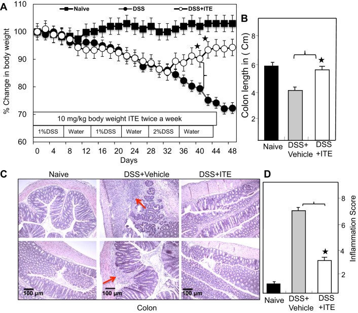
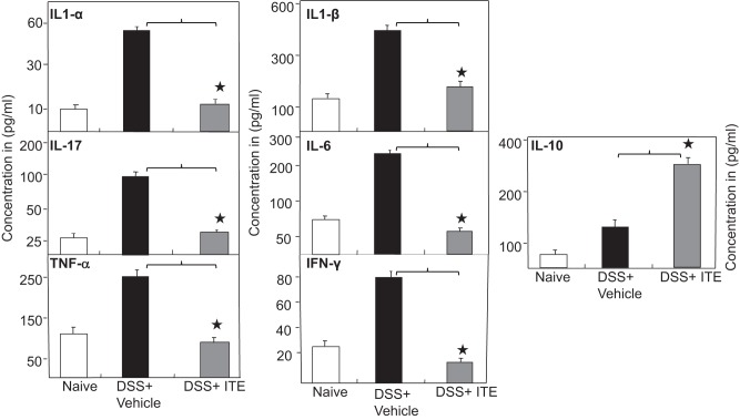
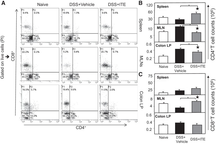
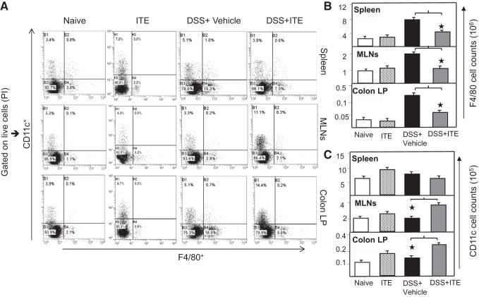
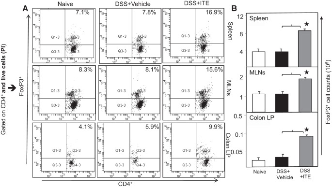
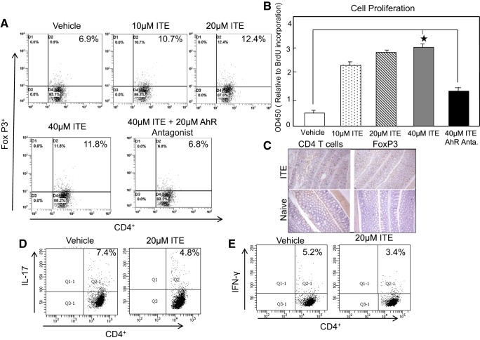
Inflammatory bowel disease (IBD) is a chronic intestinal inflammatory condition that affects millions of people with high morbidity and health care costs. The precise etiology of IBD is unknown, but clear evidence suggests that intestinal inflammation is caused by an excessive immune response to mucosal antigens. Recent studies have shown that activation of the aryl hydrocarbon receptor (AhR) induces regulatory T cells (Tregs) and suppresses autoimmune diseases. In the current study, we investigated if a nontoxic ligand of AhR, 2-(1' H-indole-3'-carbonyl)-thiazole-4-carboxylic acid methyl ester (ITE), can attenuate dextran sodium sulfate-induced colitis. Our studies demonstrated that in mice that received ITE treatment in vivo, colitis pathogenesis, including a decrease in body weight, was significantly reversed along with the systemic and intestinal inflammatory cytokines. ITE increased the expression of Tregs in spleen, mesenteric lymph nodes (MLNs), and colon lamina propria lymphocytes (cLPL) of mice with colitis when compared with controls. This induction of Tregs was reversed by AhR antagonist treatment in vitro. ITE treatment also increased dendritic cells (CD11c+) and decreased macrophages (F4/80+) from the spleen, MLNs, and cLPL in mice with colitis. ITE also reversed the systemic and intestinal frequency of CD4+ T cells during colitis and suppressed inflammatory cytokines including IFN-γ, TNF-α, IL-17, IL-6, and IL-1 as well as induced IL-10 levels. These findings suggest that ITE attenuates colitis through induction of Tregs and reduction in inflammatory CD4+ T cells and cytokines. Therefore, our work demonstrates that the nontoxic endogenous AhR ligand ITE may serve as a therapeutic modality to treat IBD. NEW & NOTEWORTHY We report the novel finding that activation of the aryl hydrocarbon receptor with the nontoxic ligand 2-(1'H-indole-3'-carbonyl)-thiazole-4-carboxylic acid methyl ester (ITE) induces regulatory T cells (Tregs) and suppresses inflammatory bowel disease (IBD). Our data suggest that ITE diminishes colitis pathology through induction of Tregs; reduces inflammatory cytokines, inflammation score, and macrophage frequency; and induces DCs resulting in amelioration of colitis. Therefore, nontoxic endogenous ITE promotes the induction of Tregs and may be useful for the treatment of IBD.

Keywords: Crohn’s disease; Th1/Th17; inflammatory bowel disease; ulcerative colitis.


Figures

Fig. 1.
Fig. 1.
Change in body weight and colon length of mice after DSS exposure. Normal BL/6 mice were given no treatment (■ naive). Others were given DSS alone (1%) in drinking water for 2 cycles of 7 days and (2%), DSS for third cycle (●), and DSS with ITE (○; 10 mg/kb body wt). After the first 7 days, DSS was replaced with a water cycle (ad libitum) for another 7 days. This was repeated for 2 more cycles of 7 days. The body weight of mice was recorded twice a week (A). The colon length was taken at the experimental end point (B). Histological sections of the colon from the groups of mice are presented. BL/6 mice receiving DSS alone (middle) showed lymphocyte infiltration and distortion of glands, whereas mice that received ITE (C, right) showed decreased lymphocyte infiltration. Naive BL/6 mice showed no cellular infiltration. The pathological changes included diffuse leukocyte infiltrates and thickening of the cLP in the area of distorted crypts in the colon. Two blind investigators unaware of this study evaluated the inflammation score (D). Representative sections from 3 separate experiments (×20 magnification) are shown. Changes in body weight were expressed as the percentage of baseline body weight. ANOVA was used to compare the change in body weight over time, colon length, and inflammation score among the groups. *P < 0.01 compared between DSS + vehicle with DSS + ITE. Data represent the mean of 3 repeated experiments involving 6 mice/group (n = 18). cLP, colon lamina propria; DSS, dextran sodium sulfate; ITE, 2-(1′H-indole-3′-carbonyl)-thiazole-4-carboxylic acid methyl ester.
Fig. 2.
Fig. 2.
Systemic levels of IL 1-α, IL-1-β, IL-6, IL-10, TNF-α, IL-17, and IFN-γ in mice after ITE treatment. After euthanasia, serum levels of IL 1-α, IL-1-β, IL-6, IL-10, TNF-α, IL-17, and IFN-γ were determined by Bio-Rad ELISA multiplex kit, which is capable of detecting >15 pg/ml of these analytes. The data presented are the mean concentrations of IL 1-α, IL-1-β, IL-6, -IL-10, TNF-α, IL-17, and IFN-γ ± SE from 3 separate repeated experiments that include 6 mice each (n = 18). The statistical significance between cytokine levels for various groups was assessed by ANOVA. Statistically significant differences (*P < 0.01) between DSS + vehicle and DSS + ITE groups. DSS, dextran sodium sulfate; ITE, 2-(1′H-indole-3′-carbonyl)-thiazole-4-carboxylic acid methyl ester.
Fig. 3.
Fig. 3.
Changes in T-cell responses after DSS exposure. Spleen cells, MLNs, and LP lymphocytes were isolated from 3 groups of 6 mice each and stained for CD4 and CD8 T-cell markers, analyzed by flow cytometry. The numbers in the bottom right quadrant indicate the total percentage of CD4 T cells and top left quadrant indicates CD8 T cells (A). The changes in the number of CD4 and CD8 T cells are shown (B and C). Representative data from three independent repeated experiment involving six mice per group (n = 18) are shown. The statistical significance between flow cytometry data for various groups was assessed by ANOVA. *Statistically significant differences between DSS + vehicle and DSS + ITE groups; *P < 0.01. Representative data of 1 of at least 3 experiments that produced similar results are shown. DSS, dextran sodium sulfate; ITE, 2-(1′H-indole-3′-carbonyl)-thiazole-4-carboxylic acid methyl ester; LP, lamina propria.
Fig. 4.
Fig. 4.
ITE treatment differentially mediates macrophages and dendritic cells (DCs). Spleen, MLNs, and cLP lymphocytes were isolated from all 4 groups of 6 mice each in 3-repeated experiments (n = 18) at the experimental end point. In this experiment, we added naive-treated with ITE alone without any exposure to determine the independent effect of ITE without colitis. Changes in the frequency and expression of macrophages (F4/80) and DCs (CD11c) from spleens, MLNs, and cLP are shown (A). Changes in the number of macrophages (F4/80) and DCs (CD11c) are shown (B and C, respectively). The statistical significance in flow cytometry data among 4 groups was assessed by ANOVA. *Statistically significant differences between DSS + vehicle and DSS + ITE groups; i.e.; *P < 0.01. Representative data of one of at least 3 experiments that produced similar results are shown. cLP, colon lamina propria; DSS, dextran sodium sulfate; ITE, 2-(1′H-indole-3′-carbonyl)-thiazole-4-carboxylic acid methyl ester; MLN, mesenteric lymph node.
Fig. 5.
Fig. 5.
ITE induces the number and frequency of Tregs. Spleen, MLNs, and LP lymphocytes were isolated from all 3 groups of 6 mice each at the experimental end point. Changes in the frequency and expression of FOXP3-expressing CD4 T cells from spleen lymphocyte, MLNs, and cLP are shown (left). The numbers in the top right quadrant indicate the total percentage of Tregs (A). Changes in the number of CD4 T cells expressing FOXP3 are shown (right) (B). Experiments involved 6 mice per group (n = 18). The statistical significance between frequencies of Tregs for each group was assessed by ANOVA. *Statistically significant differences between DSS vehicle and DSS + ITE groups; i.e.; *P < 0.01. Representative data of one of at least 3 experiments that produced similar results are shown. cLP, colon lamina propria; DSS, dextran sodium sulfate; FOXP3, forkhead box 3; ITE, 2-(1′H-indole-3′-carbonyl)-thiazole-4-carboxylic acid methyl ester; MLN, mesenteric lymph node; Tregs, regulatory T cells.
Fig. 6.
Fig. 6.
ITE treatment induces T-cells proliferation and FOXP3 expression in vitro. Lymphocytes obtained from mesenteric lymph nodes (MLNs) and cLP were further sort-purified by using a FACS-Aria (Becton Dickinson) (purity >97%) from 3 normal BL/6 mice (n = 9 in quadruplicate each time) and were cultured with anti-CD3/CD28 Abs activation in the presence of either vehicle or ITE (0, 10, 20, or 40 μm). Some cultures were also received ITE + 20 and 40 μm of AhR antagonist. The induction of in vitro CD4 + FOXP3+ (A), ex vivo IL-17 (D), and IFN-γ (E) expression was measured by flow cytometry analysis. The in vivo induction of CDT cells and FOXP3 expression was also confirmed by immunocytochemistry in the colon (C). B, top right, shows BrdU cell proliferation assay using lymphocytes as described above after 72 h of culture. Right data presented are the mean optical density (OD)450 for proliferation ± SE of quadruplicate cultures. The statistical significance between various concentration of ITE and its antagonist group was assessed using Student’s t-test. The statistical significance between in vitro flow cytometry data was assessed by ANOVA. Cell proliferation data were analyzed by using Student’s t-test. Data represent the mean of 3 independent experiments. *Statistically significant difference (*P < 0.01) between vehicle and ITE- and ITE antagonist-treated groups. Representative data of one of at least 3 experiments (quadruplicate) (n = 12) that produced similar results are shown. Abs, antibodies; AhR, aryl hydrocarbon receptor; BrdU, 5-bromo-2′-deoxy uridine; cLP, colon lamina propria; DSS, dextran sodium sulfate; FOXP3, forkhead box 3; ITE, 2-(1′H-indole-3′-carbonyl)-thiazole-4-carboxylic acid methyl ester.

References

    1. Autenrieth IB, Bucheler N, Bohn E, Heinze G, Horak I. Cytokine mRNA expression in intestinal tissue of interleukin-2 deficient mice with bowel inflammation. Gut 41: 793–800, 1997. doi: 10.1136/gut.41.6.793. - DOI - PMC - PubMed
    1. Bacchetta R, Passerini L, Gambineri E, Dai M, Allan SE, Perroni L, Dagna-Bricarelli F, Sartirana C, Matthes-Martin S, Lawitschka A, Azzari C, Ziegler SF, Levings MK, Roncarolo MG. Defective regulatory and effector T cell functions in patients with FOXP3 mutations. J Clin Invest 116: 1713–1722, 2006. doi: 10.1172/JCI25112. - DOI - PMC - PubMed
    1. Barouki R, Coumoul X, Fernandez-Salguero PM. The aryl hydrocarbon receptor, more than a xenobiotic-interacting protein. FEBS Lett 581: 3608–3615, 2007. doi: 10.1016/j.febslet.2007.03.046. - DOI - PubMed
    1. Barrett JC, Hansoul S, Nicolae DL, Cho JH, Duerr RH, Rioux JD, Brant SR, Silverberg MS, Taylor KD, Barmada MM, Bitton A, Dassopoulos T, Datta LW, Green T, Griffiths AM, Kistner EO, Murtha MT, Regueiro MD, Rotter JI, Schumm LP, Steinhart AH, Targan SR, Xavier RJ, Libioulle C, Sandor C, Lathrop M, Belaiche J, Dewit O, Gut I, Heath S, Laukens D, Mni M, Rutgeerts P, Van Gossum A, Zelenika D, Franchimont D, Hugot JP, de Vos M, Vermeire S, Louis E, Cardon LR, Anderson CA, Drummond H, Nimmo E, Ahmad T, Prescott NJ, Onnie CM, Fisher SA, Marchini J, Ghori J, Bumpstead S, Gwilliam R, Tremelling M, Deloukas P, Mansfield J, Jewell D, Satsangi J, Mathew CG, Parkes M, Georges M, Daly MJ; NIDDK IBD Genetics Consortium; Belgian-French IBD Consortium; Wellcome Trust Case Control Consortium . Genome-wide association defines more than 30 distinct susceptibility loci for Crohn’s disease. Nat Genet 40: 955–962, 2008. doi: 10.1038/ng.175. - DOI - PMC - PubMed
    1. Blanas E, Davey GM, Carbone FR, Heath WR. A bone marrow-derived APC in the gut-associated lymphoid tissue captures oral antigens and presents them to both CD4+ and CD8+ T cells. J Immunol 164: 2890–2896, 2000. doi: 10.4049/jimmunol.164.6.2890. - DOI - PubMed

Publication types