

Full Karyotype Interphase Cell Analysis
- PMID: 29672206
- PMCID: PMC6071177
- DOI: 10.1369/0022155418771613
Full Karyotype Interphase Cell Analysis
Abstract
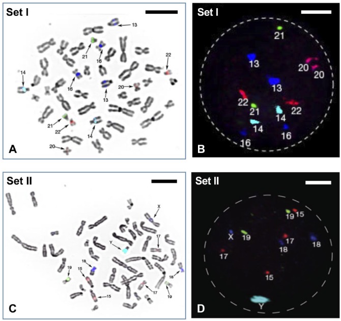
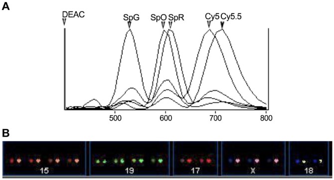
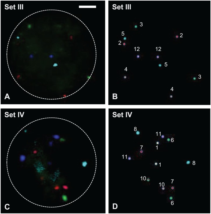
Aneuploidy seems to play not only a decisive role in embryonal development but also in tumorigenesis where chromosomal and genomic instability reflect a universal feature of malignant tumors. The cost of whole genome sequencing has fallen significantly, but it is still prohibitive for many institutions and clinical settings. No applied, cost-effective, and efficient technique has been introduced yet aiming at research to assess the ploidy status of all 24 different human chromosomes in interphases simultaneously, especially in single cells. Here, we present the selection of human probe DNA and a technique using multistep fluorescence in situ hybridization (FISH) employing four sets of six labeled FISH probes able to delineate all 24 human chromosomes in interphase cells. This full karyotype analysis approach will provide additional diagnostic potential for single cell analysis. The use of spectral imaging (SIm) has enabled the use of up to eight different fluorochrome labels simultaneously. Thus, scoring can be easily assessed by visual inspection, because SIm permits computer-assigned and distinguishable pseudo-colors to each probe during image processing. This enables full karyotype analysis by FISH of single-cell interphase nuclei.
Keywords: DNA probes; FISH; aneuploidy; chromosome enumeration; full karyotype analysis; interphase nuclei; spectral imaging.
Conflict of interest statement
Figures
Similar articles
-
An approach for quantitative assessment of fluorescence in situ hybridization (FISH) signals for applied human molecular cytogenetics.J Histochem Cytochem. 2005 Mar;53(3):401-8. doi: 10.1369/jhc.4A6419.2005. J Histochem Cytochem. 2005. PMID: 15750029
-
Small marker chromosome identification in metaphase and interphase using centromeric multiplex fish (CM-FISH).Lab Invest. 2001 Apr;81(4):475-81. doi: 10.1038/labinvest.3780255. Lab Invest. 2001. PMID: 11304566
-
Four-color FISH for the detection of low-level aneuploidy in interphase cells.Methods Mol Biol. 2014;1136:291-305. doi: 10.1007/978-1-4939-0329-0_14. Methods Mol Biol. 2014. PMID: 24633803
-
French multi-centric study of 2000 amniotic fluid interphase FISH analyses from high-risk pregnancies and review of the literature.Ann Genet. 2002 Apr-Jun;45(2):77-88. doi: 10.1016/s0003-3995(02)01118-8. Ann Genet. 2002. PMID: 12119216 Review.
-
Advantages and limitations of using fluorescence in situ hybridization for the detection of aneuploidy in interphase human cells.Mutat Res. 1995 Dec;348(4):153-62. doi: 10.1016/0165-7992(95)90003-9. Mutat Res. 1995. PMID: 8544867 Review.
Cited by
-
Digital pathology systems enabling quality patient care.Genes Chromosomes Cancer. 2023 Nov;62(11):685-697. doi: 10.1002/gcc.23192. Epub 2023 Jul 17. Genes Chromosomes Cancer. 2023. PMID: 37458325 Free PMC article. Review.
-
Characterization of Human-Induced Neural Stem Cells and Derivatives following Transplantation into the Central Nervous System of a Nonhuman Primate and Rats.Stem Cells Int. 2022 Dec 28;2022:1396735. doi: 10.1155/2022/1396735. eCollection 2022. Stem Cells Int. 2022. PMID: 36618021 Free PMC article.
-
Construction and Characterization of Immortalized Fibroblast Cell Line from Bactrian Camel.Life (Basel). 2023 Jun 7;13(6):1337. doi: 10.3390/life13061337. Life (Basel). 2023. PMID: 37374120 Free PMC article.
-
Analysis of human invasive cytotrophoblasts demonstrates mosaic aneuploidy.PLoS One. 2023 Jul 21;18(7):e0284317. doi: 10.1371/journal.pone.0284317. eCollection 2023. PLoS One. 2023. PMID: 37478076 Free PMC article.
References
-
- Schrock E, du Manoir S, Veldman T, Schoell B, Wienberg J, Ferguson-Smith MA, Ning Y, Ledbetter DH, Bar-Am I, Soenksen D, Garini Y, Ried T. Multicolor spectral karyotyping of human chromosomes. Science. 1996;273:494–7. - PubMed
-
- Kallioniemi A, Kallioniemi OP, Sudar D, Rutovitz D, Gray JW, Waldman F, Pinkel D. Comparative genomic hybridization for molecular cytogenetic analysis of solid tumors. Science. 1992;258:818–21. - PubMed
Publication types
MeSH terms
Substances
Grants and funding
LinkOut - more resources
Full Text Sources
Other Literature Sources
