. 2018 Sep;32(9):5174-5185.
doi: 10.1096/fj.201701558R. Epub 2018 Apr 19.

Macrophage-derived extracellular vesicles mediate smooth muscle hyperplasia: role of altered miRNA cargo in response to HIV infection and substance abuse

Affiliations

Macrophage-derived extracellular vesicles mediate smooth muscle hyperplasia: role of altered miRNA cargo in response to HIV infection and substance abuse

Himanshu Sharma et al. FASEB J. 2018 Sep.

Erratum in

  • corrigendum.
    [No authors listed] [No authors listed] FASEB J. 2021 Nov;35(11):e22020. doi: 10.1096/fsb2.22020. FASEB J. 2021. PMID: 34674315 Free PMC article. No abstract available.

Abstract

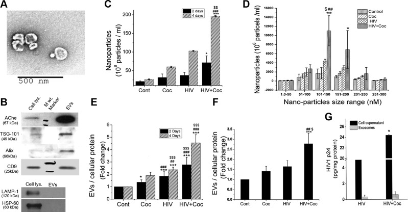
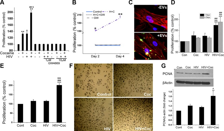
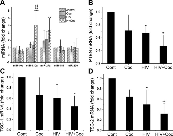
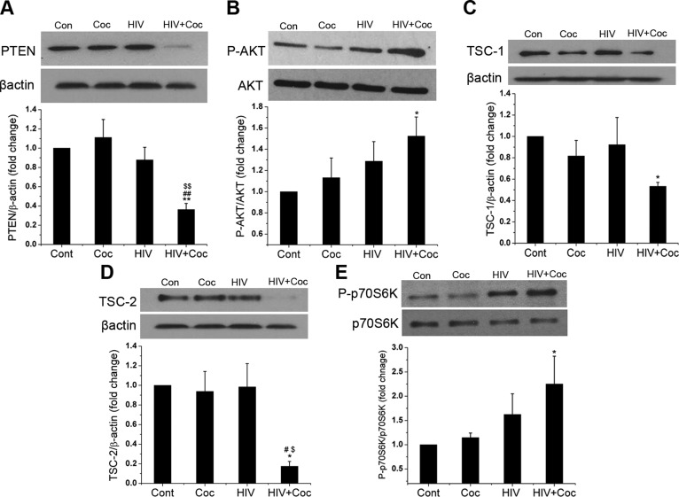
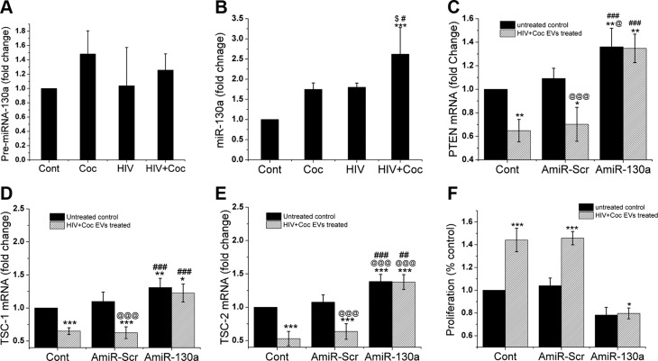
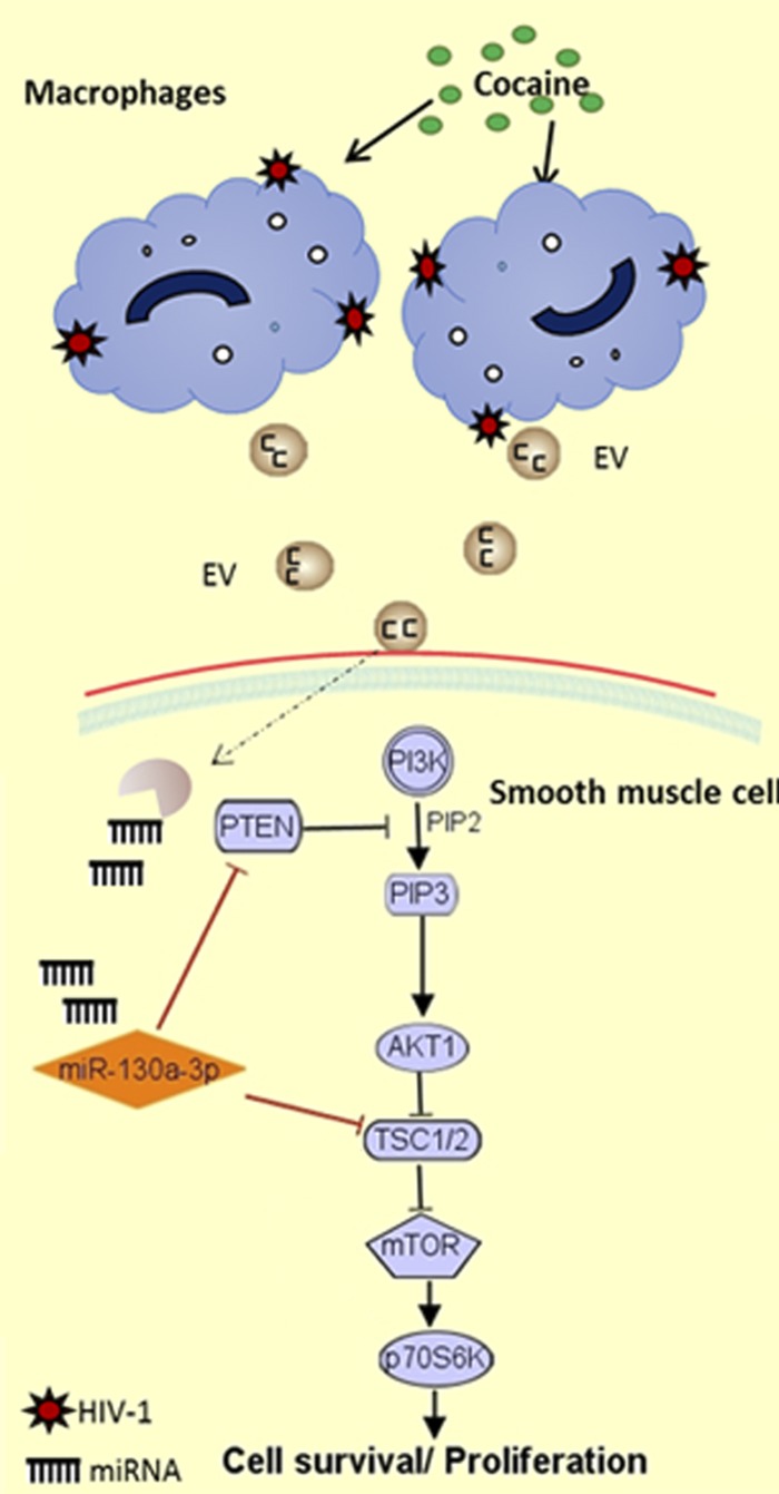
Our previous studies consistently demonstrate enhanced pulmonary vascular remodeling in HIV-infected intravenous drug users, and in simian immunodeficiency virus-infected macaques or HIV-transgenic rats exposed to opioids or cocaine. Although we reported an associated increase in perivascular inflammation, the exact role of inflammatory cells in the development of pulmonary vascular remodeling remains unknown. In this study, HIV-infected and cocaine (H+C)-treated human monocyte derived macrophages released a higher number of extracellular vesicles (EVs), compared to HIV-infected or uninfected cocaine-treated macrophages, with a significant increase in the particle size range to 100-150 nm. Treatment of primary human pulmonary arterial smooth muscle cells (HPASMCs) with these EVs resulted in a significant increase in smooth muscle proliferation. We also observed a significant increase in the miRNA-130a level in the EVs derived from H+C-treated macrophages that corresponded with the decrease in the expression of phosphatase and tensin homolog and tuberous sclerosis 1 and 2 and activation of PI3K/protein kinase B signaling in HPASMCs on addition of these EVs. Transfection of HPASMCs with antagomir-130a-ameliorated the EV-induced effect. Thus, we conclude that EVs derived from H+C-treated macrophages promote pulmonary smooth muscle proliferation by delivery of its prosurvival miRNA cargo, which may play a crucial role in the development of PAH.-Sharma, H., Chinnappan, M., Agarwal, S., Dalvi, P., Gunewardena, S., O'Brien-Ladner, A., Dhillon, N. K. Macrophage-derived extracellular vesicles mediate smooth muscle hyperplasia: role of altered miRNA cargo in response to HIV infection and substance abuse.

Keywords: HIV-PAH; cocaine; miR-130a; pulmonary vascular remodelling.


Conflict of interest statement

The authors thank the Electron Microscopy Research Laboratory (EMRL) facility and Barbara Fegley for assistance with electron microscopy of isolate extracellular vesicles; Dr. Lane Christenson for providing access to NanoSight-LM10; the Kansas Intellectual and Developmental Disabilities Research Center, U. S. National Institutes of Health (NIH) Eunice Kennedy Shriver National Institute of Child Health and Human Development Grant U54 HD 090216; and the Molecular Regulation of Cell Development and Differentiation Centers of Biomedical Research Excellence (COBRE) Grant 5P20GM104936-10 (University of Kansas Medical Center for RNA Sequencing and Bioinformatics Services). This work was supported by NIH National Institute on Drug Abuse Grants R01DA034542 and R01DA042715, and NIH National Heart, Blood, and Lung Institute R01HL129875 (to N.K.D.). The authors declare no conflicts of interest.

Figures

Figure 1
Figure 1
Increased release of EVs on cocaine treatment of HIV-infected MDMs. The human monomac-1 cell line and primary monocytes isolated from human peripheral blood mononuclear cells were differentiated into MDMs followed by HIV-1 bronchoalveolar lavage (5 ng/ml) infection, 1 µM cocaine (Coc) treatment, or both. EVs were isolated from supernatants collected at d 2 and 4 after infection. Isolated exosomes were analyzed for their purity, shape, and size distribution. A) Representative TEM micrograph showing EVs. Original magnification, ×10,000. B) Western blot showing comparison of exosomal protein expression in cells and isolated EVs. C, D) Quantification of EVs from monomac-1–derived macrophages. E, F) Changes in EV:cellular protein ratio after H+C treatment of macrophages derived from monomac-1 cells (E) and human primary monocytes (F). G) HIV-1 p24 levels in cellular supernatants and EV extracts, as measured by ELISA. Data are means ± sd from ≥3 independent experiments. *P < 0.05, **P < 0.01, ***P < 0.001 vs. control; ##P < 0.01, ###P < 0.001 vs. cocaine; $P < 0.05, $$P < 0.01, $$$P < 0.001 vs. HIV.
Figure 2
Figure 2
Increased SMC proliferation on uptake of H+C- or cocaine-treated MDM–derived EVs. A, B) Monomac-1–derived macrophages were treated with H+C in the presence or absence of GW4869 (1 or 10 μM). Supernatants collected were added on HPASMCs in 1:1 ratio with SMC medium followed by a cell proliferation assay after 2 and 4 d. C) Uptake of EVs by SMCs. EVs were labeled with fluorescent PKH67 dye (green) and added on HPASMCs (10 µg/24 well) for 2 h. Cells were later washed and fixed, followed by staining with phalloidin (red). D, E) Exosomes (2 μg) resuspended in PBS, isolated from untreated, cocaine (Coc) treated, HIV-1 infected and from both HIV-1-infected and cocaine-treated monomac-1 (D) or primary human monocyte (E) derived macrophages were added to 0.1% serum-starved HPASMCs overnight followed by replenishing the medium the next day. MTS cell proliferation assay was performed at 48 h after exosome treatment. F) Representative phase-contrast images of HPASMCs after MTS assay. G) Western blot analysis of HPASMC total cell extract after 48 h of EV treatment for proliferation marker, PCNA. Data are means ± sd from ≥3 independent experiments. *P < 0.05, **P < 0.01, ***P < 0.001 vs. control; #P < 0.05, ##P < 0.01, ###P < 0.001 vs. cocaine; $P < 0.05, $$$P < 0.001 vs. HIV.
Figure 3
Figure 3
Increased expression of miR-130a and -27a in EVs derived from H+C-treated macrophages. A) Quantitative RT-PCR analyses of miR-130a, -130a, -27a -10a, -181, and -200 in EVs derived from MDMs that have been HIV-infected, cocaine-treated (Coc), or both. BD) Quiescent HPASMCs were treated with EVs in serum-free SMC medium for 16 h followed by RNA extraction and quantitative RT-PCR analysis of PTEN (B) and TSC-1 (C) and -2 (D) mRNAs. Data are means ± sd from ≥3 independent experiments. **P < 0.01, ***P < 0.001 vs. control; ##P < 0.01 vs. cocaine; $$P < 0.01 vs. HIV.
Figure 4
Figure 4
H+C-treated macrophage–derived EVs activate PI3K/AKT signaling in HPASMCs. Quiescent HPASMCs were treated with EVs derived from H+C-, HIV-1-, or cocaine (Coc)-treated MDMs in serum-free SMC medium for 16 h followed by cell lysis with RIPA buffer for Western blot analysis of PTEN (A), p-AKT (B), TSC-1 (C), TSC-2 (D) and phospho-p70S6K (E). Means ± sem from ≥3 independent experiments. *P < 0.05, **P < 0.01 vs. control; #P < 0.05, ##P < 0.01 vs. cocaine; $P < 0.05, $$P < 0.01 vs. HIV.
Figure 5
Figure 5
Silencing of miR-130a in SMCs prevents H+C derived EVs mediated inhibition of PTEN expression and hyperproliferation. A, B) Quiescent HPASMCs were treated with exosomes derived from HIV-infected, cocaine-treated (Coc), or H+C-treated macrophages or those for 16 h followed by RNA extraction, for RT-PCR analysis of precursor (A) and mature (B) miR-130a expression. CF) HPASMC were transfected with antagomirs-130a (AmiR-130a) or scrambled miRNA (AmiR-Scr). At 24 h post-transfection cells were made quiescent for 24 h in 0.1% FBS–containing medium followed by addition of H+C EVs for 16 h to perform PTEN (C), TSC-1 (D), TSC-2 (E) mRNA analysis and for 48 h to perform cell proliferation assay (F). Data are means ± sd from ≥3 independent experiments. *P < 0.05, **P < 0.01, ***P < 0.001 vs. control; @P < 0.05, @@@P < 0.001 vs. untreated AmiR-Scr; #P < 0.05, ##P < 0.01, ###P < 0.001 vs. exosome treated AmiR-Scr.
Figure 6
Figure 6
Schematic showing miR-130a-mediated regulation of PI3K/AKT signaling on exposure to HIV-infected, cocaine-treated, and macrophage-derived EVs.

Similar articles

Cited by

References

    1. Sitbon O., Lascoux-Combe C., Delfraissy J. F., Yeni P. G., Raffi F., De Zuttere D., Gressin V., Clerson P., Sereni D., Simonneau G. (2008) Prevalence of HIV-related pulmonary arterial hypertension in the current antiretroviral therapy era. Am. J. Respir. Crit. Care Med. 177, 108–113 - PubMed
    1. L’Huillier A. G., Posfay-Barbe K. M., Pictet H., Beghetti M. (2015) Pulmonary arterial hypertension among HIV-infected children: results of a national survey and review of the literature. Front Pediatr. 3, 25. - PMC - PubMed
    1. Humbert M., Sitbon O., Chaouat A., Bertocchi M., Habib G., Gressin V., Yaici A., Weitzenblum E., Cordier J. F., Chabot F., Dromer C., Pison C., Reynaud-Gaubert M., Haloun A., Laurent M., Hachulla E., Simonneau G. (2006) Pulmonary arterial hypertension in France: results from a national registry. Am. J. Respir. Crit. Care Med. 173, 1023–1030 - PubMed
    1. Reinsch N., Buhr C., Krings P., Kaelsch H., Kahlert P., Konorza T., Neumann T., Erbel R.; Competence Network of Heart Failure (2008) Effect of gender and highly active antiretroviral therapy on HIV-related pulmonary arterial hypertension: results of the HIV-HEART study. HIV Med. 9, 550–556 - PubMed
    1. Morris A., Gingo M. R., George M. P., Lucht L., Kessinger C., Singh V., Hillenbrand M., Busch M., McMahon D., Norris K. A., Champion H. C., Gladwin M. T., Zhang Y., Steele C., Sciurba F. C. (2012) Cardiopulmonary function in individuals with HIV infection in the antiretroviral therapy era. AIDS 26, 731–740 - PMC - PubMed

Publication types

MeSH terms