. 2018 Apr 19:24:2377-2383.
doi: 10.12659/msm.909727.

Activated Eosinophils are Present in Esophageal Muscle in Patients with Achalasia of the Esophagus

Affiliations

Activated Eosinophils are Present in Esophageal Muscle in Patients with Achalasia of the Esophagus

Hong Jin et al. Med Sci Monit. .

Abstract

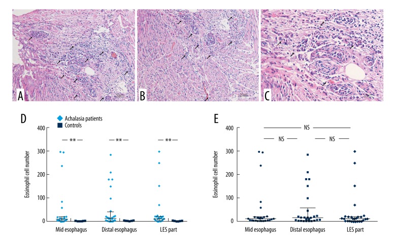
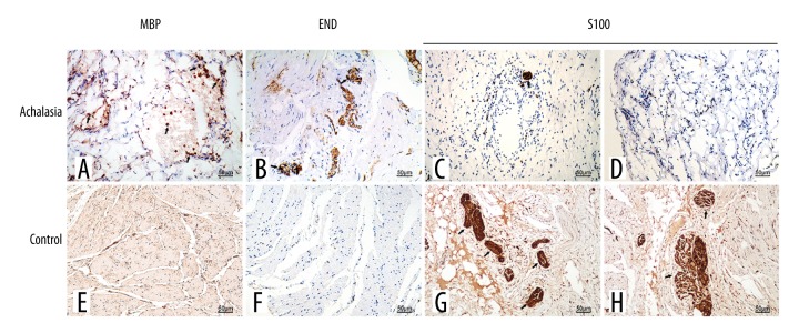
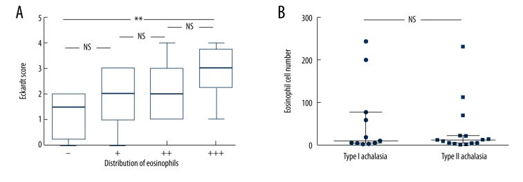
BACKGROUND The aim of this study was to undertake a histological evaluation of the presence of eosinophils in esophageal muscle in patients with achalasia before treatment with peroral endoscopic myotomy (POEM), with clinical follow-up at one year. MATERIAL AND METHODS Before treatment, esophageal biopsies including mucosa and esophageal muscle were obtained from 28 patients with achalasia. Nine patients who had undergone esophagectomy for esophageal carcinoma were included in the control group. The Eckardt Score was used to evaluate the clinical symptoms of achalasia. Histology of routinely processed tissue sections was used to perform eosinophil cell counts (0 to +++), and immunohistochemistry was used to detect expression of eosinophil major basic protein (MBP), eosinophil-derived neurotoxin (EDN), and S100 protein in cases of achalasia (n=28) and controls (n=9). The findings in patients with achalasia were compared before and one year following POEM. RESULTS Esophageal tissue from patients with achalasia showed eosinophils infiltrating into the muscularis externa in 85.7% (24/28), into the muscularis propria in 28.6% (8/28), and in 89% (25/28) there were few remaining myenteric ganglion cells, before POEM. The extent of inflammation was similar in all regions of the esophagus and between subtypes of achalasia. At one year following POEM, the Eckardt Scores between the former eosinophil (0) group and the eosinophil (+++) group were significantly different (Z=3.50, P=0.030). CONCLUSIONS Achalasia of the esophagus was associated with infiltration of the esophageal muscle by activated eosinophils and a decrease in the density of ganglion cells in the myenteric esophageal plexus.


Conflict of interest statement

Conflict of interest

None.

Figures

Figure 1
Figure 1
The muscularis externa of the esophagus in patients with achalasia showing eosinophilia. (A) Photomicrograph of the histology of the muscularis externa of the esophagus in a patient with achalasia shows severe inflammation with eosinophilia. Hematoxylin and eosin (H&E). Magnification ×200. (B) Photomicrograph of the histology of the muscularis externa of the esophagus in a patient with achalasia shows severe inflammation with eosinophilia. H&E. Magnification ×200. (C) Photomicrograph of the histology of the muscularis externa of the esophagus in a patient with achalasia shows severe inflammation with eosinophilia. H&E. Magnification ×400. (D) The number of eosinophils in regions of the esophagus from patients with achalasia, compared with controls. (E) The number of eosinophils in the muscularis layer in regions of the esophagus in patients with achalasia. ** P<0.05; NS – not significant.
Figure 2
Figure 2
Photomicrographs showing the immunohistochemical staining of the esophageal tissue for degranulating eosinophils and nerve cells. (A) Esophageal tissue from a patient with achalasia. Immunohistochemical staining shows localization of the primary antibody to eosinophil major basic protein (MBP), confirming that the cells are eosinophils. (B) Esophageal tissue from a patient with achalasia. Immunohistochemical staining shows localization of the primary antibody to eosinophil-derived neurotoxin (EDN), confirming that the cells are eosinophils. (C) Esophageal tissue from a patient with achalasia. Immunohistochemical staining for the localization of the primary antibody to S100, which shows loss of ganglion cells. (D) Esophageal tissue from a patient with achalasia. Immunohistochemical staining for the localization of the primary antibody to S100, which shows loss of ganglion cells. (E) Normal (control) esophageal tissue. Immunohistochemical staining with the primary antibody to eosinophil MBP is negative. (F) Normal (control) esophageal tissue. Immunohistochemical staining with the primary antibody to EDN is negative. (G) Normal (control) esophageal tissue. Immunohistochemical staining of the localization of the primary antibody to S100, which shows a regular distribution of ganglion cells associated with myenteric nerves. (H) Normal (control) esophageal tissue. Immunohistochemical staining of the localization of the primary antibody to S100, which shows a regular distribution of ganglion cells associated with myenteric nerves.
Figure 3
Figure 3
The Eckardt Score and the distribution of eosinophils in the esophageal muscle of patients with achalasia one year following peroral endoscopic myotomy (POEM). (A) No significant differences are found in the Eckardt Score between the groups (P>0.05), but the differences between eosinophil (0) group and the eosinophil (+++) group are statistically significant at one year following peroral endoscopic myotomy (POEM) (Z=3.50, P=0.030). (B) One year following POEM, the number of eosinophils in the esophageal muscle of patients in the different types of achalasia is similar. ** P<0.05. NS – not significant.

References

    1. Francis DL, Katzka DA. Achalasia: Update on the disease and its treatment. Gastroenterology. 2010;139:369–74. - PubMed
    1. Ghoshal UC, Daschakraborty SB, Singh R. Pathogenesis of achalasia cardia. World J Gastroenterol. 2012;18:3050–57. - PMC - PubMed
    1. Goldblum JR, Whyte RI, Orringer MB, Appelman HD. Achalasia. A morphologic study of 42 resected specimens. Am J Surg Pathol. 1994;18:327–37. - PubMed
    1. Pandolfino JE, Gawron AJ. Achalasia: A systematic review. JAMA. 2015;313:1841–52. - PubMed
    1. Landres RT, Kuster GG, Strum WB. Eosinophilic esophagitis in a patient with vigorous achalasia. Gastroenterology. 1978;74:1298–301. - PubMed