

Trait anger modulates neural activity in the fronto-parietal attention network
- PMID: 29672547
- PMCID: PMC5908080
- DOI: 10.1371/journal.pone.0194444
Trait anger modulates neural activity in the fronto-parietal attention network
Abstract
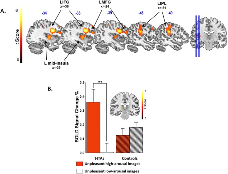
Anger is considered a unique high-arousal and approach-related negative emotion. The influence of individual differences in trait anger on the processing of visual stimuli is relevant to questions about emotional processing and remains to be explored. Using functional magnetic resonance imaging (fMRI), we explored the neural responses to standardized images, selected based on valence and arousal ratings in a group of men with high trait anger compared to those with normative to low anger scores (controls). Results show increased activation in the left-lateralized ventral fronto-parietal attention network to unpleasant images by individuals with high trait anger. There was also a group by arousal interaction in the left thalamus/pulvinar such that individuals with high trait anger had increased pulvinar activation to the high-arousal (versus low arousal) unpleasant images as compared to controls. Thus, individual differences in trait anger in men are associated with brain regions subserving executive attentional and sensory integration during the processing of unpleasant emotional stimuli, particularly to high arousal images.
Conflict of interest statement
Figures
References
-
- Carver CS, Harmon-Jones E. Anger is an approach-related affect: evidence and implications. Psychological bulletin. 2009;135:183–204. doi: 10.1037/a0013965 - DOI - PubMed
-
- Harmon-Jones E, Allen JJ. Anger and frontal brain activity: EEG asymmetry consistent with approach motivation despite negative affective valence. J Pers Soc Psychol. 1998;74:1310–6. - PubMed
-
- Spielberger CD, Gorsuch R.L., Lushene R.E. STAI Manual for the State-Trait Anxiety Inventory ("Self-Evaluation Questionnaire"). Palo Alto, CA: Consulting Psychologists Press, Inc.; 1970.
-
- Alia-Klein N, Goldstein RZ, Tomasi D, Woicik PA, Moeller SJ, Williams B, et al. Neural mechanisms of anger regulation as a function of genetic risk for violence. Emotion. 2009;9:385–96. doi: 10.1037/a0015904 - DOI - PMC - PubMed
-
- Harmon-Jones E. Contributions from research on anger and cognitive dissonance to understanding the motivational functions of asymmetrical frontal brain activity. Biological psychology. 2004;67:51–76. doi: 10.1016/j.biopsycho.2004.03.003 - DOI - PubMed
Publication types
MeSH terms
Grants and funding
LinkOut - more resources
Full Text Sources
Other Literature Sources
