

Graphical and statistical analyses of the oculocardiac reflex during a non-invasive intracranial pressure measurement
- PMID: 29672564
- PMCID: PMC5909620
- DOI: 10.1371/journal.pone.0196155
Graphical and statistical analyses of the oculocardiac reflex during a non-invasive intracranial pressure measurement
Abstract
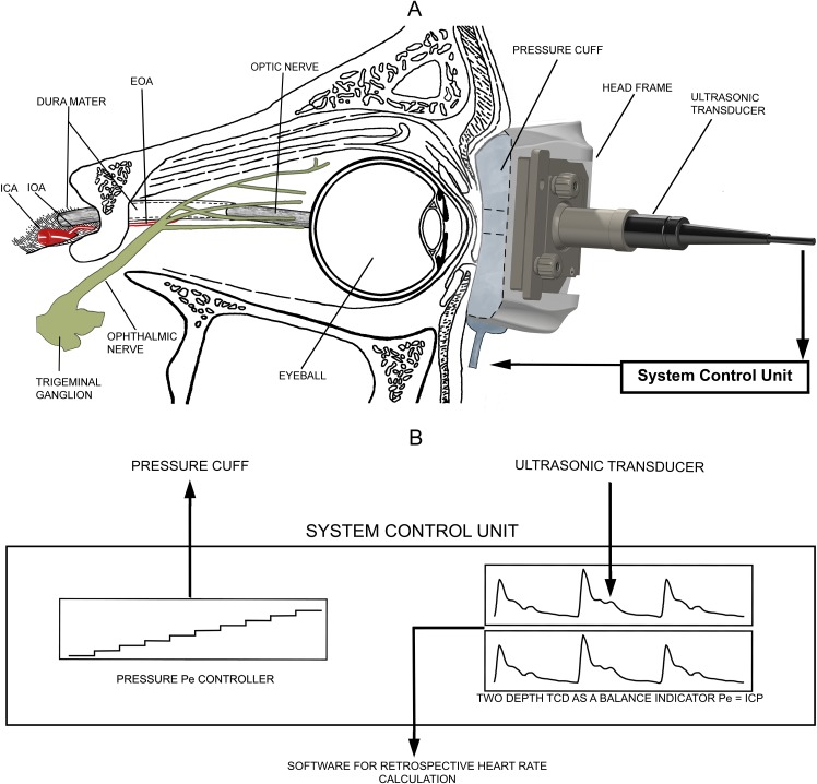
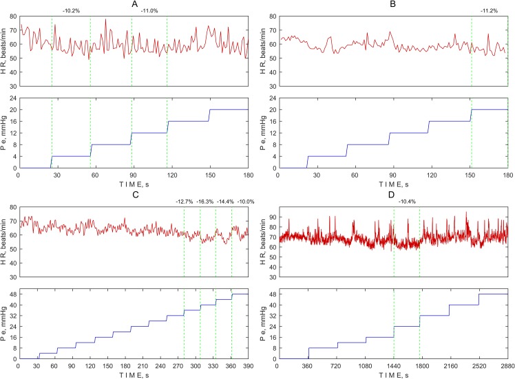
Purpose: This study aimed to examine the incidence of the oculocardiac reflex during a non-invasive intracranial pressure measurement when gradual external pressure was applied to the orbital tissues and eye.
Methods: Patients (n = 101) and healthy volunteers (n = 56) aged 20-75 years who underwent a non-invasive intracranial pressure measurement were included in this retrospective oculocardiac reflex analysis. Prespecified thresholds greater than a 10% or 20% decrease in the heart rate from baseline were used to determine the incidence of the oculocardiac reflex.
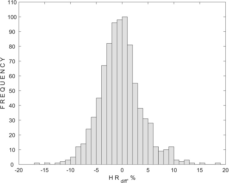
Results: None of the subjects had a greater than 20% decrease in heart rate from baseline. Four subjects had a greater than 10% decrease in heart rate from baseline, representing 0.9% of the total pressure steps. Three of these subjects were healthy volunteers, and one was a glaucoma patient.
Conclusion: The incidence of the oculocardiac reflex during a non-invasive intracranial pressure measurement procedure was very low and not associated with any clinically relevant effects.
Conflict of interest statement
Figures
References
-
- Ohashi T, Kase M, Yokoi M. Quantitative analysis of the oculocardiac reflex by traction on human extraocular muscle. Invest Ophthalmol Vis Sci. 1986;27(7): 1160–1164. - PubMed
-
- Sandu N, Cornelius J, Filis A, Nothen C, Rasper J, Kulinsky VI, et al. Cerebral hemodynamic changes during the trigeminocardiac reflex: description of a new animal model protocol. ScientificWorldJournal. 2010;10: 1416–1423. doi: 10.1100/tsw.2010.136 - DOI - PMC - PubMed
-
- Paton JF, Boscan P, Pickering AE, Nalivaiko E. The yin and yang of cardiac autonomic control: vago-sympathetic interactions revisited. Brain Res Brain Res Rev. 2005;49(3): 555–565. doi: 10.1016/j.brainresrev.2005.02.005 - DOI - PubMed
-
- Yi C, Jee D. Influence of the anaesthetic depth on the inhibition of the oculocardiac reflex during sevoflurane anaesthesia for paediatric strabismus surgery. Br J Anaesth. 2008;101(2): 234–238. doi: 10.1093/bja/aen129 - DOI - PubMed
-
- Blanc VF, Hardy JF, Milot J, Jacob JL. The oculocardiac reflex: a graphic and statistical analysis in infants and children. Can Anaesth Soc J. 1983;30(4): 360–369. - PubMed
Publication types
MeSH terms
LinkOut - more resources
Full Text Sources
Other Literature Sources
