

RUNX1-PDCD6 fusion resulting from a novel t(5;21)(p15;q22) chromosome translocation in myelodysplastic syndrome secondary to chronic lymphocytic leukemia
- PMID: 29672642
- PMCID: PMC5908135
- DOI: 10.1371/journal.pone.0196181
RUNX1-PDCD6 fusion resulting from a novel t(5;21)(p15;q22) chromosome translocation in myelodysplastic syndrome secondary to chronic lymphocytic leukemia
Abstract
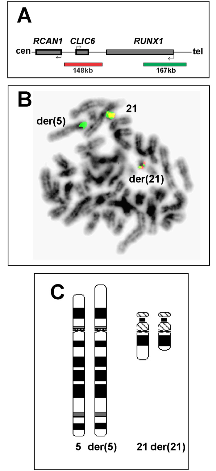
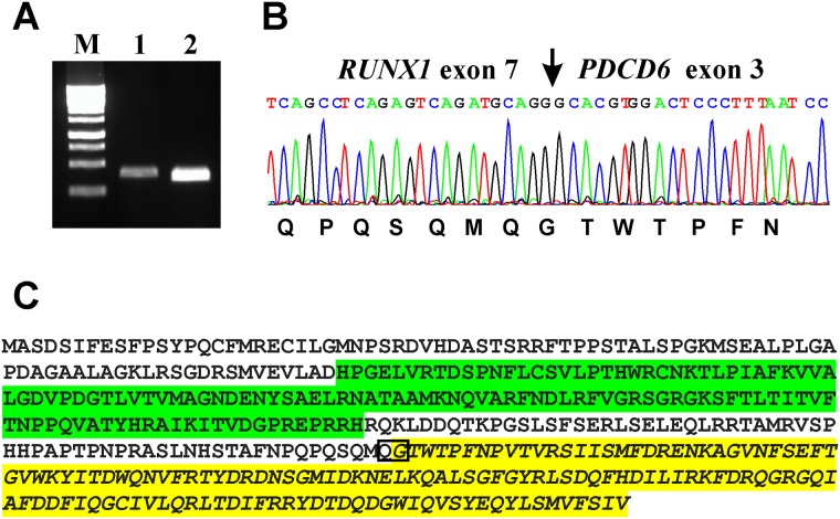
Leukemic cells often carry chromosome aberrations which generate chimeric genes of pathogenetic, diagnostic, and prognostic importance. New rearrangements giving rise to novel fusion genes define hitherto unrecognized genetic leukemia subgroups. G-banding, fluorescence in situ hybridization (FISH), and molecular genetic analyses were done on bone marrow cells from a patient with chronic lymphocytic leukemia (CLL) and secondary myelodysplasia. The G-banding analysis revealed the karyotype 46,XX,del(21)(q22)[9]/46,XX[2]. FISH on metaphase spreads with a RUNX1 break apart probe demonstrated that part of RUNX1 (from 21q22) had moved to chromosome band 5p15. RNA sequencing showed in-frame fusion of RUNX1 with PDCD6 (from 5p15), something that was verified by RT-PCR together with Sanger sequencing. Further FISH analyses with PDCD6 and RUNX1 home-made break apart/double fusion probes showed a red signal (PDCD6) on chromosome 5, a green signal on chromosome 21 (RUNX1), and two yellow fusion signals, one on der(5) and the other on der(21). Reassessment of the G-banding preparations in light of the FISH and RNA-sequencing data thus yielded the karyotype 46,XX,t(5;21)(p15;q22)[9]/46,XX[2]. The t(5;21)(p15;q22)/RUNX1-PDCD6 was detected only by performing molecular studies of the leukemic cells, but should be sought after also in other leukemic/myelodysplastic cases with del(21q).
Conflict of interest statement
Figures


Similar articles
-
RUNX1 truncation resulting from a cryptic and novel t(6;21)(q25;q22) chromosome translocation in acute myeloid leukemia: A case report.Oncol Rep. 2016 Nov;36(5):2481-2488. doi: 10.3892/or.2016.5119. Epub 2016 Sep 22. Oncol Rep. 2016. PMID: 27667292 Free PMC article.
-
RUNX1-MTG16 fusion gene in acute myeloblastic leukemia with t(16;21)(q24;q22): case report and review of the literature.Cancer Genet Cytogenet. 2008 Aug;185(1):47-50. doi: 10.1016/j.cancergencyto.2008.04.011. Cancer Genet Cytogenet. 2008. PMID: 18656694 Review.
-
t(8;21)(q22;q22) Translocation involving AML1 and ETO in B lymphoblastic leukemia [corrected].Hum Pathol. 2010 Feb;41(2):286-92. doi: 10.1016/j.humpath.2009.08.004. Epub 2009 Nov 6. Hum Pathol. 2010. PMID: 19896694
-
Coexistent t(8;21)(q22;q22) Translocation and 5q Deletion in Acute Myeloid Leukemia.J Clin Exp Hematop. 2015;55(3):181-5. doi: 10.3960/jslrt.55.181. J Clin Exp Hematop. 2015. PMID: 26763368 Review.
-
Novel MYCBP::EHD2 and RUNX1::ZNF780A Fusion Genes in T-cell Acute Lymphoblastic Leukemia.Cancer Genomics Proteomics. 2023 Jan-Feb;20(1):51-63. doi: 10.21873/cgp.20364. Cancer Genomics Proteomics. 2023. PMID: 36581344 Free PMC article.
Cited by
-
Toward personalized treatment in multiple myeloma based on molecular characteristics.Blood. 2019 Feb 14;133(7):660-675. doi: 10.1182/blood-2018-09-825331. Epub 2018 Dec 26. Blood. 2019. PMID: 30587529 Free PMC article. Review.
-
Prevalence of RUNX1 gene alterations in de novo adult acute myeloid leukemia.World J Exp Med. 2025 Mar 20;15(1):99516. doi: 10.5493/wjem.v15.i1.99516. eCollection 2025 Mar 20. World J Exp Med. 2025. PMID: 40115757 Free PMC article.
References
-
- Heim S, Mitelman F. Cancer Cytogenetics: Chromosomal and Molecular Genetic Abberations of Tumor Cells. Fourth Edition ed: Wiley-Blackwell; 2015.
-
- Sood R, Kamikubo Y, Liu P. Role of RUNX1 in hematological malignancies. Blood. 2017;129(15):2070–82. doi: 10.1182/blood-2016-10-687830 - DOI - PMC - PubMed
-
- Ichikawa M, Yoshimi A, Nakagawa M, Nishimoto N, Watanabe-Okochi N, Kurokawa M. A role for RUNX1 in hematopoiesis and myeloid leukemia. Int J Hematol. 2013;97(6):726–34. doi: 10.1007/s12185-013-1347-3 . - DOI - PubMed
-
- Cho EK, Bang SM, Ahn JY, Yoo SM, Park PW, Seo YH, et al. Prognostic value of AML 1/ETO fusion transcripts in patients with acute myelogenous leukemia. Korean J Intern Med. 2003;18(1):13–20. Epub 2003/05/23. doi: 10.3904/kjim.2003.18.1.13 . - DOI - PMC - PubMed
-
- Gandemer V, Chevret S, Petit A, Vermylen C, Leblanc T, Michel G, et al. Excellent prognosis of late relapses of ETV6/RUNX1-positive childhood acute lymphoblastic leukemia: lessons from the FRALLE 93 protocol. Haematologica. 2012;97(11):1743–50. Epub 2012/05/15. doi: 10.3324/haematol.2011.059584 . - DOI - PMC - PubMed
Publication types
MeSH terms
Substances
LinkOut - more resources
Full Text Sources
Other Literature Sources
Medical
