. 2018 Jun 1;46(10):5139-5158.
doi: 10.1093/nar/gky273.

miR-122 does not impact recognition of the HCV genome by innate sensors of RNA but rather protects the 5' end from the cellular pyrophosphatases, DOM3Z and DUSP11

Affiliations

miR-122 does not impact recognition of the HCV genome by innate sensors of RNA but rather protects the 5' end from the cellular pyrophosphatases, DOM3Z and DUSP11

Yalena Amador-Cañizares et al. Nucleic Acids Res. .

Abstract

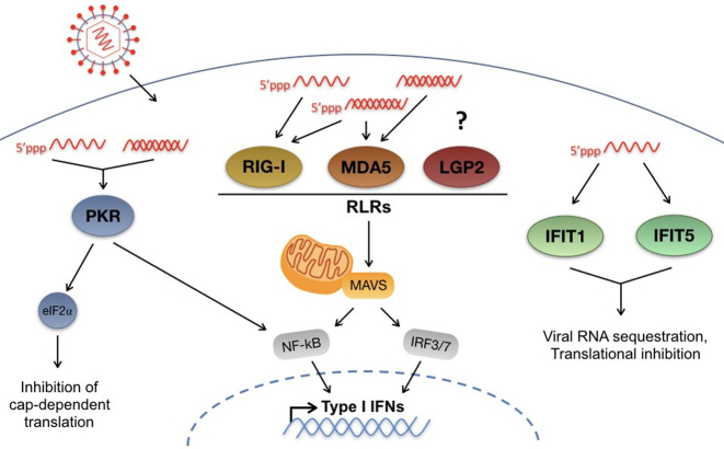
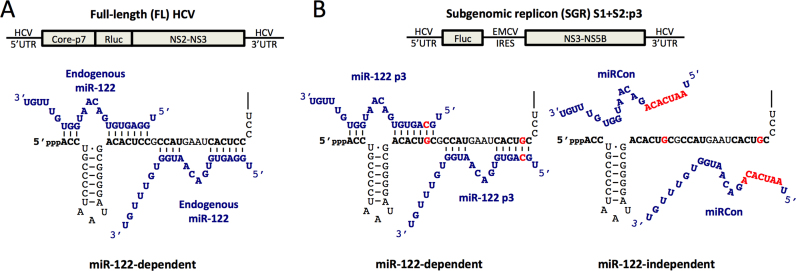
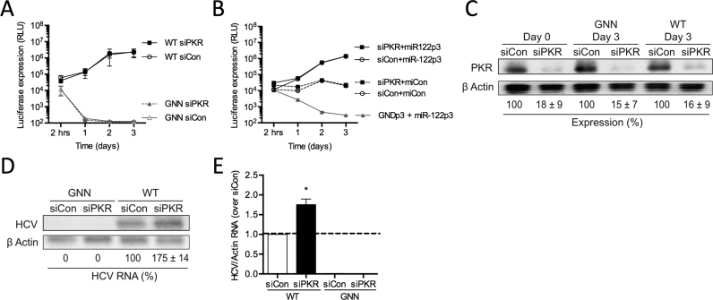
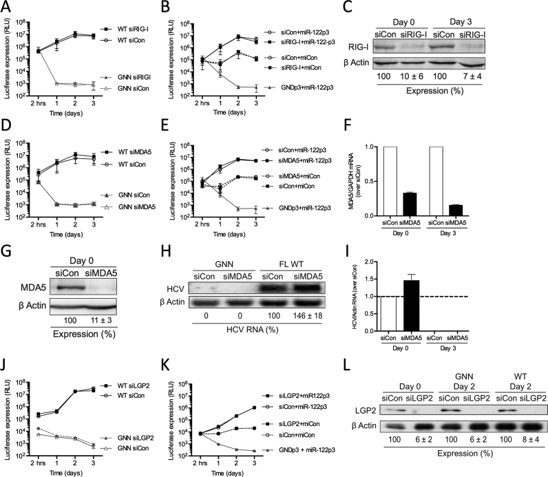
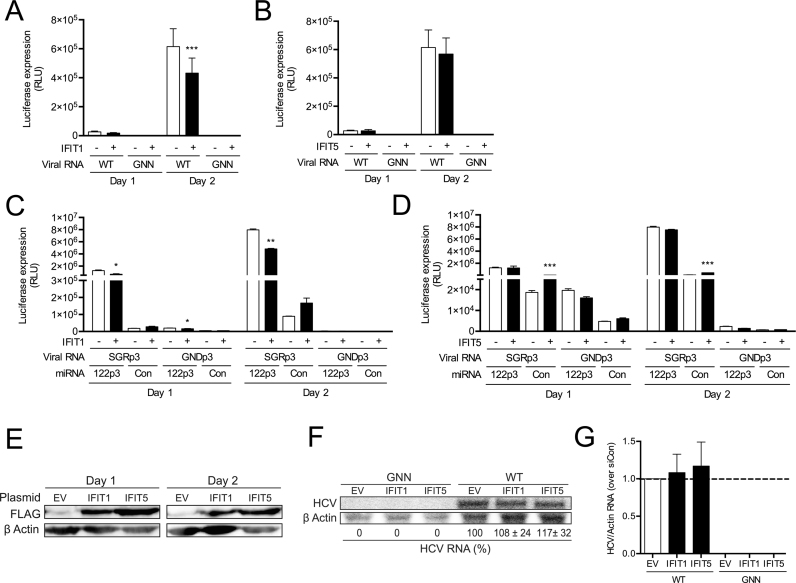
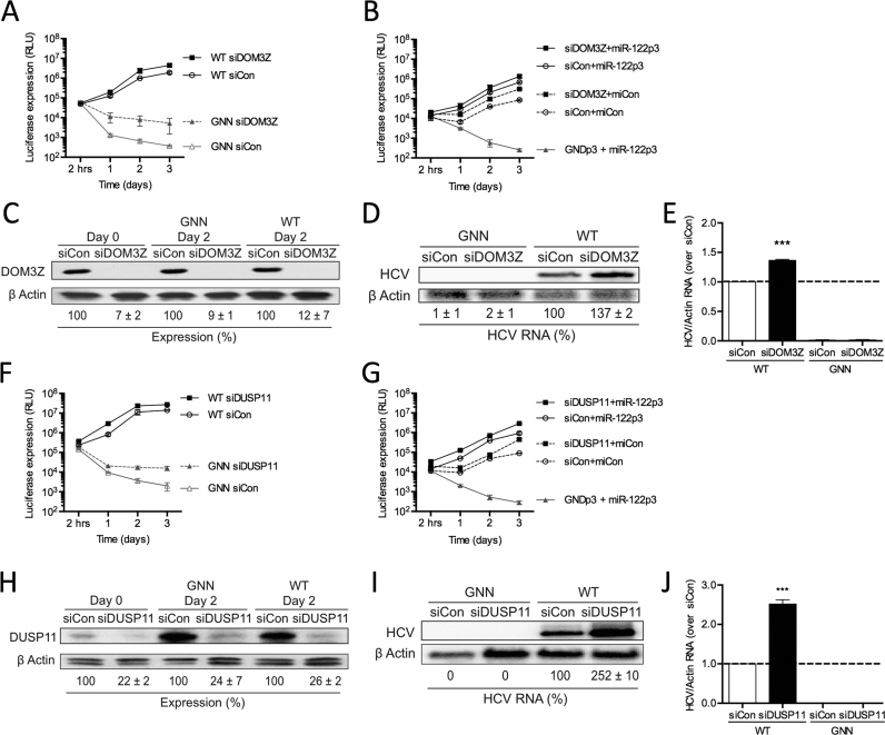
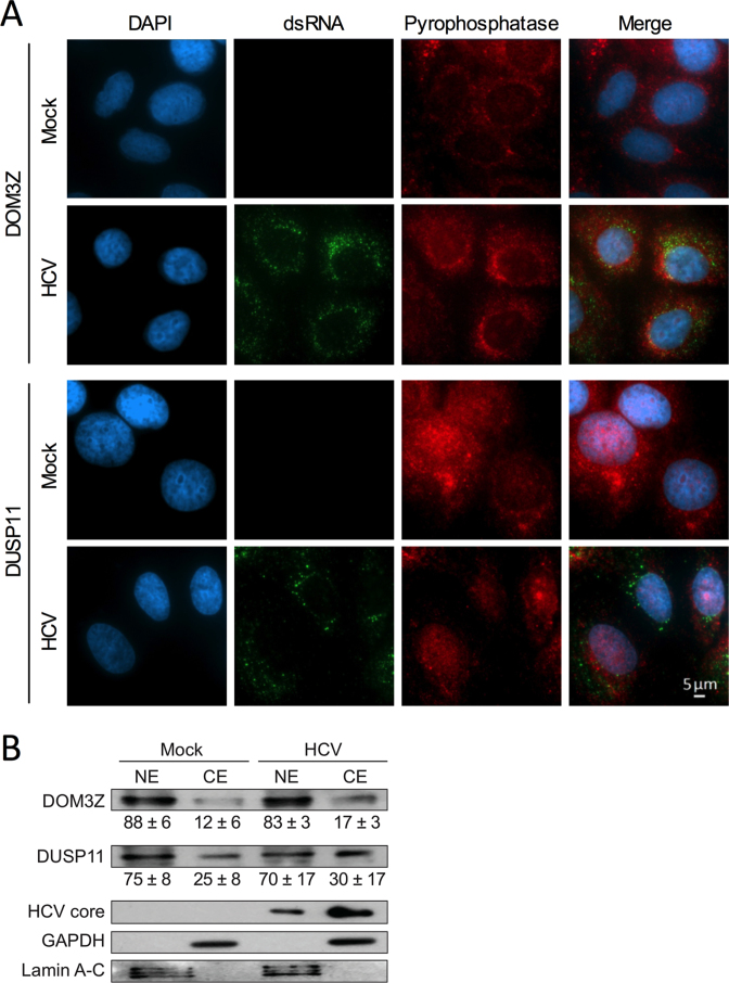
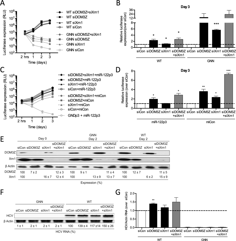
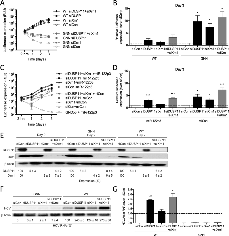
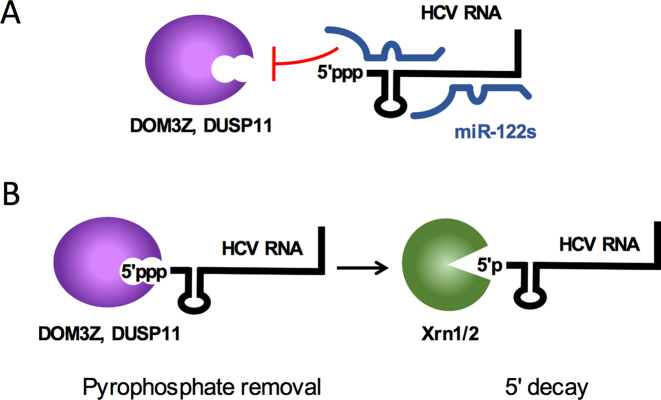
Hepatitis C virus (HCV) recruits two molecules of the liver-specific microRNA-122 (miR-122) to the 5' end of its genome. This interaction promotes viral RNA accumulation, but the precise mechanism(s) remain incompletely understood. Previous studies suggest that miR-122 is able to protect the HCV genome from 5' exonucleases (Xrn1/2), but this protection is not sufficient to account for the effect of miR-122 on HCV RNA accumulation. Thus, we investigated whether miR-122 was also able to protect the viral genome from innate sensors of RNA or cellular pyrophosphatases. We found that miR-122 does not play a protective role against recognition by PKR, RIG-I-like receptors, or IFITs 1 and 5. However, we found that knockdown of both the cellular pyrophosphatases, DOM3Z and DUSP11, was able to rescue viral RNA accumulation of subgenomic replicons in the absence of miR-122. Nevertheless, pyrophosphatase knockdown increased but did not restore viral RNA accumulation of full-length HCV RNA in miR-122 knockout cells, suggesting that miR-122 likely plays an additional role(s) in the HCV life cycle, beyond 5' end protection. Overall, our results support a model in which miR-122 stabilizes the HCV genome by shielding its 5' terminus from cellular pyrophosphatase activity and subsequent turnover by exonucleases (Xrn1/2).


Figures

Figure 1.
Figure 1.
Innate cytosolic sensors of viral RNA. Upon infection, RNA viruses access the cytosol, where they can be amplified by viral RNA-dependent RNA polymerases. Once in the cytosol, viral RNA can be recognized by several innate sensors of RNA, including: Protein Kinase R (PKR); RIG-I-like receptors (RLRs), as well as IFITs 1 and 5. PKR recognizes double-stranded (ds) RNA as well as 5′ triphosphate-containing single-stranded (ss) RNAs. Activation of PKR leads to type I IFN signaling mediated by NF-κB as well as phosphorylation of eIF2α leading to inhibition of cap-dependent translation. The RLRs, including RIG-I, MDA5 and LGP2, recognize primarily dsRNA or highly structured ssRNAs, and at least RIG-I and MDA5 have been demonstrated to be activated by 5′ triphosphate RNA, while the precise substrate for LGP2 has not been defined. Recognition of viral RNA by RLRs leads to recruitment of RIG-I and MDA-5 to the adaptor protein MAVS, which initiates downstream type I IFN signaling through NF-κB and IRF3/7. Finally, IFITs 1 and 5 have been demonstrated to interact with 5′ triphosphate containing ssRNAs leading to viral RNA sequestration and translational inhibition.
Figure 2.
Figure 2.
miR-122-dependent and miR-122-independent replication systems. (A) Cartoon diagram of Full-length (FL) Rluc HCV RNA (top) and depiction of endogenous miR-122 binding to the 5′ terminus of the 5′ UTR of FL viral RNA (bottom). (B) Diagram of subgenomic replicon (SGR) FLuc S1+S2:p3 HCV RNA (top) and depiction of miR-122-independent (miRControl, bottom left) and miR-122-dependent (miR-122p3, bottom right) replication systems. Since Huh-7 and Huh-7.5 cells endogenously express miR-122, endogenous miR-122 binding is abolished by introduction of point mutations in both of the miR-122 binding sites (S1+S2p3, indicated in red). miRControl (where the entire seed region of the miRNA is mutated, indicated in red) or miR-122p3 molecules (containing a compensatory mutation at position 3, indicated in red) are used for miR-122-independent and miR-122-dependent replication, respectively.
Figure 3.
Figure 3.
miR-122 binding does not shield the 5′ terminus of HCV RNA against PKR recognition. Huh-7.5 cells were electroporated with siPKR or siControl (siCon) at day –3 and at day 0 cells were electroporated again with the indicated siRNA, and either (A) wild-type or GNN FL HCV RNA with a firefly luciferase mRNA, or (B) S1+S2p3 SGR or S1+S2p3 GND SGR, a Renilla luciferase control mRNA, and miR-122p3 (miR-122-dependent) or miCon (miR-122-independent). Replication was measured by evaluating luciferase production at the indicated timepoints. (C) Western blot showing knockdown efficiency with antibodies against PKR and β-actin. Percent knockdown ± standard deviation relative to siCon is indicated. (D) Northern blot analysis of FL HCV RNA accumulation during PKR knockdown at day 3. (E) Densitometry quantification of northern blot analysis in (D) normalized to siCon. All data are representative of at least three independent experiments and statistical significance was determined by paired parametric t test.
Figure 4.
Figure 4.
miR-122 binding does not shield the 5′ terminus of HCV RNA against RLR recognition. Huh-7 cells were electroporated with siRIG-I or siControl (siCon) at day –3 and at day 0 cells were electroporated again with the indicated siRNA and either (A) wild-type or GNN FL HCV RNA, and a firefly luciferase control mRNA, or (B) S1+S2p3 SGR or S1+S2p3 GND SGR, a Renilla luciferase control mRNA, and miR-122p3 (miR-122-dependent) or miCon (miR-122-independent). Replication was measured by evaluating luciferase production at the indicated timepoints. (C) Western blot showing knockdown efficiency with antibodies against RIG-I and β-actin. Percent knockdown ± standard deviation relative to siCon is indicated. Huh-7 cells were electroporated with siMDA5 or siControl (siCon) at day –3, at day 0 cells were electroporated again with the indicated siRNA, and either (D) wild-type or GNN FL HCV RNA, and a firefly luciferase control mRNA or (E) S1+S2p3 SGR or S1+S2p3 GND SGR, a Renilla luciferase control mRNA, and miR-122p3 (miR-122-dependent) or miCon (miR-122-independent). Replication was measured by evaluating luciferase production at the indicated timepoints. (F) Quantitative PCR analysis indicating knockdown efficiency using MDA5-specific and GAPDH-specific TaqMan probes. MDA5 mRNA levels were calculated relative to the siCon. (G) Huh-7 cells were electroporated with siMDA5 on day –3, treated with 50 IU/ml IFN-α on day –1 and harvested for western blot at day 0 using antibodies against MDA5 and β-actin. Percent knockdown ± standard deviation relative to siCon is indicated. (H) Northern blot analysis of FL HCV RNA accumulation during MDA5 knockdown at day 3. (I) Densitometry quantification of northern blot analysis in (H) normalized to siCon. Huh-7 cells were electroporated with siLGP2 or siControl (siCon) at day –2, at day 0 cells were electroporated again with the indicated siRNA, and either (J) wild-type or GNN FL HCV RNA, and a firefly luciferase control mRNA or (K) S1+S2p3 SGR or S1+S2p3 GND SGR, a Renilla luciferase control mRNA, and miR-122p3 (miR-122-dependent) or miCon (miR-122-independent). Replication was measured by evaluating luciferase production at the indicated timepoints. (L) Western blot showing knockdown efficiency with antibodies against LGP2 and β-actin. Percent knockdown ± standard deviation relative to siCon is indicated. All data are representative of at least three independent experiments and statistical significance was determined by paired parametric t test.
Figure 5.
Figure 5.
miR-122 binding does not shield the 5′ terminus of HCV RNA against recognition of IFIT1, while IFIT5 has no effect on HCV RNA accumulation. 3XFLAG-IFIT1, 3XFLAG-IFIT5 and the corresponding empty vector (EV) were transfected into Huh7.5 cells, and two days later, plasmids plus (A, B) FL WT HCV or GNN or (C, D) S1+S2p3 SGR and the replication incompetent S1+S2p3 GND viral RNA and miRNAs were transfected again. Replication was measured by evaluating luciferase production at the indicated timepoints. (E) Western blot was used to confirm expression of IFITs 1 and 5 using antibodies against FLAG and β-actin. (F) Northern blot analysis of FL HCV RNA accumulation during IFIT1 and IFIT5 overexpression at day 2. (G) Densitometry quantification of northern blot analysis in (F) normalized to siCon. All data are representative of at least three independent experiments and statistical significance was determined by paired parametric t test.
Figure 6.
Figure 6.
Cellular pyrophosphatases DOM3Z and DUSP11 limit both miR-122-dependent and miR-122-independent viral RNA accumulation. Huh-7.5 cells were electroporated with siDOM3Z (AE) or siDUSP11 (FJ) or siControl (siCon) at day –2 and at day 0 cells were electroporated again with the indicated siRNA, and either (A, F) wild-type or GNN FL HCV RNA, and a firefly luciferase control mRNA or (B, G) S1+S2p3 SGR or S1+S2p3 GND SGR, a Renilla luciferase control mRNA, and miR-122p3 (miR-122-dependent) or miCon (miR-122-independent). Replication was measured by evaluating luciferase production at the indicated timepoints. (C, H) Western blot showing knockdown efficiency using antibodies against DOM3Z, DUSP11 and β-actin. Percent knockdown ± standard deviation relative to siCon-treated is indicated. (D, I) Northern blot analysis of FL HCV RNA accumulation during DOM3Z or DUSP11 knockdown at day 3. (E, J) Densitometry quantification of northern blot analysis of FL HCV RNA accumulation normalized to siCon. All data are representative of at least three independent experiments and statistical significance was determined by paired parametric t test.
Figure 7.
Figure 7.
DOM3Z and DUSP11 partially localize to the cytoplasm in Huh-7.5 cells. (A) Huh-7.5 cells were plated onto 8-well chamber slides and infected with JFH-1T (MOI = 0.1). After 3 days, cells were fixed and stained for dsRNA, DOM3Z (top panel) or DUSP11 (bottom panel) and DAPI. (B) Huh-7.5 cells were infected with JFH-1T (MOI = 0.1) and harvested 3 days post-infection. Following subcellular fractionation, cellular localization of the pyrophosphatases was determined by western blot with antibodies against DOM3Z and DUSP11. GAPDH and Lamin A–C were used as cytoplasmic and nuclear markers, respectively. HCV core was used to confirm HCV infection. Percent expression ± standard deviation relative to total expression is indicated. All data are representative of three independent experiments.
Figure 8.
Figure 8.
miR-122 protects the HCV genome from the pyrophosphatase DOM3Z and subsequent 5′ decay mediated by Xrn1. (A) Huh-7.5 cells were electroporated with siDOM3Z, siXrn1 or siControl (siCon) at day –2 and at day 0 cells were electroporated again with the indicated siRNA, FL WT or GNN HCV RNA, and a firefly luciferase control mRNA. Replication was measured by evaluating luciferase production at the indicated timepoints and is quantified at day 3 for FL HCV compared with siCon (B). (C) Huh-7.5 cells were electroporated with siDOM3Z, siXrn1 or siCon at day –2 and at day 0 cells were electroporated again with the indicated siRNA, S1+S2p3 SGR or S1+S2p3 GND SGR, a Renilla luciferase control mRNA, and miR-122p3 (miR-122-dependent) or miCon (miR-122-independent). Replication was measured by evaluating luciferase production at the indicated timepoints and is quantified at day 3 for S1+S2:p3 SGR compared with siCon (D). (E) Western blot showing knockdown efficiency with antibodies against DOM3Z, Xrn1 and β-actin. Percent knockdown ± standard deviation relative to siCon is indicated. (F) Northern blot analysis of FL HCV RNA accumulation during DOM3Z and Xrn1 knockdown at day 3. (G) Densitometry quantification of northern blot analysis of FL HCV RNA accumulation normalized to siCon. All data are representative of at least three independent experiments and statistical significance was determined by paired parametric t test.
Figure 9.
Figure 9.
miR-122 protects the HCV genome from the pyrophosphatase DUSP11 and subsequent 5′ decay mediated by Xrn1. (A) Huh-7.5 cells were electroporated with siDUSP11, siXrn1 or siControl (siCon) at day –2 and at day 0 cells were electroporated again with the indicated siRNA, FL WT or GNN HCV RNA, and a firefly luciferase control mRNA. Replication was measured by evaluating luciferase production at the indicated timepoints and is quantified at day 3 for FL HCV compared with siCon (B). (C) Huh-7.5 cells were electroporated with siDUSP11, siXrn1 or siCon at day –2 and at day 0 cells were electroporated again with the indicated siRNA, S1+S2p3 SGR or S1+S2p3 GND SGR, a Renilla luciferase control mRNA, and miR-122p3 (miR-122-dependent) or miCon (miR-122-independent). Replication was measured by evaluating luciferase production at the indicated timepoints and is quantified at day 3 for S1+S2:p3 SGR compared with siCon in (D). (E) Western blot showing knockdown efficiency with antibodies against DUSP11, Xrn1 and β-actin. Percent knockdown ± standard deviation relative to siCon in indicated. F) Northern blot analysis of FL HCV RNA accumulation during DUSP11 and Xrn1 knockdown at day 3. (G) Densitometry quantification of Northern blot analysis of FL HCV RNA accumulation normalized to siCon. All data are representative of at least three independent experiments and statistical significance was determined by paired parametric t test.
Figure 10.
Figure 10.
Depletion of DOM3Z and DUSP11 pyrophosphatases and Xrn1 stabilizes viral RNA, but does not restore HCV replication in miR-122 knockout (KO) cells. (A) Diagram of FL Rluc HCV RNA (top) and depiction of exogenously provided miCon or WT miR-122 binding to the 5′ terminus of the viral RNA (bottom) in miR-122 knockout cells. miR-122 KO cells were electroporated with (B) siDOM3Z, (C) siDUSP11, (D) siXrn1 and (E) siD/D/X (siDOM3Z, siDUSP11 and siXrn1) or siControl (siCon) at day –2 and at day 0 cells were electroporated again with the indicated siRNA, WT or GNN FL HCV RNA, miR-122 or miCon, and a firefly luciferase control mRNA. Replication was measured by evaluating luciferase production at the indicated timepoints. (F, G) Western blot showing knockdown efficiency with antibodies against DOM3Z, DUSP11, Xrn1, and β-actin. Percent knockdown ± standard deviation relative to siCon is indicated. All data are representative of at least three independent experiments and statistical significance was determined by paired parametric t test.
Figure 11.
Figure 11.
Model for miR-122-mediated HCV genome stability. (A) miR-122 (blue) interactions with the 5′ terminus of the HCV genome (black) prevent recognition of the 5′ triphosphate from the cellular pyrophosphatases DOM3Z and DUSP11. (B) In the absence of miR-122, the 5′ triphosphate of the HCV genome is susceptible to DOM3Z or DUSP11 pyrophosphate removal. Subsequently, 5′ monophosphorylated HCV genomic RNAs are subject to 5′ decay mediated by the cellular 5′ exonucleases, Xrn1 and 2.

References

    1. Jopling C.L. Regulation of Hepatitis C virus by microRNA-122. Biochem. Soc. Trans. 2008; 36:1220–1223. - PubMed
    1. Jopling C.L., Yi M., Lancaster A.M., Lemon S.M., Sarnow P.. Modulation of hepatitis C virus RNA abundance by a liver-specific microRNA. Science. 2005; 309:1577–1581. - PubMed
    1. Machlin E.S., Sarnow P., Sagan S.M.. Masking the 5′ terminal nucleotides of the Hepatitis C virus genome by an unconventional microRNA-target RNA complex. Proc. Natl. Acad. Sci. U.S.A. 2011; 108:3193–3198. - PMC - PubMed
    1. Lanford R.E., Hildebrandt-Eriksen E.S., Petri A., Persson R., Lindow M., Munk M.E., Kauppinen S., Orum H.. Therapeutic silencing of microRNA-122 in primates with chronic Hepatitis C virus infection. Science. 2010; 327:198–201. - PMC - PubMed
    1. Janssen H.L., Reesink H.W., Lawitz E.J., Zeuzem S., Rodriguez-Torres M., Patel K., van der Meer A.J., Patick A.K., Chen A., Zhou Y. et al. . Treatment of HCV infection by targeting microRNA. N. Engl. J. Med. 2013; 368:1685–1694. - PubMed

Publication types

MeSH terms