. 2018 Aug;27(8):1392-1406.
doi: 10.1002/pro.3428. Epub 2018 May 3.

Using disruptive insertional mutagenesis to identify the in situ structure-function landscape of the Shigella translocator protein IpaB

Affiliations

Using disruptive insertional mutagenesis to identify the in situ structure-function landscape of the Shigella translocator protein IpaB

Michael L Barta et al. Protein Sci. 2018 Aug.

Abstract

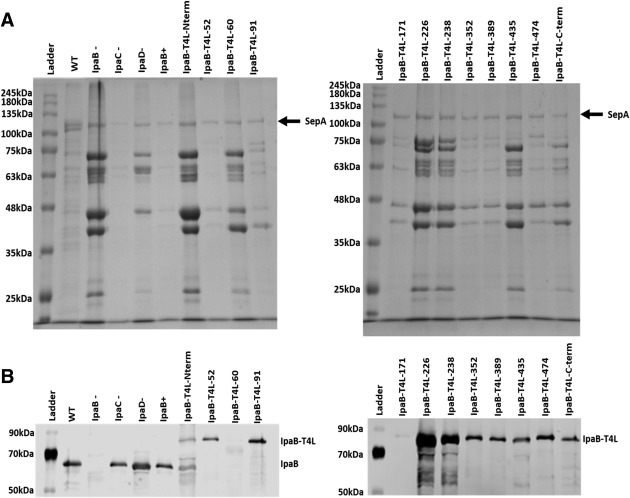
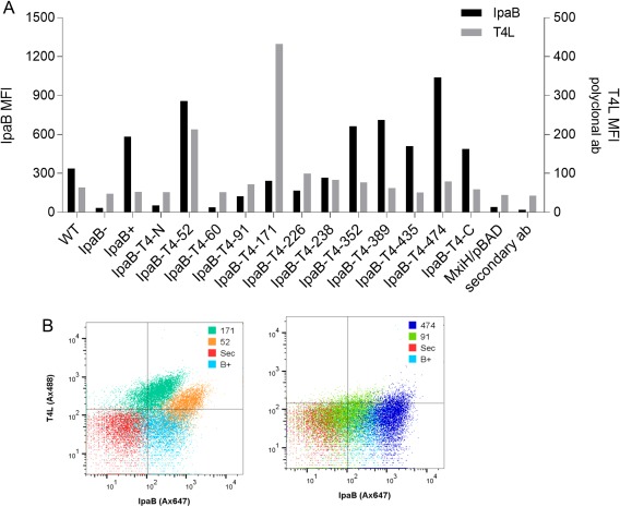
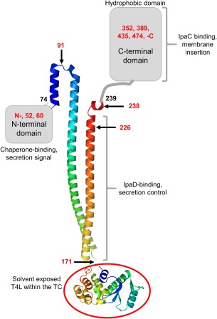
Bacterial type III secretion systems (T3SS) are used to inject proteins into mammalian cells to subvert cellular functions. The Shigella T3SS apparatus (T3SA) is comprised of a basal body, cytoplasmic sorting platform and exposed needle with needle "tip complex" (TC). TC maturation occurs when the translocator protein IpaB is recruited to the needle tip where both IpaD and IpaB control secretion induction. IpaB insertion into the host membrane is the first step of translocon pore formation and secretion induction. We employed disruptive insertional mutagenesis, using bacteriophage T4 lysozyme (T4L), within predicted IpaB loops to show how topological features affect TC functions (secretion control, translocon formation and effector secretion). Insertions within the N-terminal half of IpaB were most likely to result in a loss of steady-state secretion control, however, all but the two that were not recognized by the T3SA retained nearly wild-type hemolysis (translocon formation) and invasiveness levels (effector secretion). In contrast, all but one insertion in the C-terminal half of IpaB maintained secretion control but were impaired for hemolysis and invasion. These nature of the data suggest the latter mutants are defective in a post-secretion event, most likely due to impaired interactions with the second translocator protein IpaC. Intriguingly, only two insertion mutants displayed readily detectable T4L on the bacterial surface. The data create a picture in which the makeup and structure of a functional T3SA TC is highly amenable to physical perturbation, indicating that the tertiary structure of IpaB within the TC is more plastic than previously realized.

Keywords: IpaB; Shigella; translocon; type III secretion.


Figures

Figure 1
Figure 1
Generation of IpaB‐T4 lysozyme (T4L) insertion mutants. (A) The primary sequence of IpaB from S. flexneri is shown with the following bioinformatics predictions: secondary structure (α‐helix, red text; random coil, black text; coiled‐coil, bold), regions of disorder (underlined), and transmembrane helices (green highlight). The IpaB (residues 74–224) crystal structure is represented with solid green bars above the amino acid numbering. Locations of T4L insertions are depicted with blue squares (pegs), more information can be found in Table SI. The figure was generated using XtalPred.33 (B) The crystal structure of IpaB (residues 74–224) from S. flexneri (PDB ID: 3U0C) is shown with colors from blue (N‐terminus) to red (C‐terminus). The location of T4L insertions 91, 171, and 226 are indicated in the context of the structure.
Figure 2
Figure 2
Secretion by Shigella producing the IpaB‐T4L insertion mutants. (A) Overnight protein secretion was analyzed by SDS‐PAGE (10% polyacrylamide gel) with Coomassie blue staining after precipitation of culture supernatants. (B) Immunoblot of IpaB and the IpaB‐T4L insertion mutants present in the overnight culture supernatants (same samples as used in “A”) after probing with anti‐IpaB rabbit antibodies. Molecular mass markers (labeled Ladder) are indicated in each panel and the position of Shigella SepA (not secreted by the T3SA) is shown by an arrow in “A.”
Figure 3
Figure 3
The ability for IpaB‐T4L insertion mutants to restore virulence to S. flexneri SF620. (A) Contact‐mediated hemolysis activity was tested in sheep red blood cells for 30 min at 37°C. (B) Invasion of HeLa cells after a 1 hr incubation with the different IpaB‐expressing Shigella strains. The values shown in each panel are relative to S. flexneri SF620 expressing wild‐type IpaB (set at 100%).
Figure 4
Figure 4
Ability of IpaB‐T4L insertion mutants to release SRB from liposomes. (A) Release of SRB from liposomes results in relief of auto‐quenching and is measured as an increase in SRB fluorescence emission. Addition of IpaB (prepared in OPOE) results in a time‐dependent release of SRB. Addition of Triton X‐100 results in 100% liposome lysis. (B) Percentage of SRB release relative to total dye release was calculated and is shown as a relative percent of release caused by wild‐type IpaB. All samples contained PBS + 0.5% (v/v) OPOE with the exception of IpgC/IpaB, which was in PBS lacking detergent. OPOE alone caused no release of SRB (column 2).
Figure 5
Figure 5
Mean fluorescence intensity (MFI) of specific markers detected by flow cytometry at the surface of IpaB‐T4L insertion mutant strains grown to mid‐log phase. (A) MFI of IpaB (black bars) and T4L (grey bars) were detected using polyclonal antibodies previously generated against the purified recombinant proteins. Additional negative controls include a mxiH null mutant and a sample lacking the secondary antibody. (B) Flow cytometry analysis for IpaB and T4L labeling on the Shigella surface. Sec represents bacteria for which the secondary antibody was not added (staining negative control).
Figure 6
Figure 6
Crystal structure of IpaB74–242 at 2.10 Å. The crystal structure of S. flexneri IpaB (residues 74–239) is shown in cartoon ribbon format, colored blue (N‐terminus) to red (C‐terminus). Two copies of each polypeptide are found within the asymmetric unit with a single copy shown for clarity. The inset shows the representative model‐to‐map correlation for residues 225–239. The 2Fo – Fc weighted electron density map (contoured at 2.0 σ) is drawn as a blue cage.
Figure 7
Figure 7
Summary of the functional landscape of IpaB based on T4L insertion mutagenesis. The positions of T4L insertion sites are indicated by red numbers and key functions associated with each regions are described. Structures have not been determined for the N‐terminal domain (residues 1–73) or the C‐terminal domain which possesses a major hydrophobic component (∼residues 300–420) which contains two predicted transmembrane helices [see Fig. 1(A)]. At the bottom, the structure of T4L is shown (red circle) as it is proposed to occur for insertion at IpaB position 171. T4L is recognized by T4L anti‐sera on the surface of Shigella when inserted at positions 52 and 171 (see Fig. 5).

References

    1. Kotloff KL, Winickoff JP, Ivanoff B, Clemens JD, Swerdlow DL, Sansonetti PJ, Adak GK, Levine MM (1999) Global burden of Shigella infections: implications for vaccine development and implementation of control strategies. Bull World Health Organ 77:651–666. - PMC - PubMed
    1. Bardhan P, Faruque AS, Naheed A, Sack DA (2010) Decrease in shigellosis‐related deaths without Shigella spp.‐specific interventions, Asia. Emerg Infect Dis 16:1718–1723. - PMC - PubMed
    1. Kotloff KL, Nataro JP, Blackwelder WC, Nasrin D, Farag TH, Panchalingam S, Wu Y, Sow SO, Sur D, Breiman RF, Faruque ASG, Zaidi AKM, Saha D, Alonso PL, Tamboura B, Sanogo D, Onwuchekwa U, Manna B, Ramamurthy T, Kanungo S, Ochieng JB, Omore R, Oundo JO, Hossain A, Das SK, Ahmed S, Qureshi S, Quadri F, Adegbola RA, Antonio M, Hossain MJ, Akinsola A, Mandomando I, Nhampossa T, Acacio S, Biswas K, O'Reilly CE, Mintz ED, Berkeley LY, Muhsen K, Sommerfelt H, Robins‐Browne RM, Levine MM (2013) Burden and aetiology of diarrhoeal disease in infants and young children in developing countries (the Global Enteric Multicenter Study, GEMS): a prospective, case‐control study. Lancet 382:209–222. - PubMed
    1. DuPont HL, Levine MM, Hornick RB, Formal SB (1989) Inoculum size in shigellosis and implications for expected mode of transmission. J Infect Dis 159:1126–1128. - PubMed
    1. Killackey SA, Sorbara MT, Girardin SE (2016) Cellular aspects of Shigella pathogenesis: Focus on the manipulation of host cell processes. Front Cell Infect Microbiol 6:38. - PMC - PubMed

Publication types

LinkOut - more resources