. 2018 Apr 17;19(4):1215.
doi: 10.3390/ijms19041215.

Mechanisms of Diabetes-Induced Endothelial Cell Senescence: Role of Arginase 1

Affiliations

Mechanisms of Diabetes-Induced Endothelial Cell Senescence: Role of Arginase 1

Esraa Shosha et al. Int J Mol Sci. .

Abstract

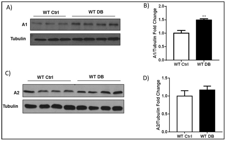
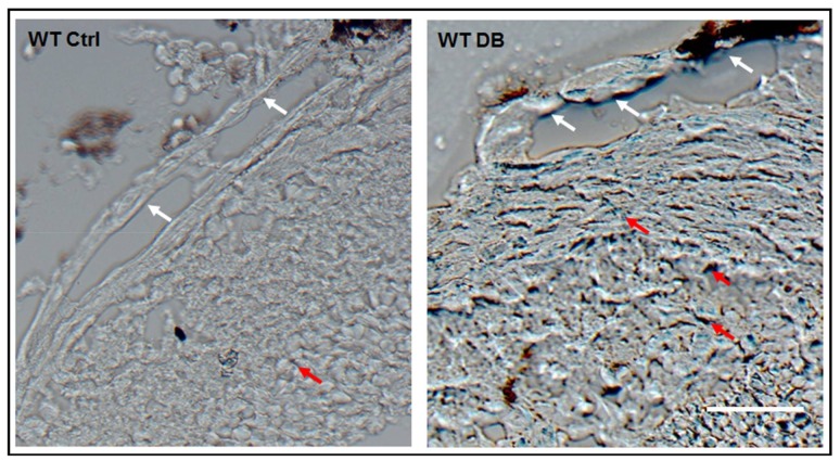
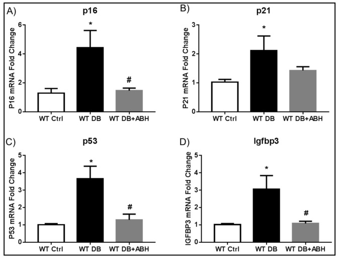
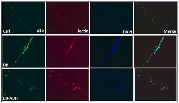
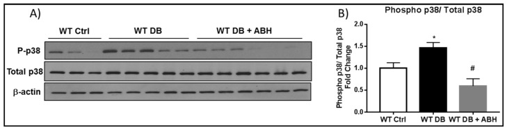
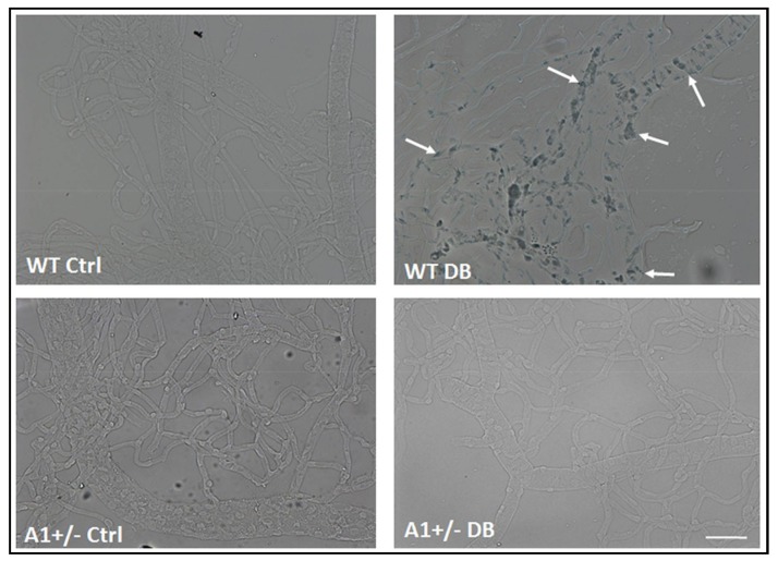
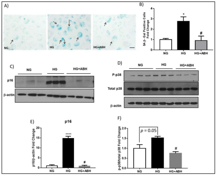
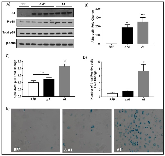
We have recently found that diabetes-induced premature senescence of retinal endothelial cells is accompanied by NOX2-NADPH oxidase-induced increases in the ureohydrolase enzyme arginase 1 (A1). Here, we used genetic strategies to determine the specific involvement of A1 in diabetes-induced endothelial cell senescence. We used A1 knockout mice and wild type mice that were rendered diabetic with streptozotocin and retinal endothelial cells (ECs) exposed to high glucose or transduced with adenovirus to overexpress A1 for these experiments. ABH [2(S)-Amino-6-boronohexanoic acid] was used to inhibit arginase activity. We used Western blotting, immunolabeling, quantitative PCR, and senescence associated β-galactosidase (SA β-Gal) activity to evaluate senescence. Analyses of retinal tissue extracts from diabetic mice showed significant increases in mRNA expression of the senescence-related proteins p16INK4a, p21, and p53 when compared with non-diabetic mice. SA β-Gal activity and p16INK4a immunoreactivity were also increased in retinal vessels from diabetic mice. A1 gene deletion or pharmacological inhibition protected against the induction of premature senescence. A1 overexpression or high glucose treatment increased SA β-Gal activity in cultured ECs. These results demonstrate that A1 is critically involved in diabetes-induced senescence of retinal ECs. Inhibition of arginase activity may therefore be an effective therapeutic strategy to alleviate diabetic retinopathy by preventing premature senescence.

Keywords: arginase; diabetes mellitus; diabetic retinopathy; endothelial cells; hyperglycemia; retina; senescence.


Conflict of interest statement

The authors declare no conflict of interest.

Figures

Figure 1
Figure 1
Diabetes induces increases in arginase 1 (A1) expression. (A) Western blot analysis and quantification (B) showing increased A1 expression in wild type (WT) diabetic retinas. ** p < 0.01 vs. WT Ctrl. n = 3–4; (C) Western blot analysis and quantification (D) show similar levels of A2 expression in WT diabetic mice and age matched controls. p = 0.4, n = 4.
Figure 2
Figure 2
Diabetes induces senescence in retinal tissue. Senescence associated β-galactosidase (SA β-Gal) activity images of frozen retinal sections showing positive signal in cells of the large vessels (white arrows) as well as in the surrounding tissues (red arrows) of the central retina. Scale bar = 20 µM.
Figure 3
Figure 3
Arginase inhibition prevents diabetes-induced alterations in retinal senescence markers. qRT-PCR showing increased mRNA levels of p16 (A); p21 (B) p53 (C); and, Igfbp3 (D) in the diabetic retinas compared to age matched controls. 2(S)-Amino-6-boronohexanoic acid (ABH) treatment significantly blocked the diabetes-induced senescence. * p < 0.05 vs. WT Ctrl, # p < 0.05 vs. WT DB. n = 4–9.
Figure 4
Figure 4
Arginase inhibition reduces p16 expression in isolated vessels. Immuno-labeling of isolated retinal vessels showing expression of the senescence protein p16 (green) in vascular endothelial cells which was co-localized with the endothelial marker (isolectin B4, red). Diabetic vessels showed increased p16 expression compared to controls. Arginase inhibition with ABH significantly reduced p16 expression. n = 3–4. Scale bar = 50 µM.
Figure 5
Figure 5
Diabetes induced- increases in phospho p38 is prevented by inhibiting arginase. (A) Western blot analysis and quantification (B) showing increased phosphorylation of p38 in WT diabetic retinal extracts compared to age matched controls. ABH treatment significantly blocked this increase. * p < 0.05 vs. WT Ctrl. # p < 0.001 vs. WT DB. n = 6–8.
Figure 6
Figure 6
A1 deletion prevents diabetes-induced senescence in retinal vessels. SA β-Gal activity images of isolated retinal vessels showing increased activity (arrows) in vessels isolated from WT diabetic mice. The vessels isolated from A1+/− diabetic retinas are negative for SA β-Gal staining. The non-diabetic control retinas from both WT and A1+/− mice are also negative for SA β-Gal staining. n = 3–5. Scale bar = 50 µM.
Figure 7
Figure 7
High glucose-induces endothelial cell senescence. (A) Senescence associated β-Gal activity (black arrows) and quantification (B) showing a significant increase in SA β-Gal positive cells with high glucose (HG, 25 mM) treatment when compared to normal glucose (NG, 5 mM). ABH treatment prevented this increase. * p < 0.05 vs. NG. # p < 0.05 vs. HG. n = 3. Scale bar = 50 µM; (C) Western blot analysis with quantification (E) showing increased p16INK4A expression in HG treated bovine retinal endothelial cells (BRECs) as compared to NG. Arginase inhibition with ABH blocked this increase. **** p < 0.0001 vs. NG. # p < 0.0001 vs. HG. n = 3. Experiments were repeated at least twice in independent batches of cells. Data are presented as a fold change of the NG; and, (D) Western blot analysis with quantification (F) showing increased phosphorylation of p38 in HG treated BRECs as compared to NG. Arginase inhibition with ABH significantly blocked this increase. # p < 0.05 vs. HG. n = 3. Experiments were repeated at least twice in independent batches of cells. Data are presented as a fold change of the NG.
Figure 8
Figure 8
Arginase 1 (A1) overexpression induces endothelial cell senescence. (A) Western blot analysis with quantification (B) showing levels of A1 protein or (C) phosphorylation of the stress marker p38 in bovine retinal endothelial cells (BRECs) transduced with 20 MOI of wild type A1 (A1), inactive mutant A1 (Δ A1), or red fluorescence protein (RFP) adenovirus vectors. The inactive mutant A1 was generated by mutation of aspartic acid 128 to glycine. (B) ** p < 0.01 vs. RFP. *** p < 0.001 vs. RFP. n = 6–8. (C) n.s., non-significant, ** p < 0.01 vs. RFP & Δ A1. n = 3–8. Experiments were repeated at least twice in independent batches of cells. Data are presented as a fold change of RFP; (E) Senescence associated β-galactosidase (SA β-Gal) activity with quantification (D) showing a significant increase in SA β-Gal positive cells with A1 overexpression as compared to RFP or the inactive mutant A1 (Δ A1). * p < 0.05 vs. RFP & Δ A1. n = 5–10. Experiments were repeated at least twice in independent batches of cells. Data are presented as a fold change of RFP. Scale bar = 50 µM.

References

    1. Oubaha M., Miloudi K., Dejda A., Guber V., Mawambo G., Germain M.A., Bourdel G., Popovic N., Rezende F.A., Kaufman R.J., et al. Senescence-associated secretory phenotype contributes to pathological angiogenesis in retinopathy. Sci. Transl. Med. 2016;8:362ra144. doi: 10.1126/scitranslmed.aaf9440. - DOI - PubMed
    1. Rojas M., Lemtalsi T., Toque H.A., Xu Z., Fulton D., Caldwell R.W., Caldwell R.B. NOX2-Induced Activation of Arginase and Diabetes-Induced Retinal Endothelial Cell Senescence. Antioxidants. 2017;6:43. doi: 10.3390/antiox6020043. - DOI - PMC - PubMed
    1. Lamoke F., Shaw S., Yuan J., Ananth S., Duncan M., Martin P., Bartoli M. Increased Oxidative and Nitrative Stress Accelerates Aging of the Retinal Vasculature in the Diabetic Retina. PLoS ONE. 2015;10:e0139664. doi: 10.1371/journal.pone.0139664. - DOI - PMC - PubMed
    1. Patel C., Rojas M., Narayanan S.P., Zhang W., Xu Z., Lemtalsi T., Jittiporn K., Caldwell R.W., Caldwell R.B. Arginase as a mediator of diabetic retinopathy. Front. Immunol. 2013;4:173. doi: 10.3389/fimmu.2013.00173. - DOI - PMC - PubMed
    1. Elms S.C., Toque H.A., Rojas M., Xu Z., Caldwell R.W., Caldwell R.B. The role of arginase I in diabetes-induced retinal vascular dysfunction in mouse and rat models of diabetes. Diabetologia. 2013;56:654–662. doi: 10.1007/s00125-012-2789-5. - DOI - PMC - PubMed

MeSH terms