

The risk of violating the posterior malleolar fracture when nailing the ipsilateral concomitant spiral distal tibial fracture
- PMID: 29673344
- PMCID: PMC5907748
- DOI: 10.1186/s12891-018-1994-x
The risk of violating the posterior malleolar fracture when nailing the ipsilateral concomitant spiral distal tibial fracture
Abstract
Background: For a distal tibial spiral fracture combined with a non-displaced posterior malleolar fragment (PMF), we proposed a hypothesis that the treating surgeon could assess the size of the PMF to determine the need for stabilizing that structure first before rodding the tibia.
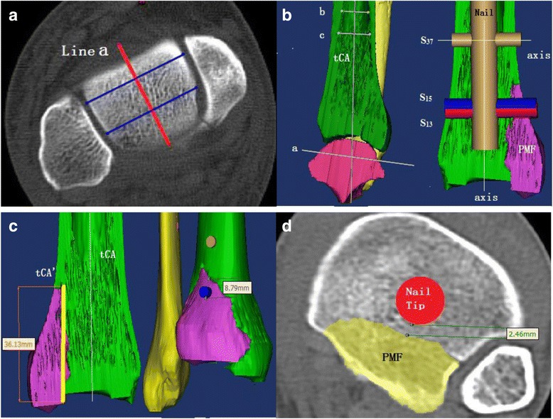
Materials and methods: Fifty 3-D models (22 females) of combined distal tibial and posterior malleolar fractures from one trauma center were reconstructed. In each case, a virtual tibial intramedullary nail (vIM nail) with three distal anteroposterior (AP) locking screws (S13, S15 and S37, the number indicating the distance from the screw to the nail tip) were inserted into the center of the tibial canal and ended on top of the distal tibial physeal scar. Contact between the screws and the PMF was defined as causing PMF displacement. The relationship between PMF secondary displacement and traumatic anatomic factors (the fragment area and height of the PMF) was explored. Then, the parameters were justified by analyzing intraoperative radiographs of 35 cases treated by nail with single locking screw (S15) design.
Results: In the analog experiment, multiple logistic regression analysis revealed that the height of the PMF could confidently predict the risk of fragment displacement (S13: odds ratio [OR] 1.18, 95% confidence interval [CI] 1.06-1.32; S15: OR 1.15, 95% CI 1.05-1.27). Regarding the height of the PMF, the receiver operating characteristic established a cut-off value of 31.2 mm for preliminary fixation of the fragment with 88.89% sensitivity and 88.89% specificity. In the operation group the nail stopped on the top of distal tibial physeal scar, no PMF secondary displacement occurred when the PMF height was less than 31.2 mm. However, the incidence of secondary displacement was 93.33% when the height of the PMF exceeded 31.2 mm.
Conclusion: When the distal tibial physeal scare was set as the limit of nail insertion depth, the height of the PMF could be used as a reliable reference predicting the risk of PMF secondary displacement caused by distal anteroposterior locking screw.
Keywords: Ankle fracture; Intramedullary nail; Morphological measurement; Tibial fracture.
Conflict of interest statement
Ethics approval and consent to participate
Approved by the Institutional Review Board (IRB)/Independent Ethics Committee (IEC) of Jiangsu Province Hospital (The First Affiliated Hospital of Nanjing Medical University) at 300 Guangzhou Road, Nanjing 210,029, China. All subjects provided informed consent in written to take part in the study.
Consent for publication
Not applicable.
Competing interests
The authors declare that they have no competing interests.
Publisher’s Note
Springer Nature remains neutral with regard to jurisdictional claims in published maps and institutional affiliations.
Figures

Similar articles
-
One quick and simple fixation method: posterior malleolus fractures in spiral tibial fractures.BMC Musculoskelet Disord. 2023 Mar 30;24(1):244. doi: 10.1186/s12891-023-06319-8. BMC Musculoskelet Disord. 2023. PMID: 36997965 Free PMC article.
-
Posterior Malleolar Fractures Associated With Tibial Shaft Fractures and Sequence of Fixation.J Orthop Trauma. 2016 Oct;30(10):568-71. doi: 10.1097/BOT.0000000000000629. J Orthop Trauma. 2016. PMID: 27164492
-
Concomitant Ankle Injuries Associated With Tibial Shaft Fractures.Foot Ankle Int. 2015 Oct;36(10):1209-14. doi: 10.1177/1071100715588381. Epub 2015 Jun 3. Foot Ankle Int. 2015. PMID: 26041543
-
[Tibial fracture with intact fibula treated by reamed nailing].Rev Chir Orthop Reparatrice Appar Mot. 2000 Feb;86(1):29-37. Rev Chir Orthop Reparatrice Appar Mot. 2000. PMID: 10669822 Review. French.
-
Intramedullary Tibial Nail Fixation of Simple Intraarticular Distal Tibia Fractures.J Orthop Trauma. 2016 Nov;30 Suppl 4:S12-S16. doi: 10.1097/BOT.0000000000000697. J Orthop Trauma. 2016. PMID: 27768627 Review.
Cited by
-
Incidence and missed diagnosis risk of occult posterior malleolar fractures associated with the tibial shaft fractures: a systematic review.J Orthop Surg Res. 2021 Jun 1;16(1):355. doi: 10.1186/s13018-021-02502-6. J Orthop Surg Res. 2021. PMID: 34074309 Free PMC article.
-
Classifications of posterior malleolar fractures: a systematic literature review.Arch Orthop Trauma Surg. 2023 Jul;143(7):4181-4220. doi: 10.1007/s00402-022-04643-7. Epub 2022 Dec 5. Arch Orthop Trauma Surg. 2023. PMID: 36469121 Free PMC article.
References
-
- Lauge-Hansen . Injuries caused by abnormal movements of the foot. Oxford: Nord. Med; 1946.
-
- Van der Werken C, Zeegers EV. Fracture of the lower leg with involvement of the posterior malleolus; a neglected combination? Injury. 1988;19:241–243. - PubMed
-
- Weber, BG: Die Verletzungen des oberen Sprunggelenkes. 1sted. Bern: Hans Huber; 1966.
-
- Böstman OM. Displaced malleolar fractures associated with spiral fractures of the tibial shaft. Clin Orthop Relat Res. 1988;(228):202–7. - PubMed
-
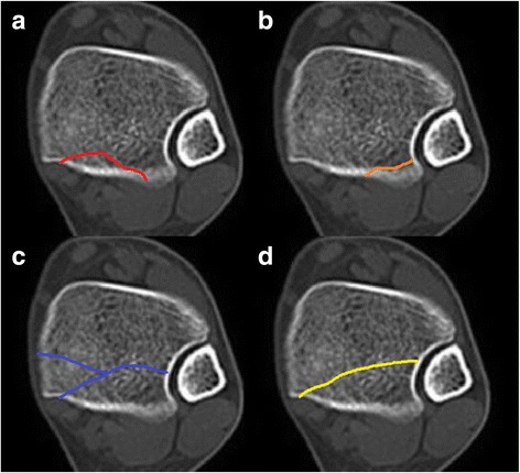
- Hou Z, Zhang L, Zhang Q, Yao S, Pan J, Irgit K, Zhang Y. The "communication line" suggests occult posterior malleolar fracture associated with a spiral tibial shaft fracture. Eur J Radiol. 2012;81:594–597. - PubMed
MeSH terms
LinkOut - more resources
Full Text Sources
Other Literature Sources
Medical
Miscellaneous
