

A multifunctional targeting probe with dual-mode imaging and photothermal therapy used in vivo
- PMID: 29673352
- PMCID: PMC5907178
- DOI: 10.1186/s12951-018-0367-9
A multifunctional targeting probe with dual-mode imaging and photothermal therapy used in vivo
Abstract
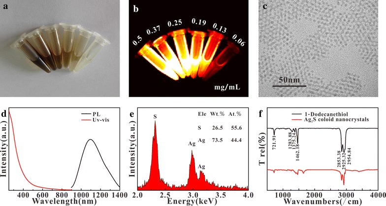
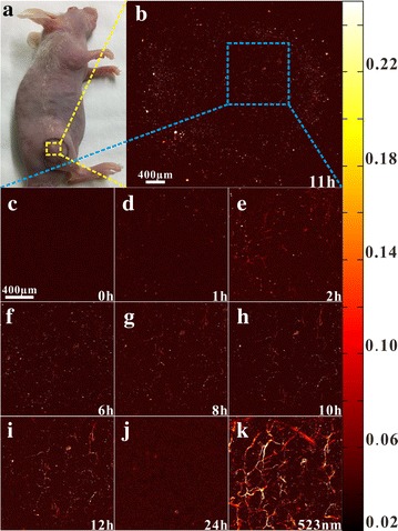
Background: Ag2S has the characteristics of conventional quantum dot such as broad excitation spectrum, narrow emission spectrum, long fluorescence lifetime, strong anti-bleaching ability, and other optical properties. Moreover, since its fluorescence emission is located in the NIR-II region, has stronger penetrating ability for tissue. Ag2S quantum dot has strong absorption during the visible and NIR regions, it has good photothermal and photoacoustic response under certain wavelength excitation.
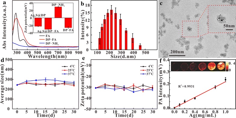
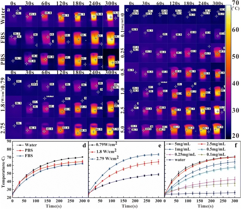
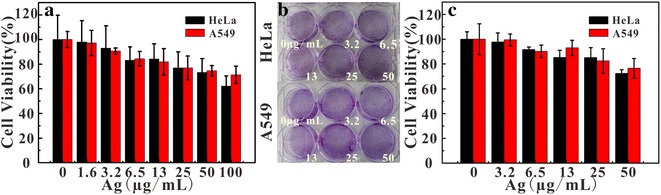
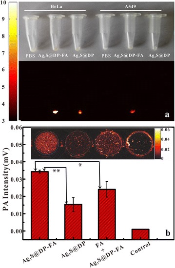
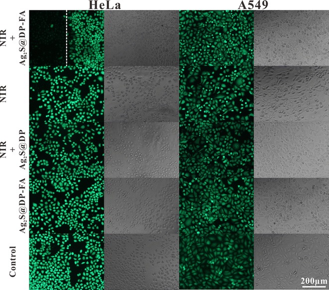
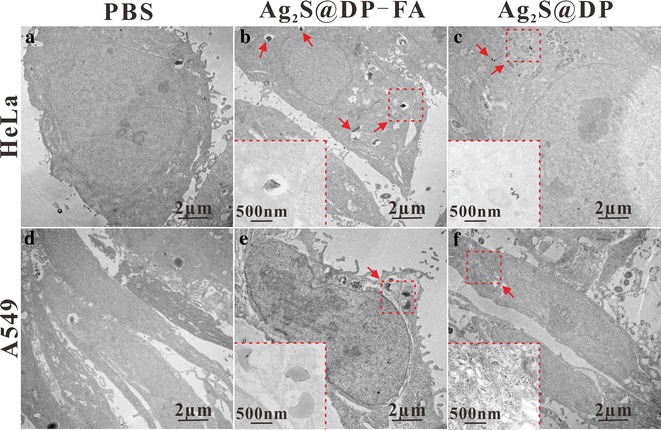
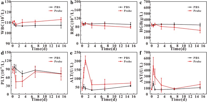
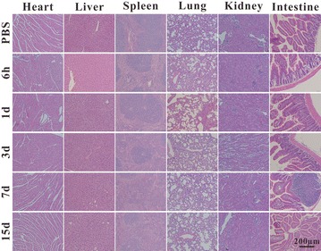
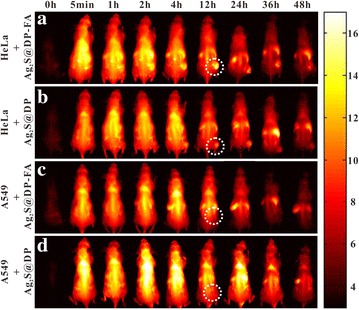
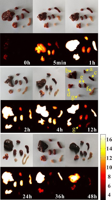
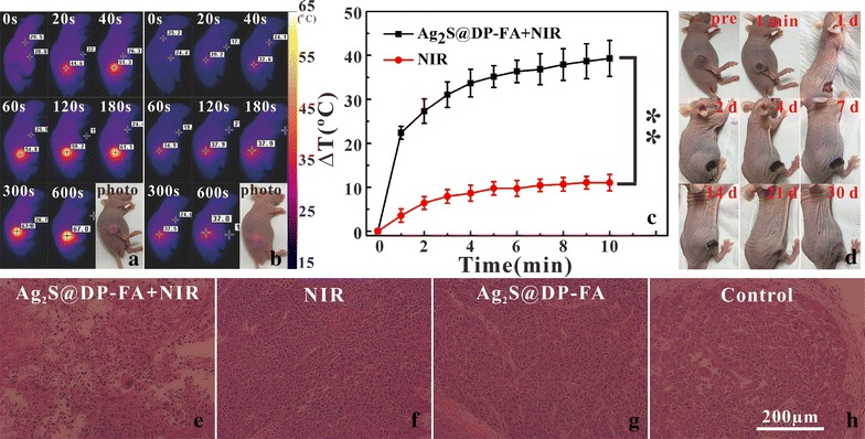
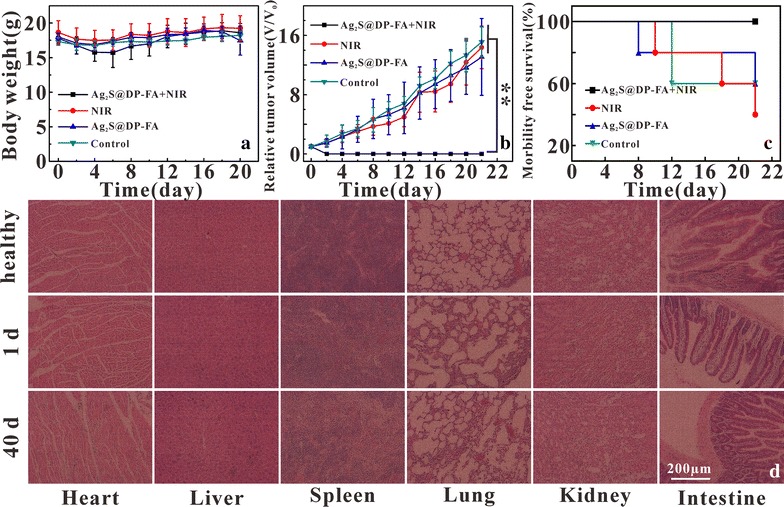
Results: 200 nm aqueous probe Ag2S@DSPE-PEG2000-FA (Ag2S@DP-FA) with good dispersibility and stability was prepared by coating hydrophobic Ag2S with the mixture of folic acid (FA) modified DSPE-PEG2000 (DP) and other polymers, it was found the probe had good fluorescent, photoacoustic and photothermal responses, and a low cell cytotoxicity at 50 μg/mL Ag concentration. Blood biochemical analysis, liver enzyme and tissue histopathological test showed that no significant influence was observed on blood and organs within 15 days after injection of the probe. In vivo and in vitro fluorescence and photoacoustic imaging of the probe further demonstrated that the Ag2S@DP-FA probe had good active targeting ability for tumor. In vivo and in vitro photothermal therapy experiments confirmed that the probe also had good ability of killing tumor by photothermal.
Conclusions: Ag2S@DP-FA was a safe, integrated diagnosis and treatment probe with multi-mode imaging, photothermal therapy and active targeting ability, which had a great application prospect in the early diagnosis and treatment of tumor.
Keywords: Ag2S; Cancer active targeting; Fluorescence imaging; Photoacoustic imaging; Photothermal therapy.
Figures
Similar articles
-
In situ aqueous synthesis of genetically engineered polypeptide-capped Ag2S quantum dots for second near-infrared fluorescence/photoacoustic imaging and photothermal therapy.J Mater Chem B. 2019 Apr 21;7(15):2484-2492. doi: 10.1039/c8tb03043j. Epub 2019 Mar 15. J Mater Chem B. 2019. PMID: 32255125
-
Folic acid modified Pluronic F127 coating Ag2S quantum dot for photoacoustic imaging of tumor cell-targeting.Nanotechnology. 2018 Feb 2;29(5):055101. doi: 10.1088/1361-6528/aa9acc. Nanotechnology. 2018. PMID: 29139396
-
A self-assembled nanoplatform based on Ag2S quantum dots and tellurium nanorods for combined chemo-photothermal therapy guided by H2O2-activated near-infrared-II fluorescence imaging.Acta Biomater. 2022 Mar 1;140:547-560. doi: 10.1016/j.actbio.2021.12.013. Epub 2021 Dec 16. Acta Biomater. 2022. PMID: 34923095
-
Nanoparticle-based Cell Trackers for Biomedical Applications.Theranostics. 2020 Jan 12;10(4):1923-1947. doi: 10.7150/thno.39915. eCollection 2020. Theranostics. 2020. PMID: 32042345 Free PMC article. Review.
-
Activated molecular probes for enzyme recognition and detection.Theranostics. 2022 Jan 1;12(3):1459-1485. doi: 10.7150/thno.66676. eCollection 2022. Theranostics. 2022. PMID: 35154500 Free PMC article. Review.
Cited by
-
Metal-Polymer Nanoconjugates Application in Cancer Imaging and Therapy.Nanomaterials (Basel). 2022 Sep 13;12(18):3166. doi: 10.3390/nano12183166. Nanomaterials (Basel). 2022. PMID: 36144953 Free PMC article. Review.
-
Synthesis and Applications of Carboxymethyl Cellulose Hydrogels.Gels. 2022 Aug 24;8(9):529. doi: 10.3390/gels8090529. Gels. 2022. PMID: 36135241 Free PMC article. Review.
-
Pretheranostic agents with extraordinaryNIRF/photoacoustic imaging performanceand photothermal oncotherapy efficacy.Acta Pharm Sin B. 2024 Dec;14(12):5370-5381. doi: 10.1016/j.apsb.2024.07.017. Epub 2024 Jul 24. Acta Pharm Sin B. 2024. PMID: 39807319 Free PMC article.
-
Fluorescence Guidance in Surgical Oncology: Challenges, Opportunities, and Translation.Mol Imaging Biol. 2019 Apr;21(2):200-218. doi: 10.1007/s11307-018-1239-2. Mol Imaging Biol. 2019. PMID: 29942988 Free PMC article. Review.
-
Renal Clearable Ru-based Coordination Polymer Nanodots for Photoacoustic Imaging Guided Cancer Therapy.Theranostics. 2019 Oct 21;9(26):8266-8276. doi: 10.7150/thno.36986. eCollection 2019. Theranostics. 2019. PMID: 31754395 Free PMC article.
References
-
- Plante M, Touhami O, Trinh XB, Renaud MC, Sebastianelli A, Grondin K, Gregoire J. Sentinel node mapping with indocyanine green and endoscopic near-infrared fluorescence imaging in endometrial cancer. A pilot study and review of the literature. Gynecol Oncol. 2015;137:443–447. doi: 10.1016/j.ygyno.2015.03.004. - DOI - PubMed
-
- Tao Z, Dang X, Huang X, Muzumdar MD, Xu ES, Bardhan NM, Song H, Qi R, Yu Y, Li T, Wei W, Wyckoff J, Birrer MJ, Belcher AM, Ghoroghchian PP. Early tumor detection afforded by in vivo imaging of near-infrared II fluorescence. Biomaterials. 2017;34:202–215. doi: 10.1016/j.biomaterials.2017.04.046. - DOI - PubMed
-
- Bashkatov AN, Genina EA, Kochubey VI, Tuchin VV. Optical properties of human skin, subcutaneous and mucous tissues in the wavelength range from 400 to 2000 nm. J Phys D Appl Phys. 2005;38:2543–2555. doi: 10.1088/0022-3727/38/15/004. - DOI
MeSH terms
Substances
Grants and funding
LinkOut - more resources
Full Text Sources
Other Literature Sources
Miscellaneous
