. 2018 Apr 19;15(1):113.
doi: 10.1186/s12974-018-1149-x.

Chorioamnionitis, neuroinflammation, and injury: timing is key in the preterm ovine fetus

Affiliations

Chorioamnionitis, neuroinflammation, and injury: timing is key in the preterm ovine fetus

Ruth Gussenhoven et al. J Neuroinflammation. .

Abstract

Background: Antenatal infection (i.e., chorioamnionitis) is an important risk factor for adverse neurodevelopmental outcomes after preterm birth. Destructive and developmental disturbances of the white matter are hallmarks of preterm brain injury. Understanding the temporal effects of antenatal infection in relation to the onset of neurological injury is crucial for the development of neurotherapeutics for preterm infants. However, these dynamics remain unstudied.

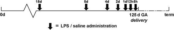
Methods: Time-mated ewes were intra-amniotically injected with lipopolysaccharide at 5, 12, or 24 h or 2, 4, 8, or 15 days before preterm delivery at 125 days gestational age (term ~ 150 days). Post mortem analyses for peripheral immune activation, neuroinflammation, and white matter/neuronal injury were performed. Moreover, considering the neuroprotective potential of erythropoietin (EPO) for perinatal brain injury, we evaluated (phosphorylated) EPO receptor (pEPOR) expression in the fetal brain following LPS exposure.

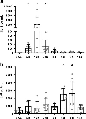
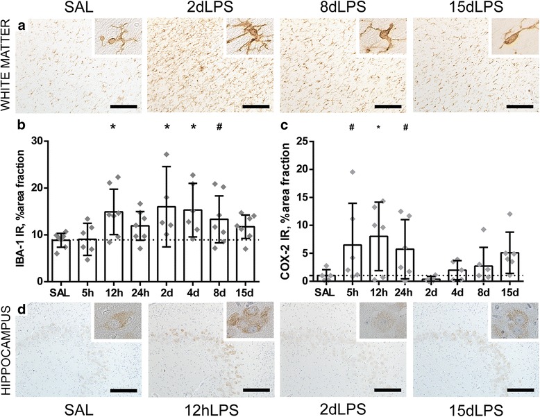
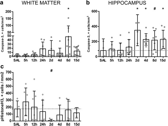
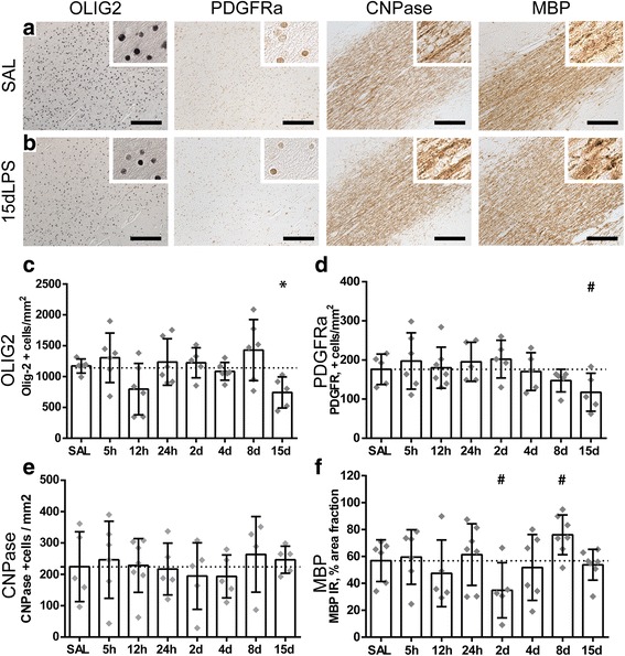
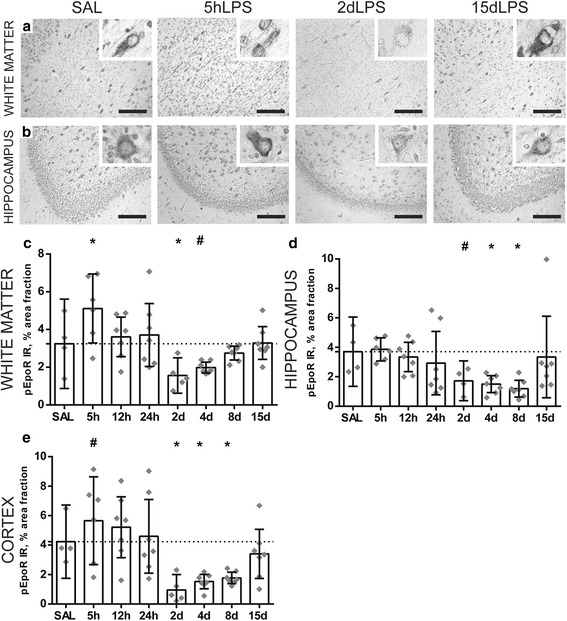
Results: Intra-amniotic exposure to this single bolus of LPS resulted in a biphasic systemic IL-6 and IL-8 response. In the developing brain, intra-amniotic LPS exposure induces a persistent microgliosis (IBA-1 immunoreactivity) but a shorter-lived increase in the pro-inflammatory marker COX-2. Cell death (caspase-3 immunoreactivity) was only observed when LPS exposure was greater than 8 days in the white matter, and there was a reduction in the number of (pre) oligodendrocytes (Olig2- and PDGFRα-positive cells) within the white matter at 15 days post LPS exposure only. pEPOR expression displayed a striking biphasic regulation following LPS exposure which may help explain contradicting results among clinical trials that tested EPO for the prevention of preterm brain injury.

Conclusion: We provide increased understanding of the spatiotemporal pathophysiological changes in the preterm brain following intra-amniotic inflammation which may aid development of new interventions or implement interventions more effectively to prevent perinatal brain damage.

Keywords: Brain injury; Chorioamnionitis; EPO receptor; Erythropoietin; Fetal; Inflammation; Preterm; Sheep.


Conflict of interest statement

Ethics approval

Animal procedures were performed with approval of the animal ethics committee of the University of Western Australia (Perth, Australia).

Competing interests

The authors declare that they have no competing interests.

Publisher’s Note

Springer Nature remains neutral with regard to jurisdictional claims in published maps and institutional affiliations.

Figures

Fig. 1
Fig. 1
Study design. Pregnant ewes received an intra-amniotic injection with 10 mg Escherichia coli-derived lipopolysaccharide (LPS) at 5, 12, or 24 h or 2, 4, 8, or 15 days (black arrows) before preterm delivery at 122 days of gestation (term ~ 150 days). Control animals received an intra-amniotic injection with an equivalent volume of 0.9% saline solution at comparable time points to LPS injections
Fig. 2
Fig. 2
ab Circulatory interleukin (IL)-6 and IL-8 concentrations illustrate a biphasic response following intra-amniotic LPS exposure. Undetectable values were assigned an arbitrary value of 1 pg/mL in order to perform statistical analysis. Statistical analysis was done with ANOVA, and values are expressed as mean ± 95% CI. Asterisk indicated p < 0.05 versus control group; number sign indicated 0.05 < p < 0.1 versus control
Fig. 3
Fig. 3
Intra-amniotic exposure to LPS induces an acute, transient cerebral inflammatory response in the preterm white matter and hippocampus. An increase of the area fraction of IBA-1 immunoreactivity (IR) was observed in the white matter at 12 h, 2 days, 4 days, and 8 days following LPS exposure compared to controls (SAL vs. 12 h LPS p = 0.012; SAL vs. 2 days LPS p = 0.006; SAL vs. 4 days LPS p = 0.005; SAL vs. 8 days LPS p = 0.088) (a, b). In the hippocampus, an increase of the area fraction of COX-2 IR was found at 5, 12, and 24 h following LPS exposure (SAL vs. 5 h LPS p = 0.055; SAL vs. 12 h LPS p = 0.016; SAL vs. 24 h LPS p = 0.096) (c, d). Representative histological figures of the IBA-1-positive microglia in animals exposed to intra-amniotic saline (SAL), 2, 8, and 15 days of LPS are shown in a. Representative histological figures of COX-2-positive neurons in the hippocampus of animals exposed to saline (SAL), 12 h, 2 days, and 15 days LPS are depicted in (d). IBA-1 IR and COX-2 IR are depicted as mean % area fraction ± 95% CI. Asterisk indicated p < 0.05 versus control (SAL); number sign indicated 0.05 < p < 0.1 versus control (SAL). Images taken at × 100 magnification (insert at × 400 magnification), scale bar = 200 μm
Fig. 4
Fig. 4
Intra-amniotic exposure to LPS results in a decrease in mitotic cells and relatively late onset of cell death in the preterm white matter and hippocampus. A significant increase of caspase-3-positive cells is observed at 8 days following LPS exposure in the white matter compared to controls (SAL vs 8 days LPS p = 0.004) (a). In the hippocampus, at 2, 4, 8, and 15 days following LPS exposure an increase in caspase-3-positive cells was found compared to controls (SAL vs 2 days LPS p = 0.002; SAL vs 4 days LPS p = 0.030; SAL vs 8 days LPS p = 0.058; SAL vs 15 days LPS p = 0.042) (b). At 2 days following LPS exposure, a decrease in pHH3+ cells was found compared to controls (SAL vs. 2 days LPS p = 0.100) (c). Caspase-3 and pHH3 are expressed as positive cells/mm2 and represented in the graphs as mean ± 95% CI. Asterisk indicated p < 0.05 versus control (SAL); number sign indicated 0.05 < p < 0.1 versus control (SAL)
Fig. 5
Fig. 5
Intra-amniotic exposure to LPS induces distinct time-dependent changes in oligodendrocyte lineage cells. A significant decrease of Olig2-positive cells was observed in animals after 15 days of LPS exposure compared to controls (SAL vs. 15 days LPS p = 0.050) (c). At the same time point, a decrease in PDGFRa-positive cells was found compared to controls (SAL vs. 15 days LPS p = 0.070) (d). No significant changes of CNPase+ cells were found following LPS exposure compared to controls (e). Area fractions (%) of MBP immunoreactivity (IR) showed a decrease at 2 days and an increase at 8 days following LPS exposure compared to controls (SAL vs. 2 days LPS, p = 0.070; SAL vs. 8 days LPS p = 0.083) (f). Representative histological figures of Olig2, PDGFRa, and CNPase-positive cells and MBP IR in animals exposed to intra-amniotic saline (SAL) and 15 days of LPS are shown in a and b respectively. Images taken at × 100 magnification (insert at × 400 magnification), scale bar = 200 μm. Asterisk indicated p < 0.05 versus control (SAL); number sign indicated 0.05 < p < 0.1 versus control (SAL)
Fig. 6
Fig. 6
Intra-amniotic exposure to LPS results in altered dendritic development in the gray matter of the fetal brain. A significant increase of the area fraction (%) of MAP-2 immunoreactivity (IR) was found in the cerebral cortex at 24 h after LPS exposure compared to controls (SAL vs 24 h LPS p = 0.036) (a, c). In the hippocampus, an increase of area fraction (%) of MAP-2 IR was observed at 5, 12, and 24 h and at 4, 8, and 15 days after LPS exposure compared to controls (SAL vs 5 h LPS p = 0.007; SAL vs 12 h LPS p = 0.073; SAL vs 24 h LPS p = 0.000; SAL vs 4 days LPS p = 0.034; SAL vs 8 days LPS p = 0.000; SAL vs 15 days LPS p = 0.057) (b, d). Representative histological figures of MAP-2 in the cerebral cortex (a) and hippocampus (b) are depicted in control animals (SAL) and animals exposed to LPS for 24 h, 2 days, and 8 days. Images taken at × 100 magnification (insert at × 400 magnification), scale bar = 200 μm. Asterisk indicated p < 0.05 versus control (SAL); number sign indicated 0.05 < p < 0.1 versus control (SAL)
Fig. 7
Fig. 7
Expression of the phosphorylated erythropoietin receptor decreases 2 days following LPS exposure. An acute increase of the area fraction (%) of pEPOR immunoreactivity (IR) was observed at 5 h after LPS exposure in the white matter (SAL vs 5 h p = 0.010) and cortex (SAL vs 5 h p = 0.100) compared to controls (c, e). At 2 days after LPS exposure, there is a significant decrease in pEPOR IR within all brain regions compared to controls: white matter (SAL vs 2 days LPS p = 0.030) (c), hippocampus (SAL vs 2 days LPS p = 0.088) (d), and cortex (SAL vs 2 days LPS p = 0.010) (e). At 4 and 8 days following LPS exposure, pEPOR expression is still decreased compared to controls in the white matter (SAL vs 4 days LPS p = 0.100), hippocampus (SAL vs 4 days LPS 0.045; SAL vs 8 days LPS p = 0.014), and cortex (SAL vs 4 days LPS 0.020.; SAL vs 8 days LPS p = 0.030). When the fetus had been exposed to 15 days of LPS, there was no decrease in pEPOR IR (ce). Representative histological figures of the pEPOR in the white matter (a) and hippocampus (b) are depicted in control animals (SAL) and animals exposed to LPS for 5 h, 2 days, and 15 days. Images taken at × 100 magnification (insert at × 400 magnification), scale bar = 200 μm. Asterisk indicated p < 0.05 versus controls (SAL), number sign indicated 0.05 < p < 0.1 versus controls (SAL)

References

    1. Rovira N, Alarcon A, Iriondo M, Ibanez M, Poo P, Cusi V, et al. Impact of histological chorioamnionitis, funisitis and clinical chorioamnionitis on neurodevelopmental outcome of preterm infants. Early Hum Dev. 2011;87(4):253–257. doi: 10.1016/j.earlhumdev.2011.01.024. - DOI - PubMed
    1. Paton MCB, McDonald CA, Allison BJ, Fahey MC, Jenkin G, Miller SL. Perinatal brain injury as a consequence of preterm birth and intrauterine inflammation: designing targeted stem cell therapies. Front Neurosci. 2017;11:200. doi: 10.3389/fnins.2017.00200. - DOI - PMC - PubMed
    1. Goncalves LF, Chaiworapongsa T, Romero R. Intrauterine infection and prematurity. Ment Retard Dev Disabil Res Rev. 2002;8(1):3–13. doi: 10.1002/mrdd.10008. - DOI - PubMed
    1. Kuypers E, Wolfs TG, Collins JJ, Jellema RK, Newnham JP, Kemp MW, et al. Intraamniotic lipopolysaccharide exposure changes cell populations and structure of the ovine fetal thymus. Reprod Sci. 2013;20(8):946–956. doi: 10.1177/1933719112472742. - DOI - PMC - PubMed
    1. Lai JCY, Rocha-Ferreira E, Ek CJ, Wang X, Hagberg H, Mallard C. Immune responses in perinatal brain injury. Brain Behav Immun. 2017;63:210–223. doi: 10.1016/j.bbi.2016.10.022. - DOI - PubMed

Substances