. 2018:603:129-150.
doi: 10.1016/bs.mie.2018.02.007. Epub 2018 Mar 15.

Giant Plasma Membrane Vesicles: An Experimental Tool for Probing the Effects of Drugs and Other Conditions on Membrane Domain Stability

Affiliations

Giant Plasma Membrane Vesicles: An Experimental Tool for Probing the Effects of Drugs and Other Conditions on Membrane Domain Stability

Zoe Gerstle et al. Methods Enzymol. 2018.

Abstract

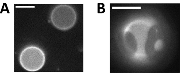
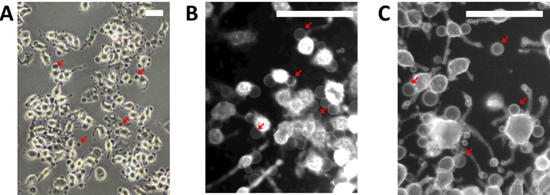
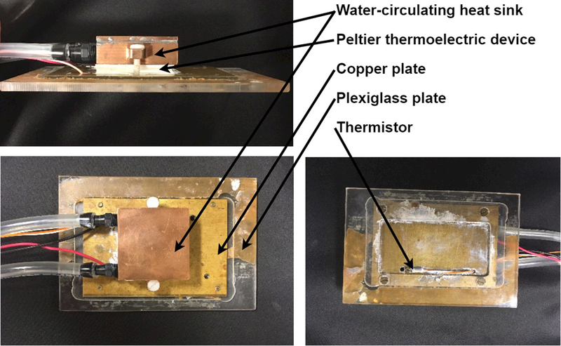
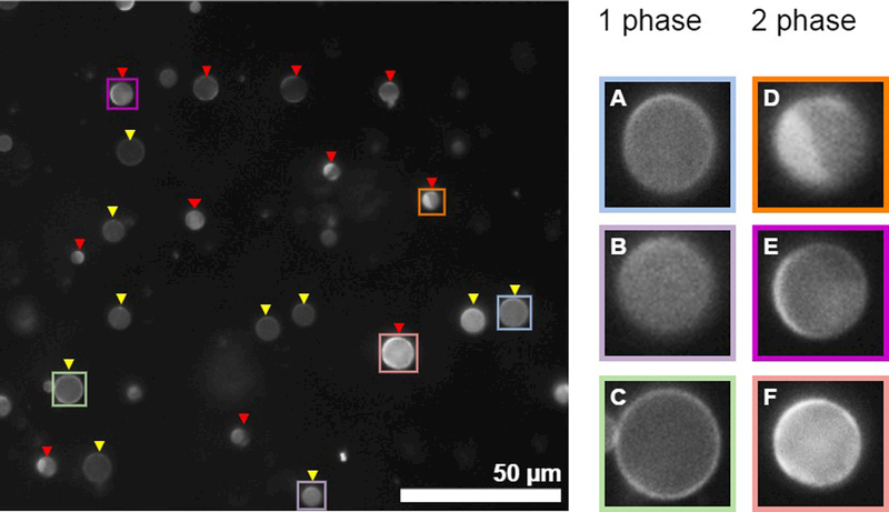
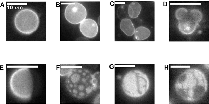
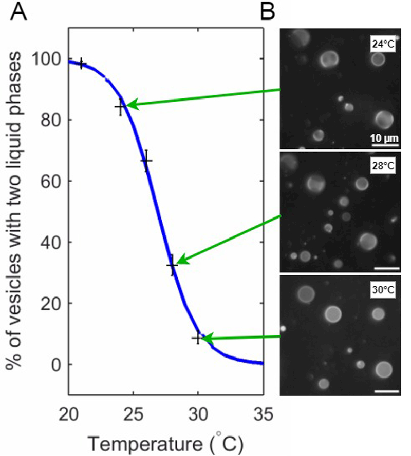
Giant plasma membrane vesicles (GPMVs) are isolated directly from living cells and provide an alternative to vesicles constructed of synthetic or purified lipids as an experimental model system for use in a wide range of assays. GPMVs capture much of the compositional protein and lipid complexity of intact cell plasma membranes, are filled with cytoplasm, and are free from contamination with membranes from internal organelles. GPMVs often exhibit a miscibility transition below the growth temperature of their parent cells. GPMVs labeled with a fluorescent protein or lipid analog appear uniform on the micron-scale when imaged above the miscibility transition temperature, and separate into coexisting liquid domains with differing membrane compositions and physical properties below this temperature. The presence of this miscibility transition in isolated GPMVs suggests that a similar phase-like heterogeneity occurs in intact plasma membranes under growth conditions, albeit on smaller length scales. In this context, GPMVs provide a simple and controlled experimental system to explore how drugs and other environmental conditions alter the composition and stability of phase-like domains in intact cell membranes. This chapter describes methods to generate and isolate GPMVs from adherent mammalian cells and to interrogate their miscibility transition temperatures using fluorescence microscopy.

Keywords: Lipid raft; Liquid-disordered; Liquid-ordered; Miscibility; Phase transition; Plasma membrane; Vesicle.


Figures

Figure 1:
Figure 1:. Giant plasma membrane vesicles (GPMVs) can contain two coexisting liquid phases.
(A) GPMVs imaged above their miscibility transition temperature (Tmix) are in a single liquid phase, as indicated by the uniform distribution of a fluorescent lipid analog across the vesicle surface. (B) GPMVs separate into coexisting liquid-ordered (Lo) and liquid-disordered (Ld) phases at a temperature below Tmix. The fluorescent dye added to the GPMVs incorporates into the Ld phase, facilitating easy distinction between the phases. Scale bars are 10μm.
Figure 2:
Figure 2:. Phase contrast (A) and fluorescent (B,C) images of cell attached GPMVs.
(A) GPMVs appear dark in phase contrast images and can be seen attached to cells or flowing above the adherent cell layer (not shown). (B) Cell attached GPMVs are difficult to visualize when cells membranes are fluorescently tagged prior to incubation with active vesiculation buffer due to trafficking of fluorophores to internal membranes. (C) GPMVs and their parent cell membranes have roughly the same fluorescent intensity when cells are labeled after incubation with active vesiculation buffer. Red arrows point to GPMVs in all panels. Scale bars are 100μm.
Figure 3:
Figure 3:. Temperature can be controlled using a simple, home-built temperature stage.
Our temperature stage consists of a water-circulating heat sink and peltier thermoelectric device (Custom Thermoelectric, Bishopville, MD) and a PID-type controller unit (Oven Industries, Mechanicsburg, PA). Water is circulated through the heat sink using a garden fountain pump (available at hardware stores) submerged in a bucket. Temperature is measured with a thermistor probe calibrated to work with the controller mounted on the copper plate close to the sample. The sample, placed between two coverslips, is adhered to the copper plate near the thermistor and the assembly is placed, sample down, on a standard Olympus inverted microscope stage and is fastened with clips to prevent drift. We have also constructed stages where the plexiglass thermal insulator was replaced by rubber or cork simply adhered to a metal plate.
Figure 4:
Figure 4:. assigning phase state of individual vesicles within a population imaged at fixed temperature.
(Left) An example image showing a field of GPMVs in which most vesicles can be unambiguously assigned to contain either 1 or liquid 2 phases. Yellow triangles point towards 1 phase vesicles while red triangles point towards 2 phase vesicles. (right) isolated view of selected vesicles shown at the Left. GPMVs shown in A-C contain a single phase. A and B show GPMVs where the image is focused on a vesicle surface, and the brightness of the fluorophore is uniform across this surface. The GPMV in C is in focus along the midplane of the vesicle, and the brightness of fluorophore is uniform around the circumference. GPMVs shown in D and E contain coexisting liquid phases. The vesicle in D where the image is focused on the vesicle surface and a clear phase boundary is observed. The vesicle in E is focused close to the midplane, and the fluorophore intensity varies along the perimeter. F is difficult to assign. However, both the surface and the circumference vary in brightness so it was classified as containing two phases. In this example, some GPMVs in the field were not labeled because they are too small or too out of focus. In general, it is most important to remain consistent regarding assignments and the characteristics of vesicles that the investigator chooses not to assign. One reason to image at least 100 vesicles is to reduce the possible contributions of user bias in assigning vesicles.
Figure 5:
Figure 5:. Images of vesicles highlighting the range of possible shapes and domain morphologies.
(A) Spherical vesicle with a single liquid phase. (B) Two single phase vesicles that each contain internal smaller vesicles (bright spots) . (C) Non-spherical vesicles imaged above Tmix. Vesicle edges undulate when vesicles are imaged in time (not shown). (D) Vesicle with bulging liquid-disordered domains. (E) Spherical vesicle exhibiting normal separation of 2 liquid phases where domains have fully coarsened through coalescence. (F) Liquid domains dispersed on the vesicle surface. In some cases domains do not coalesce, or do not coalesce on the time-scale of the measurement. (G) Vesicle that contains a gel phase domain. (H) Apparent coexistence of three phases within a single vesicle. Both gel and liquid-disordered phases exclude the fluorescent probe. The vesicle in D was imaged in the presence of detergent which increased membrane permeability, enabling the dramatic shape observed. Vesicles in G and H are GPMVs isolated from cells incubated with methyl beta cyclodextrin to reduce their cholesterol content.
Figure 6:
Figure 6:. Measurement of phase transition temperatures from GPMV images.
(A) Plot summarizing the fraction of GPMVs assigned to contain two liquid phases as a function of temperature (black crosses). The blue curve is a fit to these points by the sigmoid function stated in the main text. For this example, the best fit value for the average transition temperature Tmix is 26.9°C. (B) A small subset of the GPMV images that were assigned and used to obtain the data points shown in part A.

Similar articles

Cited by

References

    1. Allen JA, Halverson-Tamboli RA and Rasenick MM (2007). “Lipid raft microdomains and neurotransmitter signalling.” Nature Reviews Neuroscience 8(2): 128–140. - PubMed
    1. Allender DW and Schick M (2017). “The Effect of Solutes on the Temperature of Miscibility Transitions in Multi-component Membranes.” Biophysical Journal: in perss. - PMC - PubMed
    1. Baumgart T, Hammond AT, Sengupta P, Hess ST, Holowka DA, Baird BA and Webb WW (2007). “Large-scale fluid/fluid phase separation of proteins and lipids in giant plasma membrane vesicles.” Proc Natl Acad Sci U S A 104(9): 3165–3170. - PMC - PubMed
    1. Burns M, Wisser K, Wu J, Levental I and Veatch SL (2017). “Miscibility Transition Temperature Scales with Growth Temperature in a Zebrafish Cell Line.” Biophys J. - PMC - PubMed
    1. Cornell CE, McCarthy NLC, Levental KR, Levental I, Brooks NJ and Keller SL (2017). “n-Alcohol Length Governs Shift in Lo-Ld Mixing Temperatures in Synthetic and Cell-Derived Membranes.” Biophys J. - PMC - PubMed

Publication types

MeSH terms