. 2018 Apr 19;9(1):1556.
doi: 10.1038/s41467-018-03991-6.

Pathogen-derived extracellular vesicles mediate virulence in the fatal human pathogen Cryptococcus gattii

Affiliations

Pathogen-derived extracellular vesicles mediate virulence in the fatal human pathogen Cryptococcus gattii

Ewa Bielska et al. Nat Commun. .

Abstract

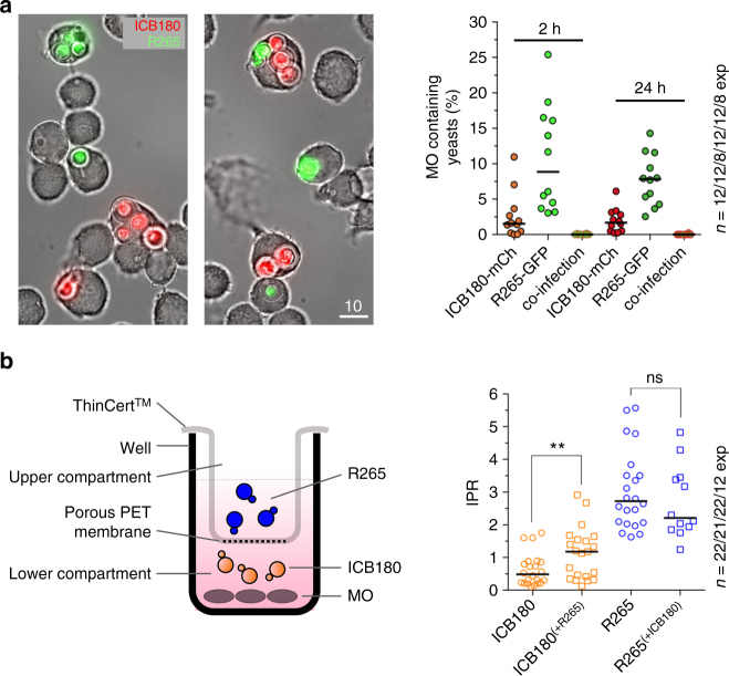
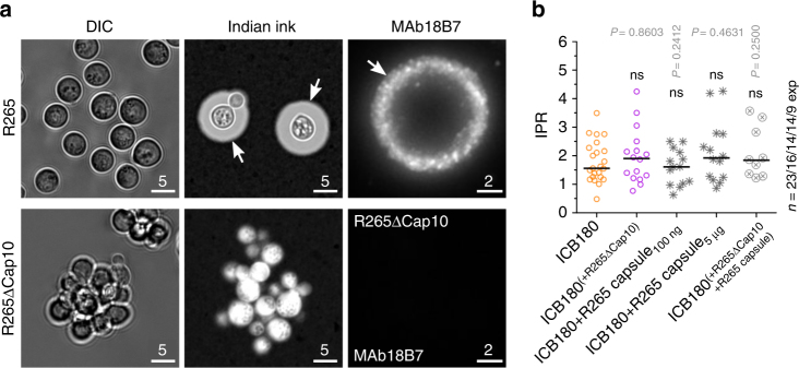
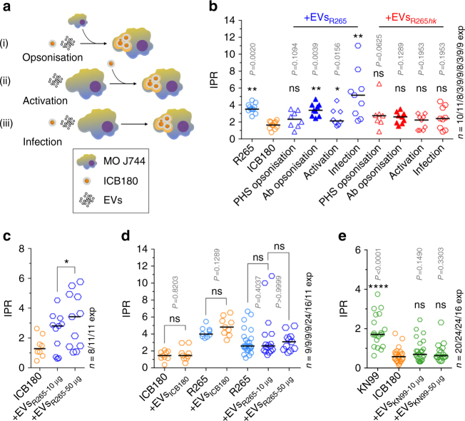
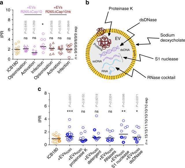
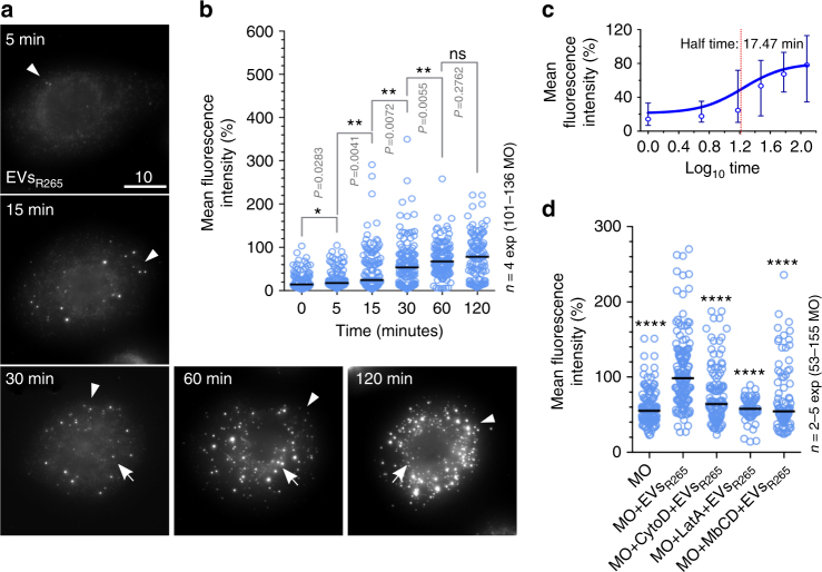
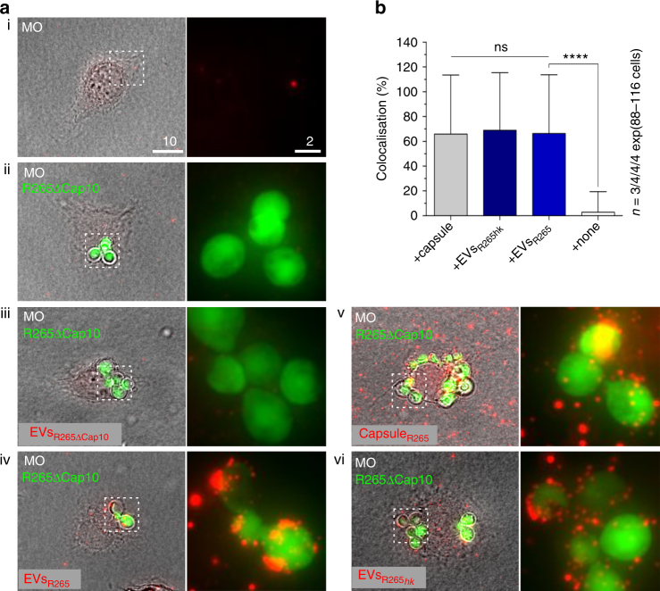
The Pacific Northwest outbreak of cryptococcosis, caused by a near-clonal lineage of the fungal pathogen Cryptococcus gattii, represents the most significant cluster of life-threatening fungal infections in otherwise healthy human hosts currently known. The outbreak lineage has a remarkable ability to grow rapidly within human white blood cells, using a unique 'division of labour' mechanism within the pathogen population, where some cells adopt a dormant behaviour to support the growth of neighbouring cells. Here we demonstrate that pathogenic 'division of labour' can be triggered over large cellular distances and is mediated through the release of extracellular vesicles by the fungus. Isolated vesicles released by virulent strains are taken up by infected host macrophages and trafficked to the phagosome, where they trigger the rapid intracellular growth of non-outbreak fungal cells that would otherwise be eliminated by the host. Thus, long distance pathogen-to-pathogen communication via extracellular vesicles represents a novel mechanism to control complex virulence phenotypes in Cryptococcus gattii and, potentially, other infectious species.


Conflict of interest statement

The authors declare no competing interests.

Figures

Fig. 1
Fig. 1
Long-distance communication can drive rapid intracellular proliferation in C. gattii. a During co-infection, different strains of C. gattii (R265-GFP shown in green; ICB180-mCherry shown in red) are rarely phagocytosed by the same macrophage. Bar: 10 μm. The number of infected macrophages containing both isolates of yeast at the same time is very low at 2 h.p.i. (2 in total 5479 tested macrophages) and at 24 h.p.i (1 in total 3235 tested macrophages). Data are presented as scattered dot plots with lines representing their medians. Data are representative of results from 8−12 independent experiments with a minimum of 150 macrophages analysed per sample per experiment. b A schematic representation of the experiment using transwell system ThinCertTM with 400 nm porous membrane that separates lower from upper compartments thereby allowing splitting of growth of two different C. gattii strains R265 (pathogenic) and ICB180 (non-pathogenic). After two initial hours of the infection the transwell system was removed and intracellular proliferation rate (IPR) of ICB180 was measured (as T0) and after following 24 h (as T24). The 2-h presence of R265 (outbreak) cryptococci in the transwell system (ICB180(+R265)) induces significantly higher intracellular proliferation of ICB180 (non-outbreak strain) within macrophages (P = 0.0038, Wilcoxon matched-pairs signed rank test), an effect that is not seen when R265 is replaced for ICB180 (R265(+ICB180); P = 0.9263, Wilcoxon matched-pairs signed rank test). Data are presented as scattered dot plots with lines representing their medians. Data are representative of results from 12–22 independent experiments with 879–5238 total yeasts counted for each sample. Wilcoxon matched-pairs signed rank test where ** (P ≤ 0.01), significant difference; ns (P > 0.05), not different
Fig. 2
Fig. 2
Capsular material is necessary but not sufficient to increase the IPR of non-outbreak cryptococci. a Differential interference contrast (DIC; left), India Ink (middle) and immunostained (MAb 18B7; right) images of wild-type R265 (top) and acapsular R265ΔCap10 (bottom), indicating the presence of the characteristic thick polysaccharide capsule in the wild-type (arrows). Note the lack of any visible capsule in the acapsular mutant. Bars: 5 and 2 μm. b IPR of a non-virulent ICB180 strain is not altered by the presence of acapsular R265ΔCap10 in the transwell assay (ICB180(+R265ΔCap10)), nor by the addition of capsular material isolated from R265 (ICB180+R265 capsule100 ng; ICB180+R265 capsule5 μg). An addition into the transwell of acapsular R265ΔCap10 strain mixed with the capsular material isolated from R265 also did not change the IPR of ICB180 (ICB180(+R265ΔCap10+R265 capsule)). Data are presented as scattered dot plots with lines representing their medians. Individual Wilcoxon matched-pairs signed rank test presented as P values above each dot plot, where ns (P > 0.05), not significantly different. Data are representative of results from at least nine independent experiments with 808–1913 total number of yeasts counted for each sample
Fig. 3
Fig. 3
EVs increase survival of cryptococci inside macrophages. a A schematic representation of the experimental assay in which extracellular vesicles (EVs) were added at different time points, either (i) to the cryptococci during opsonisation, 1 h prior to infection (‘Opsonisation’), (ii) directly to the macrophages J774 (MO J774) during 1 h of activation prior to infection (‘Activation’) or (iii) at the same time as the cryptococci are added to the macrophages (‘Infection’). b IPRs of R265 growing alone (R265), ICB180 growing alone (ICB180) and in the presence of 10 μg of EVs isolated from R265 cells (EVsR265) or heat-inactivated EVsR265 (EVsR265hk) added at different stages of infection, as described above: during yeast opsonisation by pooled human serum (‘PHS opsonisation’) or by GXM-specific antibodies—Mab 18B7 (‘Ab opsonisation’), J774 activation (‘Activation’) or during incubation with both macrophages and ICB180 yeast cells (‘Infection’). Data are presented as scattered dot plots with lines representing their medians. Data are representative of results from 3 (with biological triplicates) to 11 independent experiments with 213–1767 total number of yeasts counted for each sample. Wilcoxon paired test where * (P ≤ 0.05), significant difference; ** (P ≤ 0.01; and P = 0.0078 for infection with EVsR265), highly significant difference and ns (P > 0.05), not significantly different. c IPR values can be further increased after adding higher amounts of EVs (+EVsR265-50 μg). Data are presented as scattered dot plots with lines representing their medians. Data are representative of results from 8 to 11 independent experiments with 1504–1948 total number of yeasts counted for each sample. Wilcoxon paired test where * (P = 0.0186), significant difference. d EVs isolated from ICB180 (+EVsICB180) do not increase IPRs of ICB180 or R265 and proliferation of R265 is not enhanced by its own EVs (+EVsR265) even at higher concentration (+EVsR265-50 μg). Data are presented as scattered dot plots with lines representing their medians. Data are representative of results from 9 to 24 independent experiments with 275–7888 total number of yeasts counted for each sample. Wilcoxon paired test where ns (P > 0.05), not significantly different. e IPR of a non-virulent ICB180 strain is not altered by the presence of EVs isolated from C. neoformans virulent strain KN99 even at higher concentration of those vesicles (+EVsKN99-50 μg) added during the infection step. Data are presented as scattered dot plots with lines representing their medians. Individual Wilcoxon matched-pairs signed rank test presented as P values above each dot plot, where ns (P > 0.05), not significantly different. Data are representative of results from at least 16 independent experiments with 1742–5209 total number of yeasts counted for each sample
Fig. 4
Fig. 4
EV proteins and RNA are necessary to increase survival of cryptococci inside macrophages. a IPRs of ICB180 growing alone (ICB180) and in the presence of 10 μg of EVs isolated from acapsular strain R265ΔCap10 (EVsR265ΔCap10) or heat-inactivated EVsR265ΔCap10 (EVsR265ΔCap10hk) added at different stages of infection: during yeast opsonisation using PHS (opsonisation), J774 activation (activation) or during incubation with both macrophages and ICB180 yeast cells (infection; see also Fig. 3a). Data are presented as scattered dot plots with lines representing their medians. Data are representative of results from 8 to 9 independent experiments with 147–301 total number of yeasts counted for each sample. Wilcoxon paired test where * (P = 0.0117), significant difference; and ns (P > 0.05), not significantly different. b Schematic drawing of the EV and treatments performed towards protein degradation via proteinase K, lipids degradation via sodium deoxycholate, double-stranded DNA (dsDNA) degradation via dsDNase, single-stranded DNA (ssDNA) and single-stranded regions of RNA degradation via S1 nuclease and further RNA degradation, including RNA duplexes, via RNase cocktail of RNase A and T1. c IPR values of ICB180 are increased in the presence of 10 μg of EVs (or 50 μg—symbols with thicker borders) isolated from R265 (+EVsR265), EVsR265 treated with S1 nuclease (+EVsR265 S1 nuclease) and EVsR265 treated with dsDNase (+EVsR265 dsDNase) but not when EVs treated with proteinase K (+EVsR265 proteinase K), sodium deoxycholate (+EVsR265 detergent) or RNase cocktail (+EVsR265 RNases) were used. Data are representative of results from 10 to 15 independent experiments with 1181–2691 total number of yeasts counted for each sample. Wilcoxon paired test where * (P ≤ 0.05), significant difference; ** (P ≤ 0.01), significant difference, *** (P ≤ 0.001), significant difference and ns (P > 0.05), not significantly different
Fig. 5
Fig. 5
EVs are rapidly and actively internalised by macrophages. a Macrophages were exposed to 10 μg of EVsR265, fixed with 4% PFA after 5, 15, 30, 60 and 120 min incubation and then immunostained using capsule-specific monoclonal antibody MAb 18B7. Images are maximum projections (21 z-stacks with 0.5 μm intervals). Note the change in signal from the cell periphery (arrowheads) to the cell body (arrows) in a time-dependent manner. Bar: 10 μm. b EV uptake increases in a time-dependent manner. Data are presented as scattered dot plots with lines representing their medians. Mean fluorescent intensities were normalised using the median value at 2 h as 100%. Data are representative of results from four independent experiments with a minimum of 25 macrophages analysed per sample per experiment. Unpaired Mann−Whitney tests where * (P ≤ 0.05), significant difference; ** (P ≤ 0.01), significant difference; and ns (P > 0.05), not significantly different. c Half time determination of EVs uptake by J774 macrophages. Data are presented as a sigmoidal non-linear regression curve (four-parameter logistic curve) with medians and errors (interquartile range) obtained from four independent experiments (from Fig. 5b). d The uptake of EVs by J774 macrophages is blocked by actin polymerisation inhibitors, cytochalasin D (MO+CytoD+EVsR265) and latrunculin A (MO+LatA+EVsR265) or a lipid raft-specific inhibitor methyl-β-cyclodextrin (MO+MbCD+EVsR265), which depletes cholesterol. Data are presented as scattered dot plots with lines representing their medians. Graph showing percentages of mean fluorescent intensities from macrophages alone (MO) or incubated with MAb 18B7-immunostained EVs isolated from R265 (MO+EVsR265) and were normalised to a median value obtained for MO+EVsR265. Data are representative of results from 2 to 5 independent experiments with a minimum of 25 macrophages analysed per sample per experiment. Unpaired Mann−Whitney tests where **** (P ≤ 0.0001), highly significant difference
Fig. 6
Fig. 6
EVs added to macrophages infected with acapsular R265-GFP rapidly colocalise with the cryptococcal phagosome. a Macrophages alone (MO; i), macrophages with phagocytosed R265ΔCap10-GFP cells (ii) or macrophages containing R265ΔCap10-GFP and co-incubated with EVs isolated from R265ΔCap10 (EVsR265ΔCap10; (iii)) are not recognised by capsule-specific MAb 18B7. Those macrophages co-incubated with EVs isolated from R265 (EVsR265; (iv)), with capsule isolated from R265 (CapsuleR265; (v)) or heat-inactivated EVs isolated from R265 (EVsR265hk; (vi)) show colocalisation between R265ΔCap10-GFP cells and red signal suggesting that the EVs and capsule polysaccharides were delivered to the phagosomes. Pictures represent maximum intensity projections of 21 z-stacks obtained from 10 μm cross section through macrophages. Bars: 10 and 2 μm. b Graph showing percentage of colocalised signals of GFP labelled yeast cells R265ΔCap10 engulfed by murine macrophages and MAb 18B7-immunostained capsule (+capsule), heat-inactivated EVs isolated from R265 (EVsR265hk) or EVs isolated from R265 (+EVsR265). As a negative control intensities from macrophages infected with R265ΔCap10-GFP without EVs were used (+none). All numbers are given as means ± standard deviation (s.d.) and are representative of results from 3 to 4 independent experiments with a minimum of 25 macrophages analysed per sample per experiment. ns indicate statistically non-significant difference (Kruskal−Wallis test, P = 0.89) and quadruple asterisks indicate a highly significant difference (****, unpaired Mann−Whitney test, P 0.0001)

References

    1. Kim JY. Human fungal pathogens: why should we learn? J. Microbiol. 2016;54:145–148. doi: 10.1007/s12275-016-0647-8. - DOI - PubMed
    1. Park BJ, et al. Estimation of the current global burden of cryptococcal meningitis among persons living with HIV/AIDS. AIDS. 2009;23:525–530. doi: 10.1097/QAD.0b013e328322ffac. - DOI - PubMed
    1. Perfect JR, Bicanic T. Cryptococcosis diagnosis and treatment: what do we know now. Fungal Genet. Biol. 2015;78:49–54. doi: 10.1016/j.fgb.2014.10.003. - DOI - PMC - PubMed
    1. MacDougall L, et al. Spread of Cryptococcus gattii in British Columbia, Canada, and detection in the Pacific Northwest, USA. Emerg. Infect. Dis. 2007;13:42–50. doi: 10.3201/eid1301.060827. - DOI - PMC - PubMed
    1. Ma H, et al. The fatal fungal outbreak on Vancouver Island is characterized by enhanced intracellular parasitism driven by mitochondrial regulation. Proc. Natl. Acad. Sci. USA. 2009;106:12980–12985. doi: 10.1073/pnas.0902963106. - DOI - PMC - PubMed

Publication types

LinkOut - more resources