. 2018 Apr 19;8(1):6271.
doi: 10.1038/s41598-018-24548-z.

Celecoxib exerts protective effects in the vascular endothelium via COX-2-independent activation of AMPK-CREB-Nrf2 signalling

Affiliations

Celecoxib exerts protective effects in the vascular endothelium via COX-2-independent activation of AMPK-CREB-Nrf2 signalling

Fahad Al-Rashed et al. Sci Rep. .

Abstract

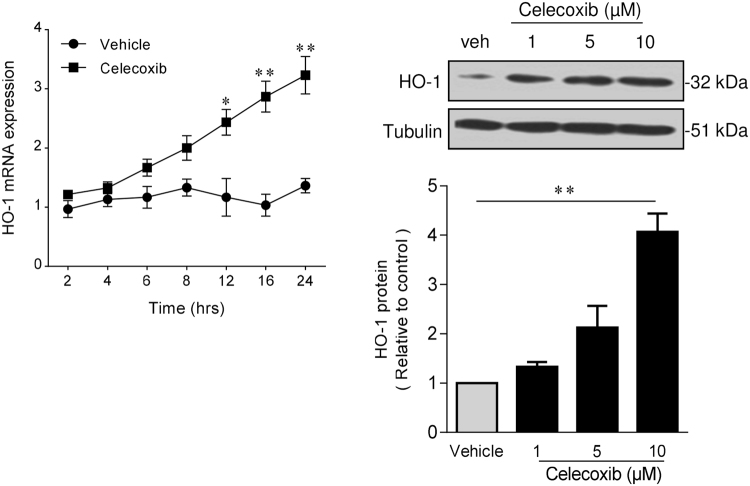
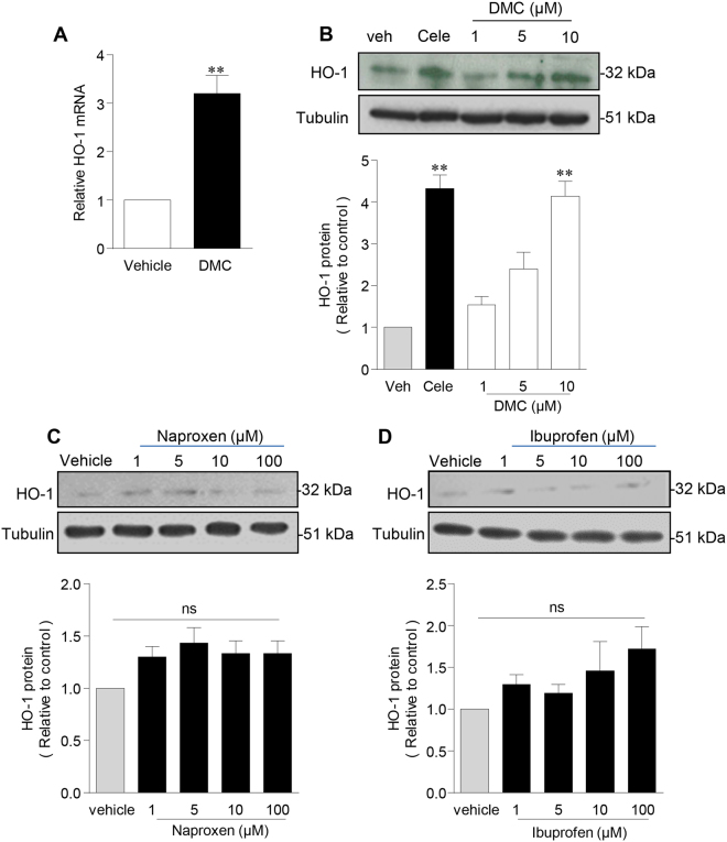
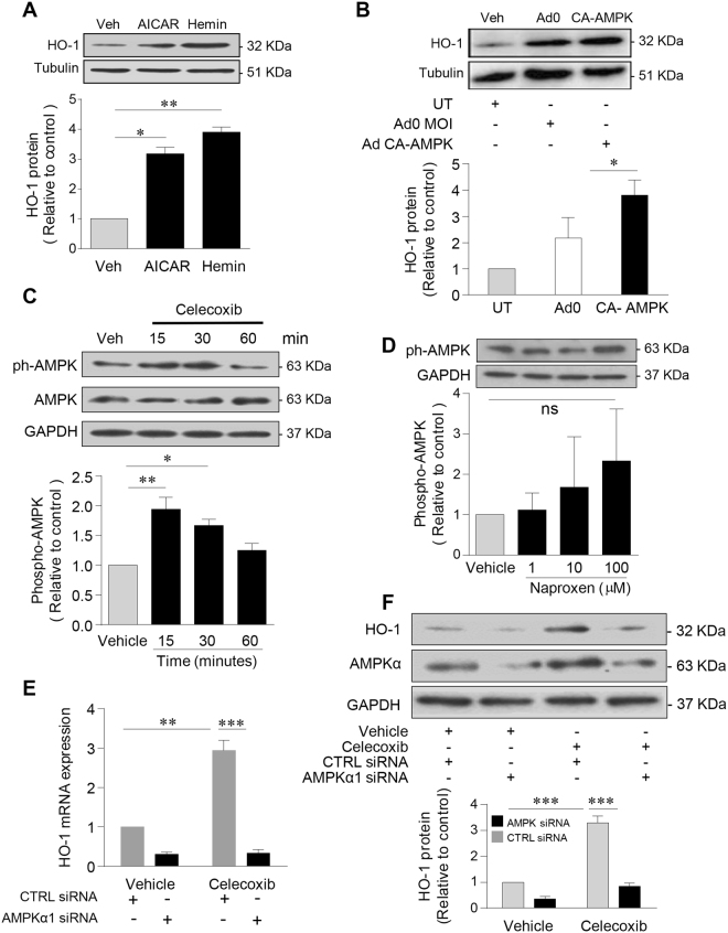
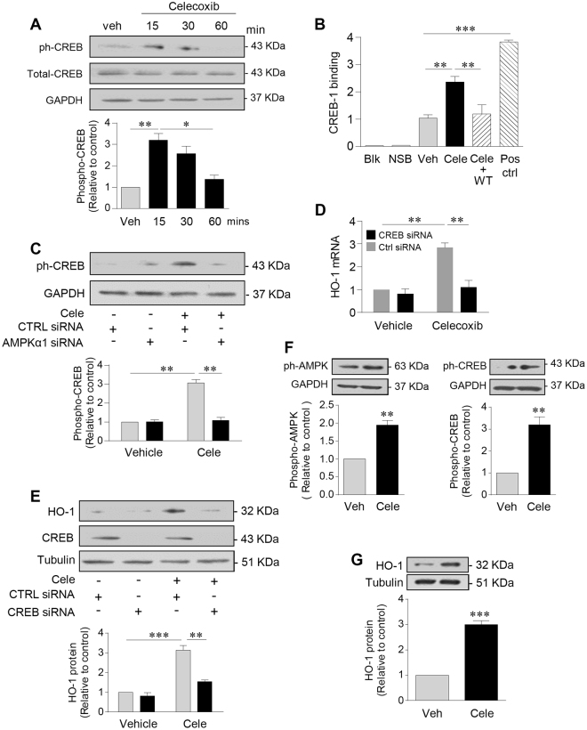
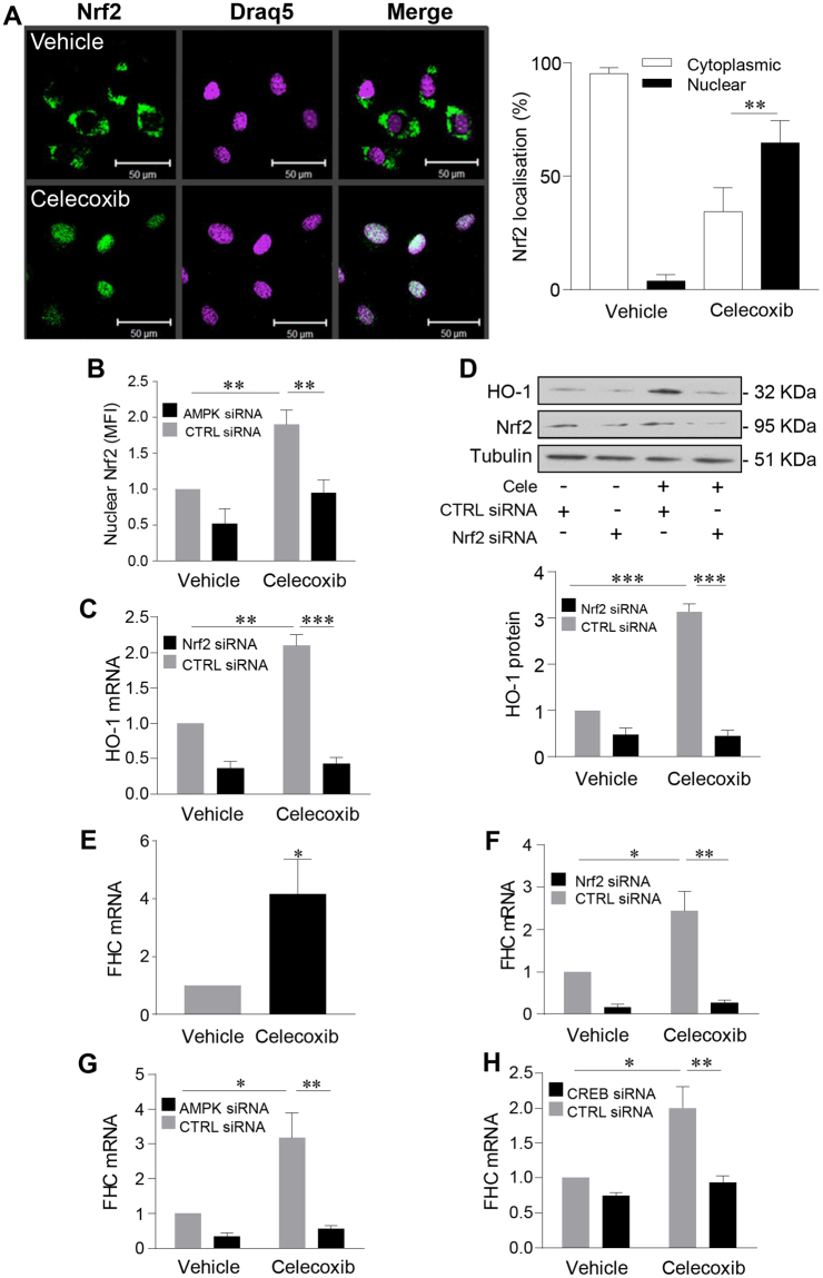
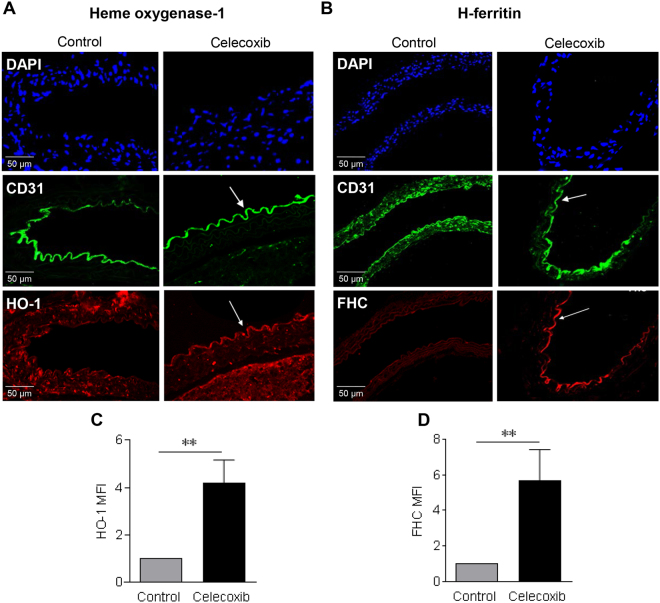
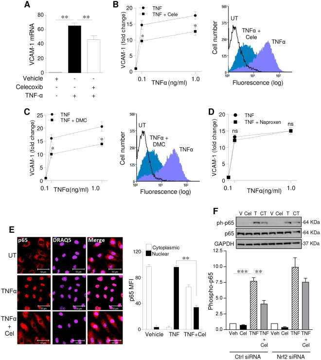
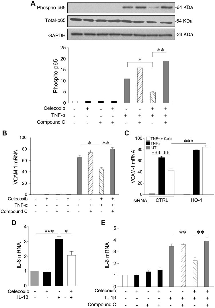
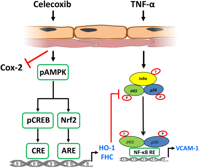
Although concern remains about the athero-thrombotic risk posed by cyclo-oxygenase (COX)-2-selective inhibitors, recent data implicates rofecoxib, while celecoxib appears equivalent to NSAIDs naproxen and ibuprofen. We investigated the hypothesis that celecoxib activates AMP kinase (AMPK) signalling to enhance vascular endothelial protection. In human arterial and venous endothelial cells (EC), and in contrast to ibuprofen and naproxen, celecoxib induced the protective protein heme oxygenase-1 (HO-1). Celecoxib derivative 2,5-dimethyl-celecoxib (DMC) which lacks COX-2 inhibition also upregulated HO-1, implicating a COX-2-independent mechanism. Celecoxib activated AMPKα(Thr172) and CREB-1(Ser133) phosphorylation leading to Nrf2 nuclear translocation. Importantly, these responses were not reproduced by ibuprofen or naproxen, while AMPKα silencing abrogated celecoxib-mediated CREB and Nrf2 activation. Moreover, celecoxib induced H-ferritin via the same pathway, and increased HO-1 and H-ferritin in the aortic endothelium of mice fed celecoxib (1000 ppm) or control chow. Functionally, celecoxib inhibited TNF-α-induced NF-κB p65(Ser536) phosphorylation by activating AMPK. This attenuated VCAM-1 upregulation via induction of HO-1, a response reproduced by DMC but not ibuprofen or naproxen. Similarly, celecoxib prevented IL-1β-mediated induction of IL-6. Celecoxib enhances vascular protection via AMPK-CREB-Nrf2 signalling, a mechanism which may mitigate cardiovascular risk in patients prescribed celecoxib. Understanding NSAID heterogeneity and COX-2-independent signalling will ultimately lead to safer anti-inflammatory drugs.


Conflict of interest statement

The authors declare no competing interests.

Figures

Figure 1
Figure 1
Celecoxib induces HO-1 in human endothelium. HUVECs were treated with celecoxib (up to 10 µM) or vehicle alone for up to 24 h, followed by: (A) RNA extraction and analysis of HO-1 by qRT-PCR, and (B) protein extraction and analysis by immunoblotting for HO-1 and α-tubulin. The histogram shows corresponding densitometry data corrected for α-tubulin. Data are expressed as the mean ± SEM (n = 4 experiments) and normalized to vehicle-treated cells, *P ≤ 0.05, **P ≤ 0.01, using a one-way ANOVA with a Bonferonni correction. Immunoblots shown have been cropped to conserve space, please see Supplementary file for original uncropped blots.
Figure 2
Figure 2
Celecoxib induction of HO-1 is COX-2-independent. HUVEC were treated with 2,5-Dimethyl-celecoxib (DMC) (up to 10 µM), celecoxib (10 µM) or vehicle alone for 24 h, followed by: (A) RNA extraction and analysis of HO-1 by qRT-PCR, and (B) protein extraction and analysis by immunoblotting for HO-1 and α-tubulin. (C and D) HUVEC were treated with vehicle alone or (C) naproxen, (D) ibuprofen (up to 100 µM) followed by immunoblotting analysis of HO-1. The histograms show corresponding densitometry data corrected for the α-tubulin and normalized to vehicle-treated cells. Data are expressed as the mean ± SEM (n = 4 experiments), *P ≤ 0.05, **P ≤ 0.01 using a one-sample t-test (A) and the one-way ANOVA with a Bonferonni correction (BD). Immunoblots shown have been cropped to conserve space, please see Supplementary file for original uncropped blots.
Figure 3
Figure 3
Celecoxib activates AMPK to induce HO-1. HUVECs were treated with: (A) vehicle alone, AICAR (1 mM for 24 h) or hemin (0.2 µM for 6 h, positive control), or (B) transfected with Ad0 (control virus), Ad-CA-AMPK (MOI 100 ifu/cell) or left untreated for 24 h, followed by protein extraction and immunoblotting for HO-1 or α-tubulin. (C) HUVECs were treated with celecoxib (10 μM) for up to 60 mins, or (D) naproxen up to 100 μM for 15 mins, prior to protein extraction and immunoblotting for phospho-AMPKThr172 and GAPDH. (E,F) HUVECs were left untransfected or transfected with control siRNA (CTRL) or AMPKα1 siRNA (50 nM) and cultured for 48 h prior to treatment with vehicle or celecoxib (10 µM) for 24 h and (E) analysis of HO-1 by qRT-PCR, or (F) protein extraction and immunoblotting for HO-1, total-AMPK and GAPDH. Data are expressed as the mean ± SEM of at least 3 separate experiments. The histograms show corresponding densitometry data corrected for α-tubulin or GAPDH and normalized to vehicle-treated cells. *P ≤ 0.05, **P ≤ 0.01, **P ≤ 0.001, using the one-way ANOVA + Bonferonni correction or a two-way ANOVA. Immunoblots shown have been cropped to conserve space, please see Supplementary file for original uncropped blots.
Figure 4
Figure 4
Celecoxib induces HO-1 in HUVEC and HAEC, by activating AMPK and CREB. (A) HUVECs were treated with vehicle (veh) or celecoxib (10 μM) for up to 60 mins, prior to lysis and immunoblotting for phospho-CREBSer133, total CREB and GAPDH. (B) Nuclear lysates, isolated from HUVEC following treatment with celecoxib 10 μM (Cele) or vehicle control for 30 mins, were analysed using a phospho-CREB transcription factor assay kit. Unstimulated Hela cells were used as a positive control (CTRL) and a wild-type oligonucleotide sequence (WT) was used for competitive binding. CREB-binding is expressed relative to vehicle-treated cells. (C,D) HUVECs were left untransfected or transfected with control siRNA (CTRL) or CREB siRNA (50 nM) and cultured for 48 h prior to treatment with vehicle or celecoxib (10 µM) for (C) 15 mins followed by immunoblotting for phospho-CREB and GAPDH, or for 24 h followed by analysis of (D) HO-1 by qRT-PCR and (E) HO-1 protein by immunoblotting. (F) HAEC were treated with celecoxib (10 μM) or vehicle for 15 mins prior to immunoblotting for: phospho-AMPKThr172, phospho-CREBSer133 and GAPDH. (G) HAECs were treated with celecoxib (10 μM) or vehicle for 24 h prior to immunoblotting for HO-1. Data are expressed as the mean ± SEM of at least 3 separate experiments. The histograms show corresponding densitometry data corrected for α-tubulin or GAPDH and normalized to vehicle-treated cells. *P ≤ 0.05, **P ≤ 0.01, **P ≤ 0.001, using the one-way with a Bonferonni correction or two-way ANOVA (AE) and a one-sample t-test. (F,G) Immunoblots shown have been cropped to conserve space, please see Supplementary file for original uncropped blots.
Figure 5
Figure 5
Celecoxib activates Nrf2 to induce HO-1 and H-ferritin. (A) HUVECs were left untreated or treated with celecoxib (10 μM) for 30 mins prior to fixation and staining with an anti-Nrf2 antibody and DRAQ-5 nuclear stain. Representative confocal images are shown along with a histogram representing pooled quantification data (n = 4). Data were analysed using Image J software and expressed as mean fluorescent intensity (MFI) of Nrf2 staining in the cytoplasm or nucleus. (B) HUVECs were transfected with control siRNA (CTRL) or AMPKα1 siRNA (50 nM) and cultured for 48 h prior to treatment with celecoxib and analysis of Nrf2 translocation as above. (C,D) HUVECs were transfected with control siRNA or pooled Nrf2 siRNAs (40 nM) and cultured for 48 h prior to celecoxib treatment (10 μM for 24 h). Changes in HO-1 were analysed by (C) qRT-PCR and (D) immunoblotting and the histogram shows corresponding densitometry data corrected for α-tubulin. (E) HUVECs were treated for 24 h with celecoxib or vehicle alone prior to analysis of H-ferritin (FHC) mRNA by qRT-PCR. (FH) HUVECs were transfected with control siRNA or: (F) Nrf2 siRNA. (G) AMPKα1 siRNA, (H) CREB siRNA, for 48 h prior to treatment with vehicle or celecoxib and analysis of FHC mRNA by qRT-PCR. All data are derived from 4 independent experiments, normalized to vehicle-treated cells and expressed as the mean ± SEM. *P ≤ 0.05, **P ≤ 0.01, **P ≤ 0.001, using the two-way ANOVA or a one-sample t-test. (E) Immunoblots shown have been cropped to conserve space, please see Supplementary file for original uncropped blots.
Figure 6
Figure 6
In vivo dosing of celecoxib upregulates aortic expression of HO-1 and H-ferritin. C57BL/6 female mice (n = 4 per group) were fed a diet containing 1000 ppm of celecoxib or matched standard laboratory diet for 48 h. Transverse sections of descending aorta were stained with: (A) an anti-HO-1 Ab or (B) an anti-H-ferritin (FHC) Ab. Anti-CD31 Ab was used to delineate endothelium and nuclei were visualized using DAPI nuclear dye. Representative confocal images (x40 magnification) of control and celecoxib-treated mice are shown. Histograms (C) HO-1 and (D) FHC show pooled quantification data from 4 independent experiments. Images were analysed by ImageJ software. Data are expressed as mean fluorescent intensity (MFI) of endothelial fluorescence ± SEM, representing MFI with antibody of interest in celecoxib or vehicle-treated mice divided by the MFI obtained with the control Ab and presented normalized to vehicle-treated animals. *P ≤ 0.05, **P ≤ 0.01, using an unpaired Student’s t-test. Images shown are at x40 magnification.
Figure 7
Figure 7
Celecoxib inhibits NF-κB activity to restrict VCAM-1 induction in vascular EC. HUVECs were pre-treated with vehicle, (A,B) celecoxib, (C) DMC or (D) naproxen (all 10 µM) for 24 h followed by TNF-α stimulation (up to 1 ng/ml) for (A) 6 h, with VCAM-1 analysed by qRT-PCR, and (B,D) 16 h followed by flow-cytometric analysis of VCAM-1, with data presented as relative fluorescence intensity (RFI), representing mean fluorescence intensity (MFI) with the VCAM-1 Ab divided by the MFI of the UT control (n = 4 experiments). (E,F) HUVECs were left untreated or treated with celecoxib for 24 h prior to addition of vehicle or TNF-α (0.1 ng/ml) for 30 minutes. (E) EC were fixed and stained with an anti-p65 antibody and DRAQ-5 nuclear stain. Representative confocal images are shown along with a histogram representing pooled quantification data (n = 3 experiments). Data were analysed using Image J software and expressed as the MFI of p65 staining in the cytoplasm or nucleus. (F) HUVECs were transfected with control siRNA (CTRL) or Nrf2 siRNA and cultured for 30 h prior to treatment with vehicle or celecoxib (10 µM) for 24 h and exposure to TNF-α (0.1 ng/ml) for 30 mins. Lysates were immunoblotted for phospho-p65Ser536, p65 and GAPDH. The fold change in phosphorylation was quantified using densitometry (n = 4 experiments), normalized with respect to GAPDH and expressed relative to the vehicle control. Data are expressed as the mean ± SEM, *P ≤ 0.05, **P ≤ 0.01, ***P ≤ 0.001, ns = non-significant using one-way or two-way ANOVA. Immunoblots shown have been cropped to conserve space, please see Supplementary file for original uncropped blots.
Figure 8
Figure 8
Celecoxib exerts anti-inflammatory effects via AMPK and induction of HO-1. HUVECs were pre-treated with Compound C (5 µM) or vehicle alone for 60 mins, prior to addition of celecoxib (10 μM) or vehicle for 24 h, followed by: (A) treatment with TNF-α (0.1 ng/ml) or vehicle for 30 mins and immunoblotting for p65, phospho-p65Ser536 and GAPDH. The histogram shows corresponding densitometry data for phospho-p65 corrected for GAPDH, or (B) treatment with TNF-α (0.1 ng/ml) or vehicle for 6 h, and analysis of VCAM-1 mRNA by qRT-PCR. (C) HUVECs were transfected with HO-1 or control (CTRL) siRNA (40 nM) for 48 h, prior to the addition of celecoxib or vehicle for 24 h and treatment with TNF-α (0.1 ng/ml) for 6 h. VCAM-1 mRNA was analysed by qRT-PCR. (D) HUVECs were left untreated or pre-treated with celecoxib for 24 h prior to addition of IL-1β (0.1 ng/ml) or vehicle for 4 h, and (E) HUVECs were left untreated or pre-treated with Compound C or vehicle alone for 60 mins, prior to addition of celecoxib for 24 h, followed by IL-1β (0.1 ng/ml) or vehicle for 4 h. Changes in IL-6 mRNA were analysed by qRT-PCR. Data in the figure are expressed as the mean ± SEM of 4 independent experiments and normalized to vehicle-treated cells. *P ≤ 0.05, **P ≤ 0.01, ***P ≤ 0.001, using one-way ANOVA with a Bonferonni correction. Immunoblots shown have been cropped to conserve space, please see Supplementary file for original uncropped blots.
Figure 9
Figure 9
Summary of COX-2-independent AMPK-CREB-Nrf2 signalling pathway activated by celecoxib in the vascular endothelium. Treatment of human endothelial cells results in COX-2 independent signalling via phosphorylation of AMPK at threonine 172 (pAMPK). This leads to activation of CREB at serine 133 (pCREB) and nuclear translocation of Nrf2. The CREB and Nrf2 pathways are thought to act in parallel, binding to the CREB response element (CRE) and the antioxidant response element (ARE) respectively to upregulate the expression of the anti-oxidant, anti-inflammatory genes heme oxygenase-1 (HO-1) and H-Ferritin (FHC). The anti-inflammatory actions of this pathway include inhibition of TNF-α-mediated activation and nuclear translocation of p65. This response attenuated the pro-inflammatory upregulation of the cellular adhesion molecule vascular cell adhesion molecule-1 (VCAM-1).

Similar articles

Cited by

References

    1. Coxib and traditional NSAID Trialists’ Collaboration. Bhala N, et al. Vascular and upper gastrointestinal effects of non-steroidal anti-inflammatory drugs: meta-analyses of individual participant data from randomised trials. Lancet. 2013;382:769–779. doi: 10.1016/S0140-6736(13)60900-9. - DOI - PMC - PubMed
    1. Patrono C. Cardiovascular effects of cyclooxygenase-2 inhibitors: A mechanistic and clinical perspective. Br J Clin Pharmacol. 2016;82:957–964. doi: 10.1111/bcp.13048. - DOI - PMC - PubMed
    1. Bresalier RS, et al. Cardiovascular events associated with rofecoxib in a colorectal adenoma chemoprevention trial. N Engl J Med. 2005;352:1092–1102. doi: 10.1056/NEJMoa050493. - DOI - PubMed
    1. Mitchell JA, Warner TD. COX isoforms in the cardiovascular system: understanding the activities of non-steroidal anti-inflammatory drugs. Nat Rev Drug Discov. 2006;5:75–86. doi: 10.1038/nrd1929. - DOI - PubMed
    1. Trelle S, et al. Cardiovascular safety of non-steroidal anti-inflammatory drugs: network meta-analysis. BMJ. 2011;342:c7086. doi: 10.1136/bmj.c7086. - DOI - PMC - PubMed

Publication types

MeSH terms