. 2018 Apr 19;9(1):1558.
doi: 10.1038/s41467-018-04013-1.

The molecular basis of JAK/STAT inhibition by SOCS1

Affiliations

The molecular basis of JAK/STAT inhibition by SOCS1

Nicholas P D Liau et al. Nat Commun. .

Abstract

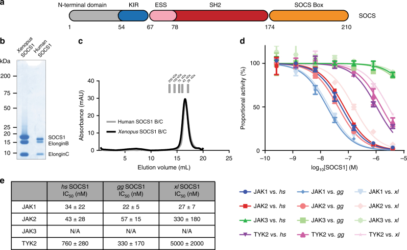
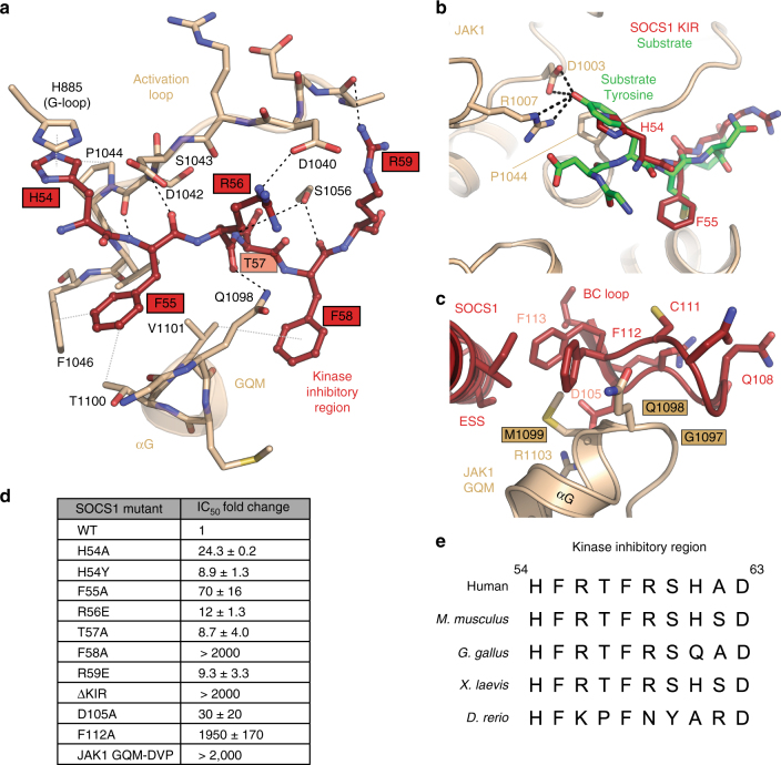
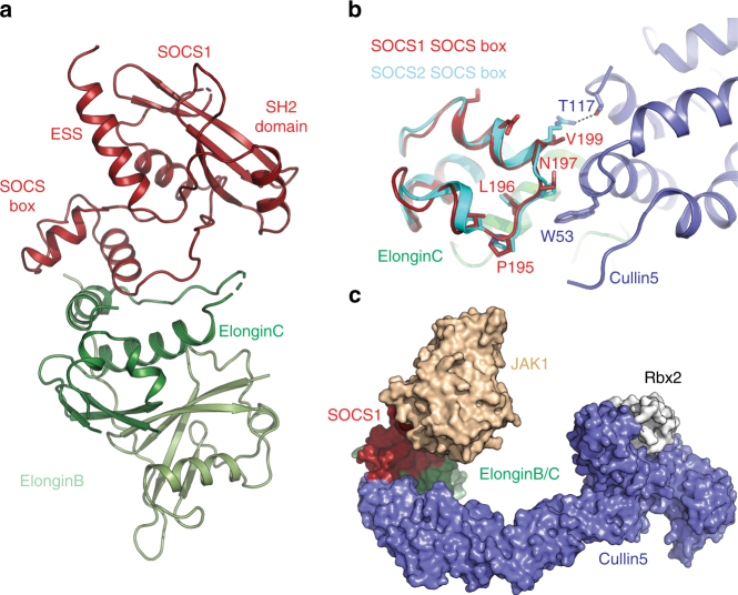
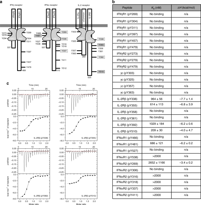
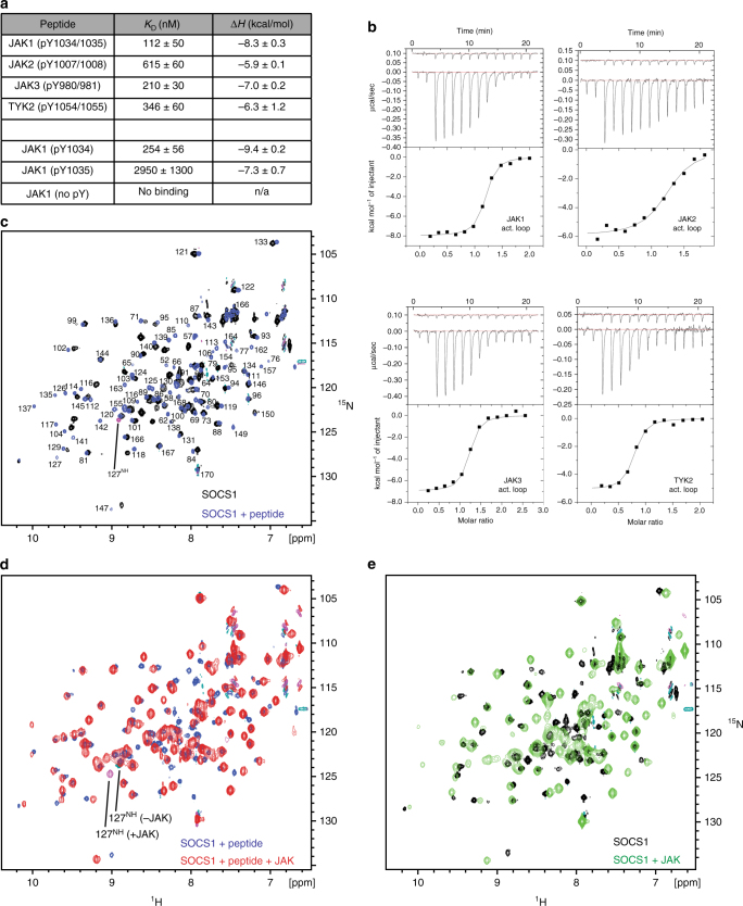
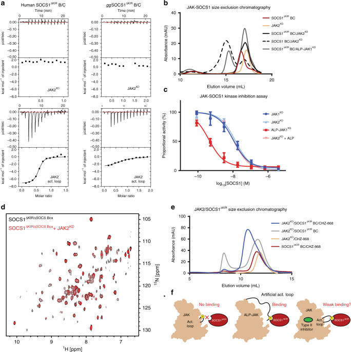
The SOCS family of proteins are negative-feedback inhibitors of signalling induced by cytokines that act via the JAK/STAT pathway. SOCS proteins can act as ubiquitin ligases by recruiting Cullin5 to ubiquitinate signalling components; however, SOCS1, the most potent member of the family, can also inhibit JAK directly. Here we determine the structural basis of both these modes of inhibition. Due to alterations within the SOCS box domain, SOCS1 has a compromised ability to recruit Cullin5; however, it is a direct, potent and selective inhibitor of JAK catalytic activity. The kinase inhibitory region of SOCS1 targets the substrate binding groove of JAK with high specificity and thereby blocks any subsequent phosphorylation. SOCS1 is a potent inhibitor of the interferon gamma (IFNγ) pathway, however, it does not bind the IFNγ receptor, making its mode-of-action distinct from SOCS3. These findings reveal the mechanism used by SOCS1 to inhibit signalling by inflammatory cytokines.


Conflict of interest statement

The authors declare no competing interests.

Figures

Fig. 1
Fig. 1
SOCS1 is a direct inhibitor of JAK kinase activity. a Schematic representation of the SOCS1 domain architecture. SOCS1 consists of an unstructured N-terminal region, followed by the kinase inhibitory region (KIR), an extended SH2 subdomain (ESS), a SH2 domain and a SOCS box domain. b Purified recombinant SOCS1 in complex with Elongins B and C shown on Coomassie stained SDS-PAGE gel. c Gel filtration analysis of purified recombinant SOCS1 in complex with Elongins B and C. d Kinase inhibition assays indicate that SOCS1 from Homo sapiens (hs), Gallus gallus (gg) and Xenopus laevis (xl) inhibits the kinase domains of JAK1, JAK2 and TYK2, but not JAK3. e IC50 values of SOCS1 and SOCS3 constructs against JAKs as determined by kinase inhibition assays. The error bars shown in d represent the range of the data from two technical replicates whilst errors given in e are the standard error of the mean from 3 independent experiments
Fig. 2
Fig. 2
The structure of a SOCS1/JAK1/ADP complex. a Cartoon representation of the JAK1 (beige) /SOCS1 (red) /ADP (green) complex. The SOCS1 kinase inhibitory region (KIR), the extended SH2 subdomain (ESS) as well as the SH2 domain proper (but not its phosphotyrosine-binding groove) all make substantial contacts with the C-lobe of the JAK1 kinase domain. Notably, JAK1 Tyr 1034 and 1035 in the activation loop are unphosphorylated in this structure. b Details of key SOCS1 interactions centred around the JAK1 GQM motif (electrostatic surface view). c JAK1 from the JAK1/SOCS1/ADP complex structure (beige) is shown overlaid with a JAK1/ADP structure (white, PDB: 5KHW). Small perturbations in the JAK1 activation loop and GQM motif can be seen between the two structures as well as a larger perturbation in the glycine-rich loop. The JAK1 activation loop is seen in the active conformation both in the presence and absence of SOCS1, despite being unphosphorylated in the SOCS1 bound structure. d Ribbon diagram of the structure of SOCS1 in the presence (red) and absence (white) of JAK1. The major conformational change to SOCS1 upon JAK1 binding is an ordering of the kinase inhibitory region. e The SOCS1 KIR is shown overlaid with the SOCS3 KIR (green) (PDB ID: 4GL9). Residue numbering of SOCS1 orthologues in all figures refers to the analogous residue in the human sequence
Fig. 3
Fig. 3
The kinase inhibitory region acts as a pseudosubstrate. a Molecular details of the interaction between the SOCS1 kinase inhibitory region (red) and JAK1 (beige). The interaction is driven by six continuous residues in the SOCS1 KIR (His54 to Arg59), these primarily interact with the activation loop, GQM motif and glycine-rich loop of JAK1. Black dashed lines indicate hydrogen-bonds whilst grey dotted lines are van der Waals contacts. b A model of a peptide substrate (green) bound to the JAK1 kinase domain (based on the insulin receptor kinase/substrate structure (PDB: 1IRK)) indicates that the SOCS1 KIR acts as a pseudosubstrate with His54 mimicking the substrate tyrosine. c The GQM motif (Gly1097, Gln1098, Met1099) in JAK1 is a primary interaction site with SOCS1 and contacts the KIR, extended SH2 subdomain and SH2 domain (particularly the BC loop). d IC50 values of SOCS1 KIR mutants indicate that each residue of the KIR contributes to the affinity of the interaction. Errors are standard error of the mean from two independent experiments. e Sequence alignment of the KIR of SOCS1 from various species shows a high level of sequence conservation
Fig. 4
Fig. 4
Structure of the SOCS1–Elongin B/C complex. a Structure of Xenopus laevis SOCS1 (red) in complex with Elongin B (pale green) and Elongin C (green). b The SOCS1 SOCS Box is shown overlaid with the structure of SOCS2 (cyan) bound to Cullin5 (blue). SOCS1 Asn197 takes the place of terminal proline typically seen in the canonical Cullin5 binding motif (LPφP). This asparagine clashes with Trp53 of Cullin5. Likewise, the Arg186 (SOCS2)-Thr117 (Cul5) hydrogen bond is lost in SOCS1 which has a valine (Val200) in place of the arginine. c Model of the JAK1-SOCS1-Elongin B/C-Cullin5-Rbx2 (white) complex based upon the SOCS1/JAK1 (this manuscript), SOCS2/ElonginB/ElonginC/Cullin5 NTD (PDB: 4JGH) and Cul5 CTD/Rbx2 structures (PDB: 3DPL). The SOCS1/JAK1 interaction orients JAK1 toward rbx2, which is the site of activated-ubiquitin (ubiquitin-E2) binding
Fig. 5
Fig. 5
SOCS1 does not bind to any pTyr sites on the interferon gamma receptor. a Schematic diagrams of the IFN-α, ɣ and IL-2 receptors. All intracellular tyrosines are shown. b Table showing the binding affinities of human SOCS1 for each IFN-ɣ and IL-2 receptor phosphopeptide. Errors represent standard deviation from three independent experiments. c Representative ITC curves are shown for the four IL-2R phosphopeptides that bind to SOCS1 with sub-micromolar affinity
Fig. 6
Fig. 6
SOCS1 binds to the activation loop peptides from all four JAKs. a Table showing the affinity and enthalpy of each SOCS1/JAK activation loop peptide interaction as determined by isothermal titration calorimetry. Human SOCS1 binds to phosphorylated peptides representing all four JAK activation loops. The first phosphotyrosine of the JAK1 activation loop (pTyr1034) is the key residue for SOCS1 binding. Errors represent standard error of the mean from three independent experiments. b Representative ITC binding data for the four JAK activation loop peptides (doubly-phosphorylated) binding to SOCS1 used to generate a. c NMR analysis of phosphopeptide binding. 1H–15N SOFAST HMQC spectra of ggSOCS1 in the presence (blue) and absence (black) of the JAK1 activation loop are shown overlaid. Both spectra were assigned, the labels indicate the positions of amide resonances from the bound form of the protein. d NMR analysis of JAK binding in the presence of exogenous phosphopeptide. 1H–15N SOFAST HMQC spectra of ggSOCS1/activation loop complex in the presence (red) and absence (blue) of the JAK1 kinase domain are shown overlaid. The sidechain epsilon NH resonance of Arg127 from both JAK-bound and unbound forms are indicated. These are of opposite phase to backbone amide resonances. The presence of the Arg127 resonance in that section of spectra indicates that the phosphopeptide-binding groove is occupied in both complexes. e NMR analysis of JAK binding in the absence of exogenous phosphopeptide. 1H–15N SOFAST HMQC spectra of ggSOCS1 in the presence (green) and absence (black) of the JAK1 kinase domain are shown overlaid. The line-broadening in the green spectra indicated the formation of a SOCS1/JAK complex and the lack of the Arg127 sidechain resonance in either spectra indicates the phosphotyrosine-binding groove is unoccupied
Fig. 7
Fig. 7
Steric hindrance prevents SOCS1 from binding the JAK activation loop in the intact kinase domain. a ITC binding data indicates both human SOCS1ΔKIR B/C (left) and ggSOCS1ΔKIR B/C (right) do not bind to the JAK2 kinase domain but do bind to JAK2 activation loop peptides. b Size exclusion chromatography (using a Superdex 200 10/300 column) data indicate that SOCS1ΔKIR B/C does not form a complex with JAK2. As positive controls, wild-type SOCS1 B/C does form a complex with JAK2, while SOCS1ΔKIR B/C does form a complex with JAK1 when the activation loop peptide (ALP) motif is artificially fused to the N-terminus of JAK. (ALP-JAK1KD). c Kinase inhibition assays show that SOCS1 inhibits JAK2 with similar IC50 values in the absence and presence of activation loop peptide. Conversely, SOCS1 displays a lower IC50 against ALP-JAK1KD. Error bars represent the range of the data from two technical replicates. d NMR data indicate that ggSOCS1ΔKIRΔSOCSbox does not bind the phosphorylated JAK2 kinase domain. 1H–15N SOFAST HMQC spectra of 15N-labelled ggSOCS1ΔKIRΔSOCSbox in the presence (red) and absence (black) of the phosphorylated JAK2 kinase domain (unlabelled) are shown overlaid. e Size exclusion chromatography data (using a Superdex 75 10/30 column) indicate that SOCS1ΔKIR B/C forms a complex with JAK2 in the presence of 5 µM CHZ-868 Type II inhibitor, but not in its absence. f Schematic summary of SOCS1 SH2 domain interactions. Under normal conditions, SOCS1ΔKIR does not bind to the JAK activation loop due to steric hinderance. In the presence of a Type II inhibitor, the SOCS1 SH2 domain weakly associates with the JAK activation loop

References

    1. Hilton DJ, et al. Twenty proteins containing a C-terminal SOCS box form five structural classes. Proc. Natl Acad. Sci. USA. 1998;95:114–119. doi: 10.1073/pnas.95.1.114. - DOI - PMC - PubMed
    1. Babon JJ, et al. The SOCS box domain of SOCS3: structure and interaction with the elonginBC-cullin5 ubiquitin ligase. J. Mol. Biol. 2008;381:928–940. doi: 10.1016/j.jmb.2008.06.038. - DOI - PMC - PubMed
    1. Kamizono S, et al. The SOCS box of SOCS-1 accelerates ubiquitin-dependent proteolysis of TEL-JAK2. J. Biol. Chem. 2001;276:12530–12538. doi: 10.1074/jbc.M010074200. - DOI - PubMed
    1. Yasukawa H, et al. The JAK-binding protein JAB inhibits Janus tyrosine kinase activity through binding in the activation loop. EMBO J. 1999;18:1309–1320. doi: 10.1093/emboj/18.5.1309. - DOI - PMC - PubMed
    1. Endo TA, et al. A new protein containing an SH2 domain that inhibits JAK kinases. Nature. 1997;387:921–924. doi: 10.1038/43213. - DOI - PubMed

Publication types

MeSH terms