Review
. 2018 Jul;19(7):451-463.
doi: 10.1038/s41580-018-0009-y.

Microtubule dynamics: an interplay of biochemistry and mechanics

Affiliations
Review

Microtubule dynamics: an interplay of biochemistry and mechanics

Gary J Brouhard et al. Nat Rev Mol Cell Biol. 2018 Jul.

Abstract

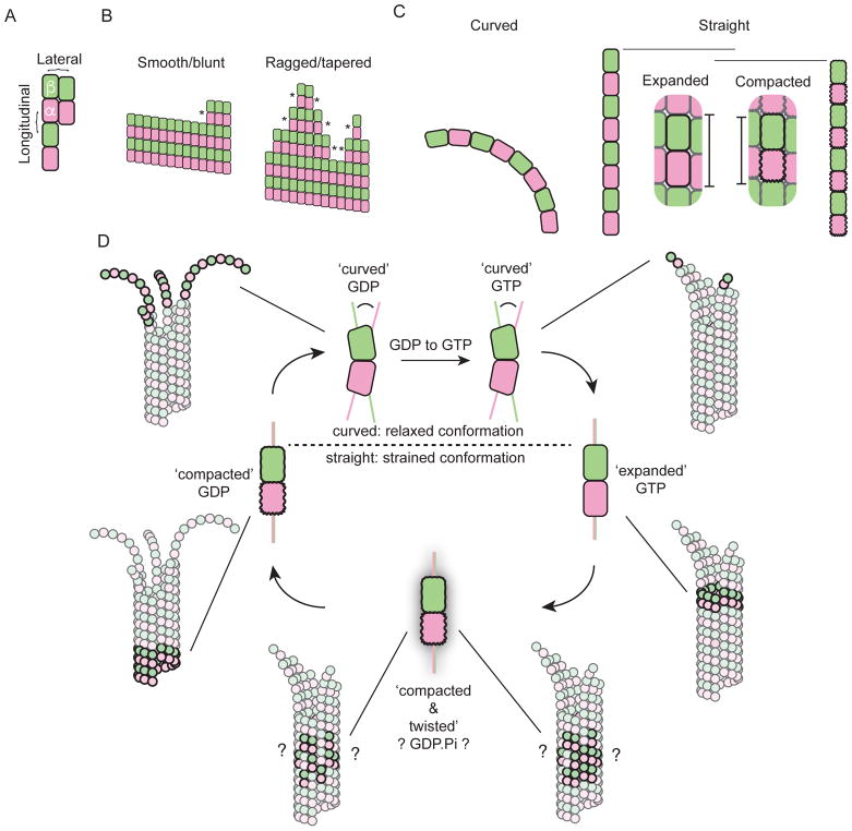
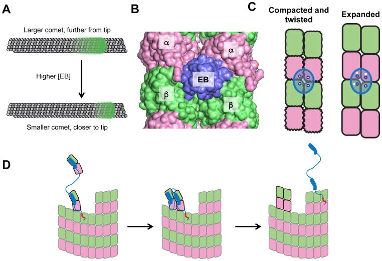
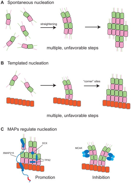
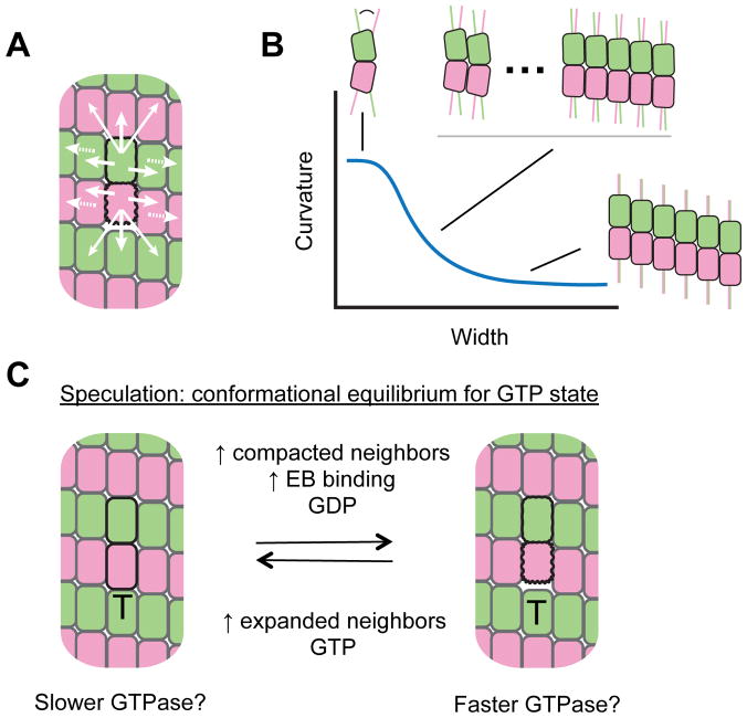
Microtubules are dynamic polymers of αβ-tubulin that are essential for intracellular organization, organelle trafficking and chromosome segregation. Microtubule growth and shrinkage occur via addition and loss of αβ-tubulin subunits, which are biochemical processes. Dynamic microtubules can also engage in mechanical processes, such as exerting forces by pushing or pulling against a load. Recent advances at the intersection of biochemistry and mechanics have revealed the existence of multiple conformations of αβ-tubulin subunits and their central role in dictating the mechanisms of microtubule dynamics and force generation. It has become apparent that microtubule-associated proteins (MAPs) selectively target specific tubulin conformations to regulate microtubule dynamics, and mechanical forces can also influence microtubule dynamics by altering the balance of tubulin conformations. Importantly, the conformational states of tubulin dimers are likely to be coupled throughout the lattice: the conformation of one dimer can influence the conformation of its nearest neighbours, and this effect can propagate over longer distances. This coupling provides a long-range mechanism by which MAPs and forces can modulate microtubule growth and shrinkage. These findings provide evidence that the interplay between biochemistry and mechanics is essential for the cellular functions of microtubules.


Figures

Figure 1
Figure 1
Tubulin biochemistry, conformation, and mechanics. A. Cartoon illustrating the fundamental longitudinal (vertical) and lateral (horizontal) interactions between αβ-tubulins (pink and green) that make up the microtubule lattice. B. Two dimensional schematics of blunt (few ‘corner’ sites, *) and ragged/tapered (more corner sites, *) microtubule ends. Taper refers to the extension of some protofilaments beyond others; ragged describes an uneven or rough distribution of protofilament lengths. C. Cartoons of longitudinal assemblies of αβ-tubulin illustrating the three major conformations in structural detail. (left) Tubulin adopts a curved conformation in single protofilaments and when unpolymerized. (right) Tubulin adopts straight ‘expanded’ or ‘compacted’ conformations in the body of the microtubule. The horizontal bars and the inset indicate that the compacted conformation is somewhat ‘shorter’ than the expanded one. D. The tubulin conformation cycle is a mechanical cycle. Straight conformations of αβ-tubulin are strained but stabilized by interactions with the microtubule lattice (not illustrated here). Release of that strain during depolymerization can do mechanical work. The dark/bold subunits in the thumbnail cartoons of growing or shrinking microtubules indicate the region of the microtubules where the different conformations occur. The ‘glow’ indicates the compacted and twisted state, and the ?’s indicate uncertainty about whether this conformation reflects a specific nucleotide state and about the distribution of these sites in the microtubule.
Figure 2
Figure 2
Regulatory proteins and the tubulin conformation cycle. A. EBs form a ‘comet’ (green glow) near but somewhat behind the growing microtubule end (top). Increasing the concentration of EB (arrow) reduces the size of the comet and moves it closer to the growing end (bottom). B. EB (purple) binds at a vertex of 4 αβ-tubulin dimers (α: pink; β: green) in the lattice. C. EB (purple oval; the 4 empty blue circles illustrate the arrangement of EB’s tubulin-binding epitopes) binds most tightly to a ‘compacted and twisted’ conformation of αβ-tubulin. Red circles indicate the EB-contacting surfaces on the lattice (white circles). EB binds poorly to the expanded lattice because the EB-contacting epitopes are improperly spaced (mismatch between white and blue circles). D. Conformation-based mechanism of a microtubule polymerase. Linked TOG domains like those in Stu2 bind selectively to curved tubulin and increase the rate of tubulin:microtubule associations by concentrating unpolymerized tubulin near the microtubule end via a tethering mechanism (left). The tethered TOG-bound tubulin associates faster with the microtubule end, and lateral interactions between TOG-bound tubulins on the microtubule end (middle) leads to straightening, which releases the TOGs (right) for another round of incorporation. The red segment indicates a basic region that mediates lattice binding.
Figure 3
Figure 3
Barriers to spontaneous and templated nucleation. A. Spontaneous nucleation involves multiple, unfavorable steps. Unpolymerized tubulin is curved (right) and must straighten to form small oligomers (middle). The growth of these oligomers (left) requires increased straightening for newly added tubulins and of those already in the oligomer. B. Templated nucleation also involves multiple, unfavourable steps. A blunt template (right) presents fewer high-affinity “corner” sites than a tapered growing end; curved tubulin binding to the blunt template may also have to straighten more than at the partially curved microtubule end (middle). The transition of the blunt template into a growing microtubule end requires many tubulins to straighten (left). C. MAPs regulate microtubule nucleation by altering the conformation of tubulin. Nucleation-promoting MAPs like XMAP215, TPX2, and DCX help form a nascent plus end (left). Nucleation-inhibiting MAPs like MCAK prevent this formation (right).
Figure 4
Figure 4
Mechanical coupling in the microtubule lattice. A. Cartoon illustrating a rationale for mechanical coupling in the lattice. A compacted tubulin (dark outline) is shown in a majority expanded lattice (light outlines). The mismatch between expanded and compacted conformations is likely resolved through conformational changes near the mismatch (white arrows). The periodic nature of the lattice means that such accommodation could propagate beyond nearest-neighbors (dashed arrows). B. At the microtubule end or in nucleation intermediates, tubulin curvature is thought to vary with width of the taper, affecting the strength of tubulin:tubulin interactions. Colored lines on the αβ-tubulin cartoons provide a visual reference for the amount of curvature. This provides another example of long-range mechanical coupling. C. Model for conformational control of GTPase that was inspired by observations about EB proteins. The model speculates that expanded tubulins may have slower GTPase than compacted tubulins. ‘T’ on the cartoons indicates GTP nucleotide state. Combined with long-range coupling, such a model is expected to display threshold-type behaviours and could give rise to cooperative, positive-feedback enhancement of GTPase activity in the stabilizing cap.

Similar articles

Cited by

References

    1. Desai A, Mitchison TJ. Annu Rev Cell Dev Biol. 1997;13:83–117. - PubMed
    1. Carlier MF, Pantaloni D. Biochemistry. 1981;20:1918–1924. - PubMed
    1. Akhmanova A, Steinmetz MO. Tracking the ends: a dynamic protein network controls the fate of microtubule tips. Nature reviews. Molecular cell biology. 2008;9:309–322. doi: 10.1038/nrm2369. - DOI - PubMed
    1. Akhmanova A, Steinmetz MO. Control of microtubule organization and dynamics: two ends in the limelight. Nature reviews. Molecular cell biology. 2015;16:711–726. doi: 10.1038/nrm4084. - DOI - PubMed
    1. Howard J, Hyman AA. Microtubule polymerases and depolymerases. Current opinion in cell biology. 2007;19:31–35. doi: 10.1016/j.ceb.2006.12.009. - DOI - PubMed

Publication types

MeSH terms