Review
. 2018 Apr 5:11:105.
doi: 10.3389/fnmol.2018.00105. eCollection 2018.

SALM/Lrfn Family Synaptic Adhesion Molecules

Affiliations
Review

SALM/Lrfn Family Synaptic Adhesion Molecules

Eunkyung Lie et al. Front Mol Neurosci. .

Abstract

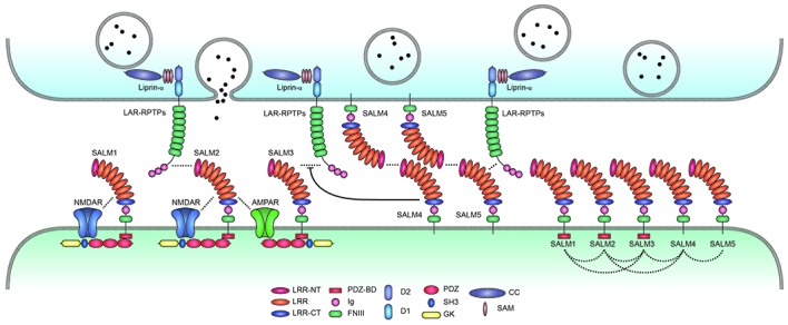
Synaptic adhesion-like molecules (SALMs) are a family of cell adhesion molecules involved in regulating neuronal and synapse development that have also been implicated in diverse brain dysfunctions, including autism spectrum disorders (ASDs). SALMs, also known as leucine-rich repeat (LRR) and fibronectin III domain-containing (LRFN) proteins, were originally identified as a group of novel adhesion-like molecules that contain LRRs in the extracellular region as well as a PDZ domain-binding tail that couples to PSD-95, an abundant excitatory postsynaptic scaffolding protein. While studies over the last decade have steadily explored the basic properties and synaptic and neuronal functions of SALMs, a number of recent studies have provided novel insights into molecular, structural, functional and clinical aspects of SALMs. Here we summarize these findings and discuss how SALMs act in concert with other synaptic proteins to regulate synapse development and function.

Keywords: Lrfn; PSD-95; SALM; adhesion molecules; synaptic.


Figures

Figure 1
Figure 1
Domain structure of Synaptic adhesion-like molecules (SALMs) and LAR-RPTPs. (A) Domain structure of SALMs 1–5. Note that the PDZ domain-binding motif (PDZ-BD) is present in SALMs 1–3 but not in SALM4 or SALM5. FNIII, fibronectin III domain; Ig, immunoglobulin domain; LRR, leucine-rich repeats; NT and CT, N-terminal and C-terminal LRR. Note that the number of LRRs in this diagram is seven, although it was suggested to be six in early studies based on amino acid sequence analyses (Ko et al., ; Morimura et al., ; Wang et al., ; Nam et al., 2011). Recent X-ray crystallographic studies have identified seven LRRs in SALM5 (Lin et al., 2018) and eight LRRs in SALM2 and SALM5 (Goto-Ito et al., 2018), which may reflect different ways of defining LRRs. (B) Domain structure of LAR-RPTPs (LAR, PTPσ and PTPδ). D1 and D2, membrane-proximal and -distal tyrosine phosphatase domains of LAR-RPTPs; meA/B/C; mini-exon A/B/C.
Figure 2
Figure 2
Trans-synaptic, cis-, and cytoplasmic interactions of SALMs. SALMs interact trans-synaptically with presynaptic LAR-RPTPs (LAR, PTPσ and PTPδ), in cis with AMPA/NMDA receptors and other SALM proteins, and cytoplasmically with the postsynaptic scaffolding protein PSD-95 (in the case of SALMs 1–3 but not SALM4 or SALM5). Protein interactions are indicated by the close proximity of the indicated proteins/domains or by dotted lines. Whether SALMs directly interact with NMDA/AMPA receptors remains to be determined. The trans-synaptic interactions between postsynaptic SALM3/5 and presynaptic LAR-RPTPs are known to promote presynaptic differentiation, although the function of the newly identified SALM2–LAR-RPTP (PTPδ) interaction is unclear. SALM4 interacts in cis with SALM3 to suppress the binding of SALM3 to presynaptic LAR-RPTPs and SALM3-dependent presynaptic differentiation. Postsynaptic SALM5 can also interacts with presynaptic SALM5 in a homophilic manner, which may interfere with the trans-synaptic interaction between presynaptic LAR-RPTPs and postsynaptic SALM5. The cis-interactions between different postsynaptic SALMs are based on both in vitro and in vivo results, and may be mediated by the SALM–SALM dimerization revealed by X-ray crystallographic studies. Although not shown here, some LAR-RPTPs are thought to be present and function at postsynaptic sites, in addition to presynaptic sites.
Figure 3
Figure 3
X-ray crystal structure of SALM5 in complex with PTPδ in a 2:2 heterotetrameric format. (A) Side view of the structure (surface representation). (B) Top-down view of the structure (ribbon diagram). These images were borrowed without modification from Figures 1B,C of a recent report on the crystal structure of SALM5 in complex with PTPδ (Lin et al., 2018), which are under a Creative Commons Attribution 4.0 International License (http://creativecommons.org/licenses/by/4.0/).

References

    1. Aricescu A. R., McKinnell I. W., Halfter W., Stoker A. W. (2002). Heparan sulfate proteoglycans are ligands for receptor protein tyrosine phosphatase sigma. Mol. Cell. Biol. 22, 1881–1892. 10.1128/mcb.22.6.1881-1892.2002 - DOI - PMC - PubMed
    1. Bemben M. A., Shipman S. L., Nicoll R. A., Roche K. W. (2015). The cellular and molecular landscape of neuroligins. Trends Neurosci. 38, 496–505. 10.1016/j.tins.2015.06.004 - DOI - PMC - PubMed
    1. Bereczki E., Branca R. M., Francis P. T., Pereira J. B., Baek J. H., Hortobágyi T., et al. . (2018). Synaptic markers of cognitive decline in neurodegenerative diseases: a proteomic approach. Brain 141, 582–595. 10.1093/brain/awx352 - DOI - PMC - PubMed
    1. Biederer T., Scheiffele P. (2007). Mixed-culture assays for analyzing neuronal synapse formation. Nat. Protoc. 2, 670–676. 10.1038/nprot.2007.92 - DOI - PubMed
    1. Biederer T., Stagi M. (2008). Signaling by synaptogenic molecules. Curr. Opin. Neurobiol. 18, 261–269. 10.1016/j.conb.2008.07.014 - DOI - PMC - PubMed

LinkOut - more resources