

Neuronal Calcium Signaling in Metabolic Regulation and Adaptation to Nutrient Stress
- PMID: 29674958
- PMCID: PMC5895653
- DOI: 10.3389/fncir.2018.00025
Neuronal Calcium Signaling in Metabolic Regulation and Adaptation to Nutrient Stress
Abstract
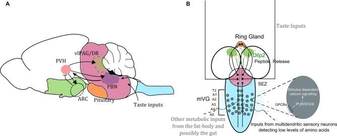
All organisms can respond physiologically and behaviorally to environmental fluxes in nutrient levels. Different nutrient sensing pathways exist for specific metabolites, and their inputs ultimately define appropriate nutrient uptake and metabolic homeostasis. Nutrient sensing mechanisms at the cellular level require pathways such as insulin and target of rapamycin (TOR) signaling that integrates information from different organ systems like the fat body and the gut. Such integration is essential for coordinating growth with development. Here we review the role of a newly identified set of integrative interneurons and the role of intracellular calcium signaling within these neurons, in regulating nutrient sensing under conditions of nutrient stress. A comparison of the identified Drosophila circuit and cellular mechanisms employed in this circuit, with vertebrate systems, suggests that the identified cell signaling mechanisms may be conserved for neural circuit function related to nutrient sensing by central neurons. The ideas proposed are potentially relevant for understanding the molecular basis of metabolic disorders, because these are frequently linked to nutritional stress.
Keywords: AgRP neurons; Drosophila melanogaster; glutamatergic neurons; hypothalamus; mNSC; neuronal control of metabolism.
Figures
Similar articles
-
Hypothalamic carnitine metabolism integrates nutrient and hormonal feedback to regulate energy homeostasis.Mol Cell Endocrinol. 2015 Dec 15;418 Pt 1:9-16. doi: 10.1016/j.mce.2015.08.002. Epub 2015 Aug 8. Mol Cell Endocrinol. 2015. PMID: 26261054 Review.
-
Drosophila larval to pupal switch under nutrient stress requires IP3R/Ca(2+) signalling in glutamatergic interneurons.Elife. 2016 Aug 5;5:e17495. doi: 10.7554/eLife.17495. Elife. 2016. PMID: 27494275 Free PMC article.
-
Nutrient Sensing via Gut in Drosophila melanogaster.Int J Mol Sci. 2022 Feb 28;23(5):2694. doi: 10.3390/ijms23052694. Int J Mol Sci. 2022. PMID: 35269834 Free PMC article. Review.
-
A Multicomponent Neuronal Response Encodes the Larval Decision to Pupariate upon Amino Acid Starvation.J Neurosci. 2018 Nov 21;38(47):10202-10219. doi: 10.1523/JNEUROSCI.1163-18.2018. Epub 2018 Oct 9. J Neurosci. 2018. PMID: 30301757 Free PMC article.
-
Membrane tension sensing formin-binding protein 1 is a neuronal nutrient stress-responsive Golgiphagy receptor.Metabolism. 2025 Jan;162:156040. doi: 10.1016/j.metabol.2024.156040. Epub 2024 Sep 26. Metabolism. 2025. PMID: 39341273
Cited by
-
Bariatric surgery-induced weight loss and associated genome-wide DNA-methylation alterations in obese individuals.Clin Epigenetics. 2022 Dec 18;14(1):176. doi: 10.1186/s13148-022-01401-9. Clin Epigenetics. 2022. PMID: 36528638 Free PMC article.
-
Metabolic Adaptation in Epilepsy: From Acute Response to Chronic Impairment.Int J Mol Sci. 2024 Sep 6;25(17):9640. doi: 10.3390/ijms25179640. Int J Mol Sci. 2024. PMID: 39273587 Free PMC article.
-
TRP Channels Interactome as a Novel Therapeutic Target in Breast Cancer.Front Oncol. 2021 Jun 10;11:621614. doi: 10.3389/fonc.2021.621614. eCollection 2021. Front Oncol. 2021. PMID: 34178620 Free PMC article.
-
Average firing rate rather than temporal pattern determines metabolic cost of activity in thalamocortical relay neurons.Sci Rep. 2019 May 6;9(1):6940. doi: 10.1038/s41598-019-43460-8. Sci Rep. 2019. PMID: 31061521 Free PMC article.
-
Generation of GCaMP6s-Expressing Zebrafish to Monitor Spatiotemporal Dynamics of Calcium Signaling Elicited by Heat Stress.Int J Mol Sci. 2021 May 24;22(11):5551. doi: 10.3390/ijms22115551. Int J Mol Sci. 2021. PMID: 34074030 Free PMC article.
References
-
- Banerjee S., Joshi R., Venkiteswaran G., Agrawal N., Srikanth S., Alam F., et al. (2006). Compensation of inositol 1,4,5-trisphosphate receptor function by altering sarco-endoplasmic reticulum calcium ATPase activity in the Drosophila flight circuit. J. Neurosci. 26, 8278–8288. 10.1523/JNEUROSCI.1231-06.2006 - DOI - PMC - PubMed
Publication types
MeSH terms
Substances
LinkOut - more resources
Full Text Sources
Other Literature Sources
Molecular Biology Databases