. 2018 Mar 11:2018:1598178.
doi: 10.1155/2018/1598178. eCollection 2018.

Age-Related Differences in Cortical and Subcortical Activities during Observation and Motor Imagery of Dynamic Postural Tasks: An fMRI Study

Affiliations

Age-Related Differences in Cortical and Subcortical Activities during Observation and Motor Imagery of Dynamic Postural Tasks: An fMRI Study

A Mouthon et al. Neural Plast. .

Abstract

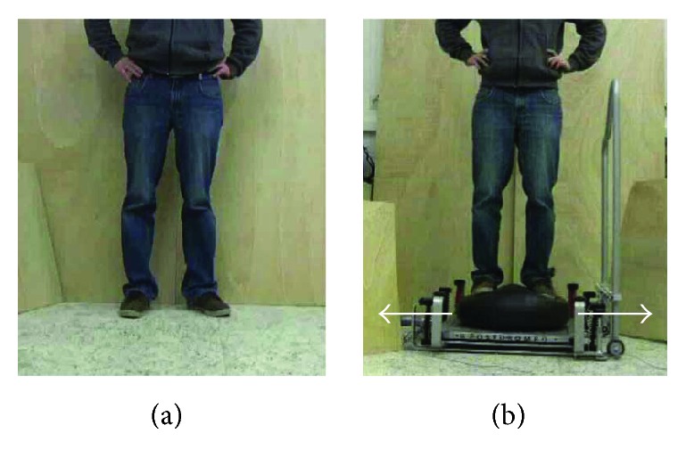
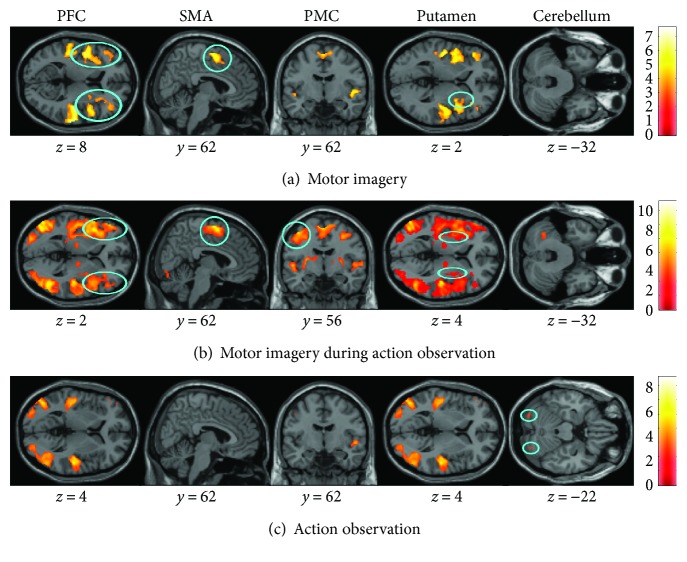
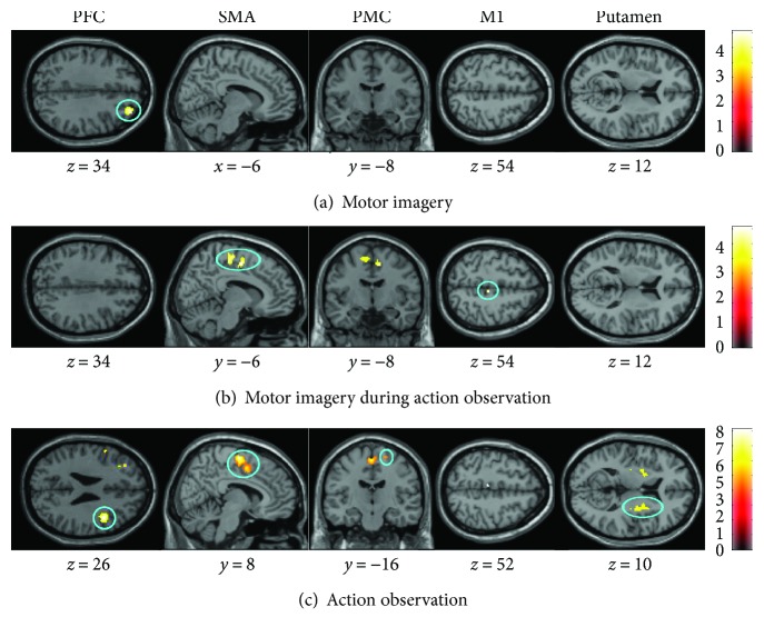
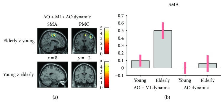
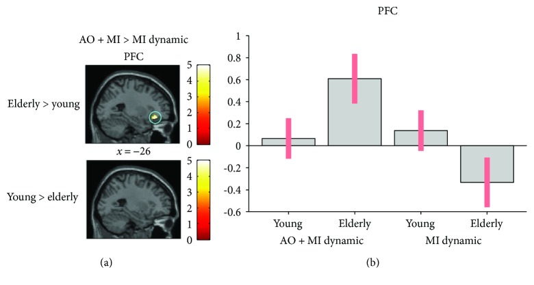
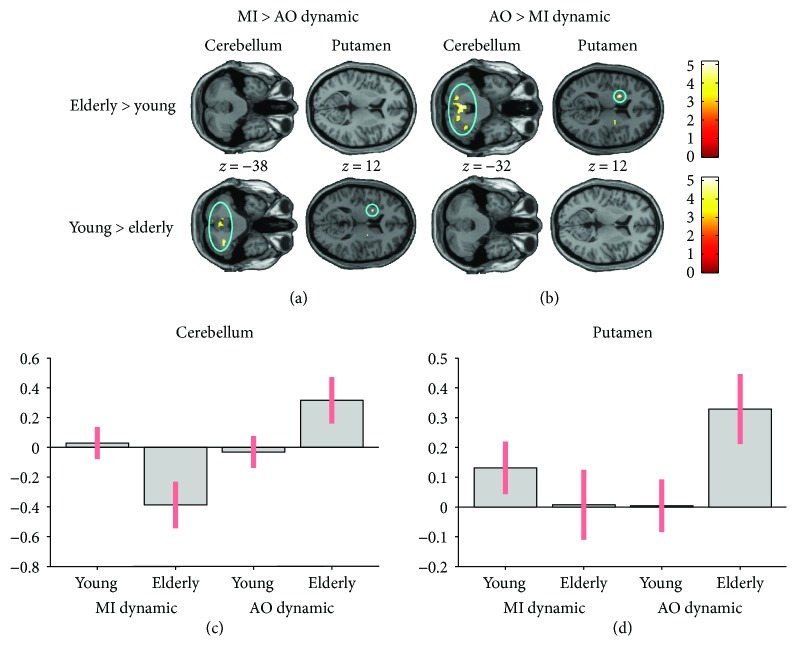
Age-related changes in brain activation other than in the primary motor cortex are not well known with respect to dynamic balance control. Therefore, the current study aimed to explore age-related differences in the control of static and dynamic postural tasks using fMRI during mental simulation of balance tasks. For this purpose, 16 elderly (72 ± 5 years) and 16 young adults (27 ± 5 years) were asked to mentally simulate a static and a dynamic balance task by motor imagery (MI), action observation (AO), or the combination of AO and MI (AO + MI). Age-related differences were detected in the form of larger brain activations in elderly compared to young participants, especially in the challenging dynamic task when applying AO + MI. Interestingly, when MI (no visual input) was contrasted to AO (visual input), elderly participants revealed deactivation of subcortical areas. The finding that the elderly demonstrated overactivation in mostly cortical areas in challenging postural conditions with visual input (AO + MI and AO) but deactivation in subcortical areas during MI (no vision) may indicate that elderly individuals allocate more cortical resources to the internal representation of dynamic postural tasks. Furthermore, it might be assumed that they depend more strongly on visual input to activate subcortical internal representations.


Figures

Figure 1
Figure 1
Balance tasks displayed during the experiment. (a) For the static balance task, a person was displayed standing on stable ground. (b) For the dynamic balance task, a person was shown compensating for a mediolateral perturbation while standing on a free-swinging platform (from [47]).
Figure 2
Figure 2
Common brain activity in young and elderly adults detected by a conjunction analysis. The three mental simulation conditions were contrasted with the baseline (mental simulation > baseline). The figure presents shared activities for the dynamic balance task during (a) motor imagery (MI), (b) motor imagery during action observation (AO + MI), and (c) action observation (AO). Colored circles underline significant common brain activity. Whole brain results are presented with p < 0.001 at the voxel level, extended by a p < 0.05 FWE corrected at the cluster level. Colored bars display the significance level of the contrast. Spatial coordinates (x, y, and z) are provided in MNI space.
Figure 3
Figure 3
Age-related differences in brain activity. The three mental simulation conditions were contrasted between age group by means of an ROI analysis. Presented are significantly different activities for the dynamic balance task during (a) motor imagery (MI), (b) motor imagery during action observation (AO + MI), and (c) action observation (AO). Colored circles highlight significantly greater brain activity in elderly adults. Activations are presented with p < 0.001 at the voxel level, extended by a p < 0.05 FWE corrected at the cluster level. Colored bars indicate the significance level of the contrasts. Spatial coordinates (x, y, and z) are provided in MNI space.
Figure 4
Figure 4
Age-related differences in brain activity when motor imagery during action observation (AO + MI) is contrasted with action observation (AO), revealed by an ROI analysis. (a) presents differences in activity in the SMA (p = 0.02) and PMC (p = 0.02) between groups. (b) shows the activation level of a representative voxel (8, −24, and 58) of the SMA. Colored circles highlight significantly stronger brain activity. Activations are presented with p < 0.001 at the voxel level, extended by a p < 0.05 FWE corrected at the cluster level. Colored bars indicate the significance level of the contrasts. Spatial coordinates (x, y, and z) are provided in MNI space.
Figure 5
Figure 5
Age-related differences in brain activity when motor imagery during action observation (AO + MI) is contrasted with motor imagery alone (MI), revealed by an ROI analysis. (a) shows higher brain activity for the dynamic task in the PFC (p = 0.05) in older adults when compared to young adults. (b) shows the activation level of a representative voxel (−28, 34, and −14) of the PFC depending on the group and the mental simulation condition (AO + MI or MI). Colored circles highlight significantly stronger brain activity in elderly adults. Activations are displayed with p < 0.001 at the voxel level, extended by a p < 0.05 FWE corrected at the cluster level. Colored bars indicate the significance level of the contrasts. Spatial coordinates (x, y, and z) are provided in MNI space.
Figure 6
Figure 6
Age-related differences in the interactions between brain activity during motor imagery (MI) and action observation (AO), revealed by an ROI analysis. (a) shows higher brain activity in the cerebellum (p = 0.02) and putamen (p = 0.05) in young adults compared to older adults, when a condition with no visual support (MI) is contrasted with one with visual support (AO) for the dynamic task. Inversely, (b) presented greater cerebral activity in the cerebellum (p = 0.02) and putamen (p = 0.008) in elderly adults compared to young adults, when a condition with visual support (AO) is contrasted with one with no visual support (MI). (c) and (d) represent the activation of the cerebellum and the putamen depending on the group and the mental simulation condition. The plots for the cerebellum and the putamen are based on voxels (−2, −60, and −34 and 30, −18, and 2, resp.). Colored circles highlight significantly stronger brain activity in young adults. Activations are displayed with p < 0.001 at the voxel level, extended by a p < 0.05 FWE corrected at the cluster level. Colored bars indicate the significance level of the contrasts. Spatial coordinates (x, y, and z) are provided in MNI space.

Similar articles

Cited by

References

    1. Maki B. E., McIlroy W. E. Postural control in the older adult. Clinics in Geriatric Medicine. 1996;12(4):635–658. - PubMed
    1. Muir S. W., Berg K., Chesworth B., Klar N., Speechley M. Quantifying the magnitude of risk for balance impairment on falls in community-dwelling older adults: a systematic review and meta-analysis. Journal of Clinical Epidemiology. 2010;63(4):389–406. doi: 10.1016/j.jclinepi.2009.06.010. - DOI - PubMed
    1. Papegaaij S., Taube W., Baudry S., Otten E., Hortobágyi T. Aging causes a reorganization of cortical and spinal control of posture. Frontiers in Aging Neuroscience. 2014;6:p. 28. doi: 10.3389/fnagi.2014.00028. - DOI - PMC - PubMed
    1. Luccarini P., Gahery Y., Pompeiano O. Injection of a cholinergic agonist in the dorsolateral pontine tegmentum of cats affects the posturokinetic responses to cortical stimulation. Neuroscience Letters. 1990;114(1):75–81. doi: 10.1016/0304-3940(90)90431-8. - DOI - PubMed
    1. Sherrington C. S. Flexion-reflex of the limb, crossed extension-reflex, and reflex stepping and standing. The Journal of Physiology. 1910;40(1-2):28–121. doi: 10.1113/jphysiol.1910.sp001362. - DOI - PMC - PubMed

Publication types

LinkOut - more resources